. 2008 Jun;28(12):3932-42.
doi: 10.1128/MCB.02191-07. Epub 2008 Apr 7.

MED1 phosphorylation promotes its association with mediator: implications for nuclear receptor signaling

Affiliations

MED1 phosphorylation promotes its association with mediator: implications for nuclear receptor signaling

Madesh Belakavadi et al. Mol Cell Biol. 2008 Jun.

Abstract

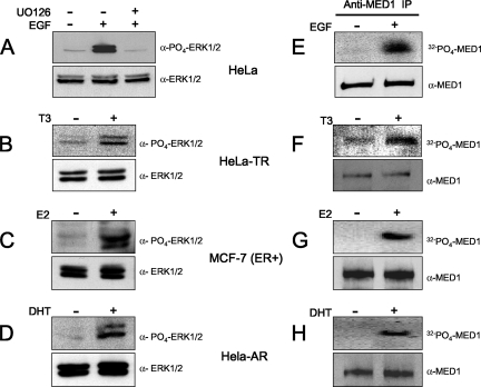
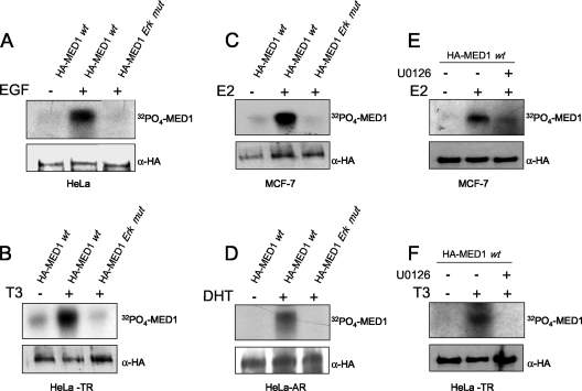
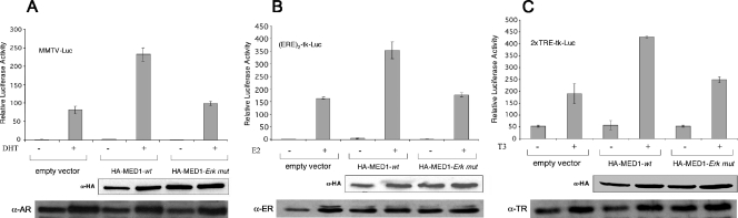
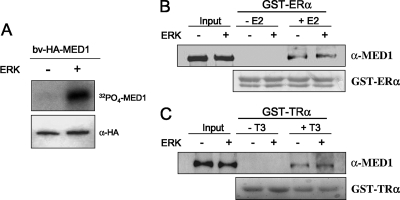
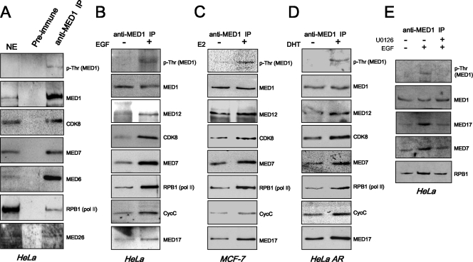
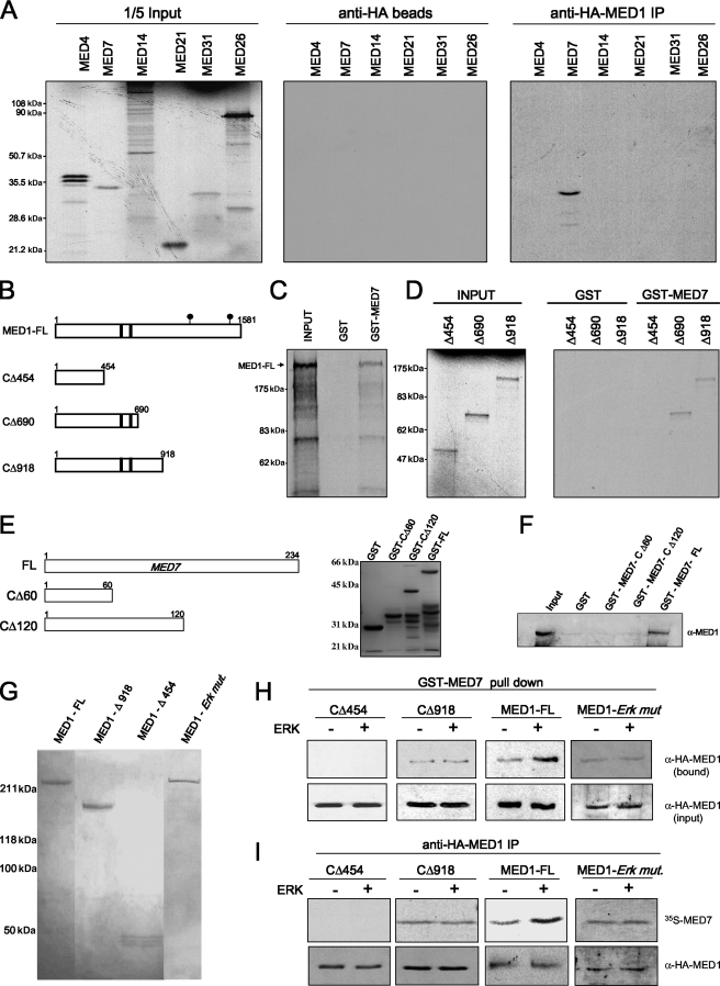
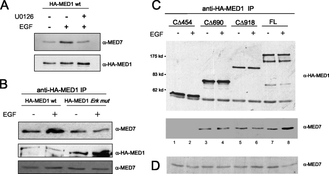
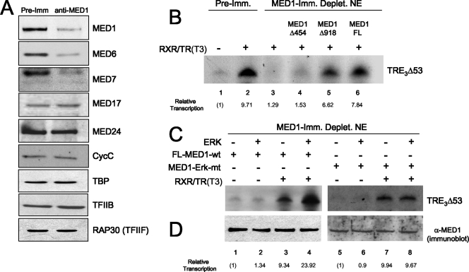
Mediator is a conserved multisubunit complex that acts as a functional interface between regulatory transcription factors and the general RNA polymerase II initiation apparatus. MED1 is a pivotal component of the complex that binds to nuclear receptors and a broad array of other gene-specific activators. Paradoxically, MED1 is found in only a fraction of the total cellular Mediator complexes, and the mechanisms regulating its binding to the core complex remain unclear. Here, we report that phosphorylation of MED1 by mitogen-activated protein kinase-extracellular signal-regulated kinase (MAPK-ERK) promotes its association with Mediator. We show that MED1 directly binds to the MED7 subunit and that ERK phosphorylation of MED1 enhances this interaction. Interestingly, we found that both thyroid and steroid hormones stimulate MED1 phosphorylation in vivo and that MED1 phosphorylation is required for its nuclear hormone receptor coactivator activity. Finally, we show that MED1 phosphorylation by ERK enhances thyroid hormone receptor-dependent transcription in vitro. Our findings suggest that ERK phosphorylation of MED1 is a regulatory mechanism that promotes MED1 association with Mediator and, as such, may facilitate a novel feed-forward action of nuclear hormones.


Figures

FIG. 1.
FIG. 1.
Steroid and thyroid hormones stimulate MED1 phosphorylation via activated MAPK-ERK signaling. (A to D) HeLa, HeLa-TR, MCF-7, and HeLa-AR cells were serum starved and then treated with EGF, U0126, T3, E2, or DHT (+) or untreated (−), as indicated (see Materials and Methods). The whole-cell lysate was then probed by immunoblotting for ERK1/2 expression (bottom) and ERK1/2 phosphorylation (top). (E to H) HeLa, HeLa-TR, MCF-7, and HeLa-AR cells were serum starved and then pulsed with 32Pi with or without hormones, as indicated. Parallel reactions lacking 32Pi were performed side by side. Native MED1 was immunoprecipitated with anti-MED1 antibodies and resolved by SDS-PAGE. Reactions containing 32Pi were detected by autoradiography (top), while reactions lacking 32Pi were detected by anti-MED1 immunoblotting (bottom).
FIG. 2.
FIG. 2.
Steroid and thyroid hormones stimulate phosphorylation of ectopic MED1. HeLa, HeLa-TR, MCF-7, and HeLa-AR cells were transfected with (+) pSG5-HA-MED1-wt (wild type) (HA-MED1 wt) or pSG5-HA-ERK mutant (HA-MED1 Erk mut), as indicated at the top of each panel. The cells were serum starved and then pulsed with 32Pi with or without specific hormones or U0126, as indicated (see Materials and Methods). Parallel reactions lacking 32Pi were performed side by side. HA-MED1 was immunoprecipitated with anti-HA antibodies and resolved by SDS-PAGE. Reactions containing 32Pi were detected by autoradiography (top), while reactions lacking 32Pi were detected by anti-HA immunoblotting (bottom).
FIG. 3.
FIG. 3.
MED1 phosphorylation is required for NR coactivator activity. (A and B) HeLa-AR or MCF-7 cells were transfected with MMTV-Luc or (ERE)2-tk-Luc, respectively, along with pSG5-HA-MED1-wt (HA-MED1-wt) or -ERK mutant (HA-MED1-Erk mut) or empty vector, as shown. (C) HeLa cells were transfected with pBK-CMV-FLAG-TRα and 2 × TRE-tk-Luc, along with pSG5-HA-MED1-wt or -ERK mutant or empty vector as shown. For panels A to C, the cells were subsequently cultured in charcoal/dextran-stripped FBS with or without DHT, E2, or T3, as indicated. Whole-cell lysate was assayed for luciferase activity and for MED1 and NR expression via immunoblotting (bottom). All luciferase assays were performed in triplicate and normalized against β-galactosidase (pSV-βgal) expression. The results are presented as the mean ± standard error.
FIG. 4.
FIG. 4.
MED1 phosphorylation is not required for NR binding. (A) ERK phosphorylation of MED1 in vitro. Purified bv-HA-MED1 was incubated with [γ-32P]ATP in the presence (+) or absence (−) of ERK1, as indicated. The reactions were resolved by SDS-PAGE and exposed for autoradiography. As a loading control, parallel reactions lacking [γ-32P]ATP were probed by immunoblotting with anti-HA antibodies. (B and C) bv-HA-MED1 was incubated with nonisotopic ATP in kinase buffer containing (+) or lacking (−) purified ERK1 (as in panel A) and then incubated with GST-ER or GST-TR with or without ligand, as indicated. Bound protein was detected by immunoblotting using anti-MED1 antibodies; 50% of the input was loaded in the first two lanes as a control. As a loading control for equivalent GST fusion protein, the nitrocellulose membranes were stained with Ponceau S, thus revealing the GST-ER and GST-TR polypeptides in each reaction (bottom).
FIG. 5.
FIG. 5.
MED1 phosphorylation promotes its association with the Mediator complex. (A) Isolation of native Mediator complexes containing phosphorylated MED1 and RNA Pol II. Nuclear extract prepared from HeLa cells cultured in FBS was incubated with anti-MED1 or preimmune antibodies. The bound complexes were then eluted and probed by immunoblotting with the antibodies indicated on the right of the panels. HeLa nuclear extract (NE) (10 μg) was loaded as a control. IP, immunoprecipitation. (B to E) HeLa, MCF-7, and HeLa-AR cells were serum starved and then stimulated with (+) or without (−) EGF, E2, DHT, or U0126, as indicated. Nuclear extracts were then prepared and incubated with anti-MED1 antibodies. The bound complexes were first normalized for equivalent MED1 concentrations via anti-MED1 immunoblotting. The normalized blots were then stripped and reprobed for RNA Pol II and other Mediator subunits, as indicated. For panels A to D, the immunoblot signal generated by the anti-phosphothreonine antibody precisely overlapped the band generated by the anti-MED1 antibody on the same blot.
FIG. 6.
FIG. 6.
MED1 interacts with MED7 in vitro. (A) [35S]methionine-labeled Mediator subunits were incubated with bv-FL-HA-MED1. HA pull-down was carried out, and the bound proteins were detected by autoradiography. IP, immunoprecipitation. (B) Schematic representation of FL-MED1 and MED1 C-terminal deletion mutants. The black bars indicate LXXLL motifs; the black circles indicate ERK phosphorylation sites. (C and D) GST or GST-MED7 was incubated with [35S]methionine-labeled FL-MED1 (MED1-FL) (C) or C-terminal deletion mutants (D). GST pull-down assays were carried out, and the bound proteins were detected by autoradiography. (E) Schematic representation and corresponding Coomassie blue-stained gel showing each GST-MED7 fusion protein. (F) GST-MED7 FL, GST-MED7-Δ1 or -Δ2, or GST alone was incubated with FL-bv-HA-MED1. GST pull-down assays were carried out, and the bound proteins were detected via anti-MED1 immunoblotting. (G) Purified FL-bv-HA-MED1 (MED1 - FL) and -HA-MED1-ERK mutant (MED1 - Erk mut.) and C-terminal MED1 deletion mutants stained with Coomassie blue (panel B shows a schematic representation). (H and I) FL or truncated bv-HA-MED1 proteins were preincubated in kinase buffer containing (+) or lacking (−) ERK1 and then incubated with GST-MED7 (H) or [35S]methionine-labeled MED7 (I). In panel H, GST pull-down assays were carried out, and bound MED1 protein was detected via anti-HA immunoblotting. In panel I, HA pull-down assays were carried out, and bound MED7 protein was blotted to a membrane and detected by autoradiography; the same blot was stripped and reprobed with anti-HA antibodies to determine precipitated HA-MED1 levels.
FIG. 7.
FIG. 7.
ERK phosphorylation enhances MED1 association with MED7 in vivo. (A and B) COS-7 cells were transfected with pCIN4-MED7 and either pSG5-HA-MED1-wt or pSG5-HA-MED1-ERK mutant (HA-MED1 Erk mut). The cells were serum starved and then treated with EGF or U0126 (+) or untreated (−), as indicated. Whole-cell lysate was then incubated with anti-HA antibodies, and the precipitated proteins were detected by anti-MED7 and anti-HA immunoblotting. Equal amounts of whole-cell extract, taken prior to the anti-HA immunoprecipitation (IP) in panel B, were probed by immunoblotting with anti-MED7 antibodies. (C) COS-7 cells were transfected with pCIN4-MED7, together with either pSG5-HA-MED1 (FL), -Δ454, -Δ690, or -Δ918. The cells were serum starved and then treated with EGF or untreated. Whole-cell lysate was incubated with anti-HA antibodies, and the precipitated proteins were detected by anti-MED7 and anti-HA immunoblotting. (D) Equal amounts of whole-cell extract, taken prior to the anti-HA immunoprecipitation in panel C, were probed by immunoblotting with anti-MED7 antibodies.
FIG. 8.
FIG. 8.
MED1 phosphorylation enhances TR-dependent transcription in vitro. (A) Immunodepletion of MED1 from HeLa cell nuclear extract. Shown is an immunoblot of HeLa nuclear extracts incubated with either anti-MED1 antibodies or preimmune serum (Pre-Imm). The specific antibodies used for immunoblotting are indicated on the right. (B) Purified recombinant MED1 restores TR-mediated transcription in MED1-depleted nuclear extract. Transcription was measured in vitro by incubating the TRE3Δ53 reporter gene in MED1-depleted (or preimmune mock-depleted) (Deplet.) HeLa cell nuclear extracts, together with purified baculovirus-expressed TRα, RXRα, and T3, as described previously (9). Purified bv-FL-MED1 (FL), -MED1-Δ454, or -MED1-Δ918 was added to reactions as indicated. (C) MED1 phosphorylation enhances TR-dependent transcription. In vitro transcription assays were performed as for panel B, except that either FL-MED1 wild type (FL-MED1-wt) or MED1-ERK mutant (MED1-Erk-mt) protein was preincubated in kinase buffer containing (+) or lacking (−) ERK1. The entire mock or true kinase reaction mixtures were then added to the transcription reactions. (D) As a control, parallel ERK (+ or −) kinase reactions were set up side by side and assayed via anti-MED1 immunoblotting.

Similar articles

Cited by

References

    1. Belakavadi, M., and J. D. Fondell. 2006. Role of the mediator complex in nuclear hormone receptor signaling. Rev. Physiol. Biochem. Pharmacol. 15623-43. - PubMed
    1. Chadick, J. Z., and F. J. Asturias. 2005. Structure of eukaryotic Mediator complexes. Trends Biochem. Sci. 30264-271. - PubMed
    1. Cheskis, B. J. 2004. Regulation of cell signalling cascades by steroid hormones. J. Cell Biochem. 9320-27. - PubMed
    1. Conaway, J. W., L. Florens, S. Sato, C. Tomomori-Sato, T. J. Parmely, T. Yao, S. K. Swanson, C. A. Banks, M. P. Washburn, and R. C. Conaway. 2005. The mammalian Mediator complex. FEBS Lett. 579904-908. - PubMed
    1. Crawford, S. E., C. Qi, P. Misra, V. Stellmach, M. S. Rao, J. D. Engel, Y. Zhu, and J. K. Reddy. 2002. Defects of the heart, eye, and megakaryocytes in peroxisome proliferator activator receptor-binding protein (PBP) null embryos implicate GATA family of transcription factors. J. Biol. Chem. 2773585-3592. - PubMed

Publication types

LinkOut - more resources