. 2008 Jun;28(12):4068-79.
doi: 10.1128/MCB.02114-07. Epub 2008 Apr 7.

The neogenin intracellular domain regulates gene transcription via nuclear translocation

Affiliations

The neogenin intracellular domain regulates gene transcription via nuclear translocation

David Goldschneider et al. Mol Cell Biol. 2008 Jun.

Abstract

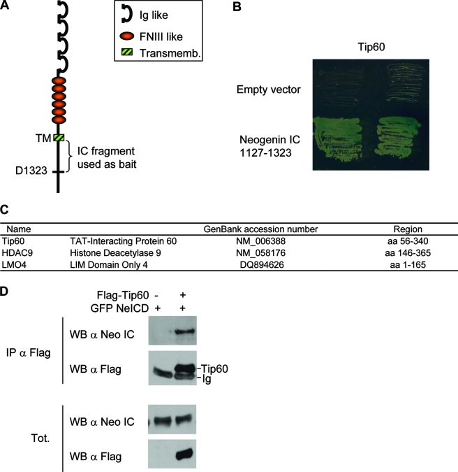
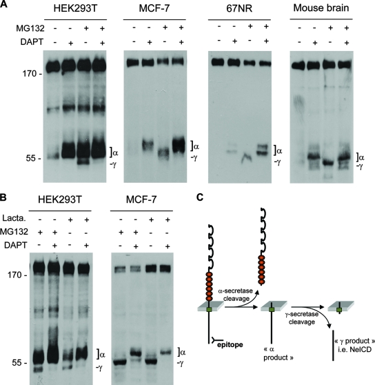
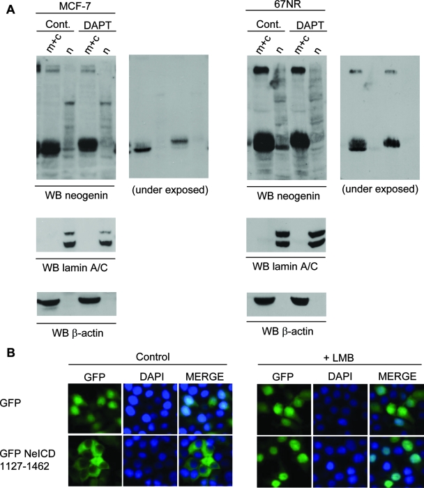
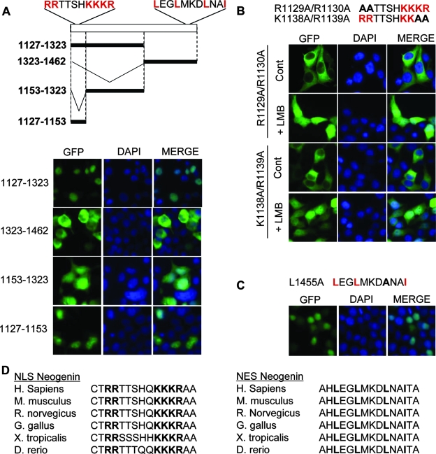
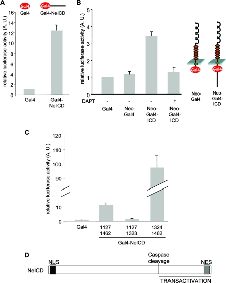
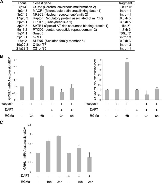
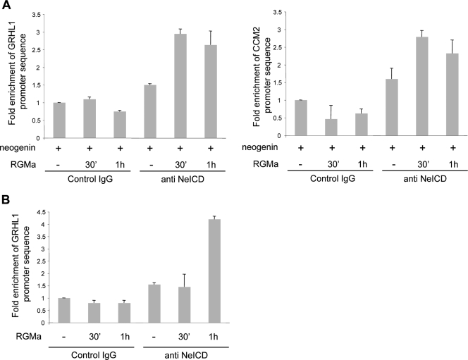
Neogenin is a multifunctional receptor implicated in axon navigation, neuronal differentiation, morphogenesis, and cell death. Very little is known about signaling downstream of neogenin. Because we found that the neogenin intracellular domain (NeICD) interacts with nuclear proteins implicated in transcription regulation, we investigated further whether neogenin signals similarly to the Notch receptor. We show here that neogenin is cleaved by gamma-secretase, an event that releases the complete NeICD. We also describe that NeICD is located at the nucleus, a feature regulated through a balance between nuclear import and export. NeICD triggers gene reporter transactivation and associates with nuclear chromatin. Direct transcriptional targets of NeICD were determined and were shown to be up-regulated in the presence of neogenin ligand. Together, we reveal here a novel aspect of neogenin signaling that relies on the direct implication of its intracellular domain in transcriptional regulation.


Figures

FIG. 1.
FIG. 1.
The NeICD interacts with nuclear proteins including TIP60. (A) Scheme of neogenin showing the structure of the protein consisting of Ig-like and fibronectin III-like domains for the extracellular part, a transmembrane domain (TM and Transmemb.), and an intracellular domain (IC). The fragment used as bait for the two-hybrid screen is also shown. (B) Neogenin aa 1127 to 1323 interact with TIP60 in yeast. AH109 yeast cells cotransformed with TIP60 fused to the transactivating domain of Gal4 together with the Gal4 DNA binding domain alone (empty vector) or fused to neogenin aa 1127 to 1323 were grown in selective medium, as described in Materials and Methods. (C) Some of the putative interactors of neogenin aa 1127 to 1323 revealed by the two-hybrid screen and known to display a nuclear activity. (D) Coimmunoprecipitation (IP) performed in HEK293T cells cotransfected with a neogenin-expressing vector together with a Flag-tagged TIP60-expressing construct. Anti-Flag M2 antibody was used for the pull down, which was revealed using a neogenin immunoblot (WB).
FIG. 2.
FIG. 2.
Neogenin is cleaved by γ-secretase. (A) Neogenin immunoblot from lysates of neogenin-transfected HEK293T cells, from lysate of MCF-7 cells or 67NR cells, or from brains from mouse embryos at embryonic day 13.5 and treated for 6 h with 2 μM DAPT and/or the proteasome inhibitor MG132 (1 μM). Positions of the neogenin fragments cleaved by γ-secretase (γ) or α-secretase/ADAM metalloprotease (α), i.e., as this fragment is inhibited by treatment with TAPI-1 (see Fig. S1 in the supplemental material), are indicated. (B) Treatments of neogenin-transfected HEK293T or MCF-7 cells with two proteasome inhibitors show the same neogenin profiles (lactacystin [Lacta.] was used at 10 μM for 6 h). (C) Hypothetical model for the secretases cleavage of neogenin according to our results and the literature on other secretase-cleaved receptors.
FIG. 3.
FIG. 3.
The NeICD displays a nuclear localization. (A) Cellular fractionation of MCF-7 or 67NR cells treated or not treated with 2 μM DAPT and/or the proteasome inhibitor MG132 (1 μM) for 6 h. Neogenin immunoblotting (WB) was performed on cellular fractions, the nucleus (n) and the membrane and cytosol (m+c); two exposition times are shown. Please note that a shorter exposition of the blot allows a better visualization of the migration difference between γ and α products in cytosolic fractions. A lamin A/C immunoblot was used as a control for nuclear fraction, while a β-actin immunoblot was used to control the cytosolic fraction. (B) HEK293T cells transfected with GFP-NeICD, further incubated with the inhibitor LMB (1 μM for 1 h), and monitored for fluorescence localization. DAPI staining was used to label the nucleus.
FIG. 4.
FIG. 4.
The NeICD displays an NLS and an NES to regulate its nuclear localization. HEK23T cells were transfected with the indicated constructs, further incubated or not with the inhibitor LMB (1 μM for 1 h), and monitored for fluorescence localization. DAPI staining was used to label the nucleus. (A, top) Scheme representing the different mutants of neogenin IC used in the following panels. (Bottom) Same as in Fig. 3B but with the indicated deletion mutants. (B) The neogenin NLS is in the N-terminal region of NeICD. Data are the same as described above (A) but with the indicated point mutants. (C) The NES is the C-terminal region of NeICD. Data are the same as described above (A), with the neogenin IC L1455A mutant. (D) Sequence alignments showing conservation of NESs and NLSs in the different species indicated. H. sapiens, Homo sapiens; M. musculus, Mus musculus; R. norvegicus, Rattus norvegicus; G. gallus, Gallus gallus; X. tropicalis, Xenopus tropicalis; D. rerio, Danio rerio.
FIG. 5.
FIG. 5.
The NeICD displays a gene-transactivating activity. (A) The NeICD promotes reporter gene transcription. The Gal4-expressing construct or Gal4 DNA binding domain fused to the NeICD as indicated (top) was cotransfected with a Gal4-lucerifase reporter plasmid into HEK293T cells, and luminescence signals were recorded. The relative luciferase activity index is shown as the ratio between the neogenin construct and the control Gal4 vector. Standard errors of the means are indicated. (B) γ-Secretase-dependent NeICD release from full-length neogenin promotes reporter gene transcription. The Gal4 DNA binding domain was inserted into full-length neogenin. The resulting construct, NeoGal4ICD, was cotransfected with a Gal4 luciferase reporter plasmid into HEK293T cells. The NeoGal4 construct lacking the intracellular domain was used as a control. DAPT treatment (2 μM) was performed for 24 h. (C) Same as described above (A), with the indicated deletion mutants of NeICD-Gal4. (D) Scheme representing different functional domains of the NeICD. A.U., arbitrary units.
FIG. 6.
FIG. 6.
The NeICD behaves as a transactivator of gene transcription. (A) Different genes in which the NeICD has been shown to interact within their proximities (12 of over 25 clones analyzed). ChIP was performed on the NeICD, and immunoprecipitated DNA was cloned and sequenced. (B) GRHL1 and CCM2 Q-RT-PCR performed on mock- versus neogenin-transfected HEK293T cells treated or not treated for 3 or 6 h with RGMa in the presence or absence of DAPT. CCM2 and GRHL1 mRNA levels are regulated by neogenin/RGM signaling in neogenin-transfected HEK293T cells. Normalization was done on β2-microglobulin. The CCM2 or GRHL1 level is presented for each condition as a ratio between the normalized neogenin sample to the mock-transfected sample. (C) Same as described above (B), but the GRHL1 mRNA level was determined in RGMa/DAPT-treated MCF7 cells. Normalization was also done with β2-microglobulin, and the GRHL1 level is presented as a ratio between GRHL1 and β2-microglobulin.
FIG. 7.
FIG. 7.
The NeICD binds target promoters after RGMa stimulation. (A) The NeICD binds GRHL1 and CCM2 promoters upon RGMa stimulation in neogenin-transfected HEK293T cells. Cross-linked chromatin was isolated from untreated and RGMa-treated neogenin-transfected HEK293T cells and precipitated with an NeICD-specific antibody or a control antibody. The precipitated chromatin was used as a template for quantitative PCR. (B) The endogenous NeICD binds the GRHL1 promoter upon RGMa stimulation. Data are the same as described above (A), but the experiment was performed with MCF7 cells untreated or treated with RGMa.

Similar articles

Cited by

References

    1. Bernet, A., L. Mazelin, M. M. Coissieux, N. Gadot, S. L. Ackerman, J. Y. Scoazec, and P. Mehlen. 2007. Inactivation of the UNC5C Netrin-1 receptor is associated with tumor progression in colorectal malignancies. Gastroenterology 1331840-1848. - PMC - PubMed
    1. Bray, S. J. 2006. Notch signalling: a simple pathway becomes complex. Nat. Rev. Mol. Cell Biol. 7678-689. - PubMed
    1. Bredesen, D. E., P. Mehlen, and S. Rabizadeh. 2005. Receptors that mediate cellular dependence. Cell Death Differ. 121031-1043. - PubMed
    1. Campbell, D. S., and C. E. Holt. 2003. Apoptotic pathway and MAPKs differentially regulate chemotropic responses of retinal growth cones. Neuron 37939-952. - PubMed
    1. Cao, X., and T. C. Südhof. 2001. A transcriptively [sic] active complex of APP with Fe65 and histone acetyltransferase Tip60. Science 293115-120. - PubMed

Publication types

LinkOut - more resources