. 2008 Jun;28(11):3804-16.
doi: 10.1128/MCB.00709-07. Epub 2008 Apr 7.

Cyclic AMP (cAMP)-mediated stimulation of adipocyte differentiation requires the synergistic action of Epac- and cAMP-dependent protein kinase-dependent processes

Affiliations

Cyclic AMP (cAMP)-mediated stimulation of adipocyte differentiation requires the synergistic action of Epac- and cAMP-dependent protein kinase-dependent processes

Rasmus Koefoed Petersen et al. Mol Cell Biol. 2008 Jun.

Abstract

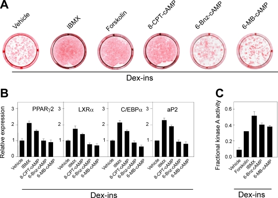
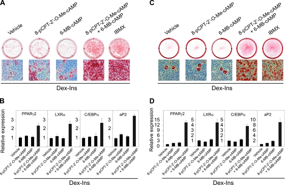
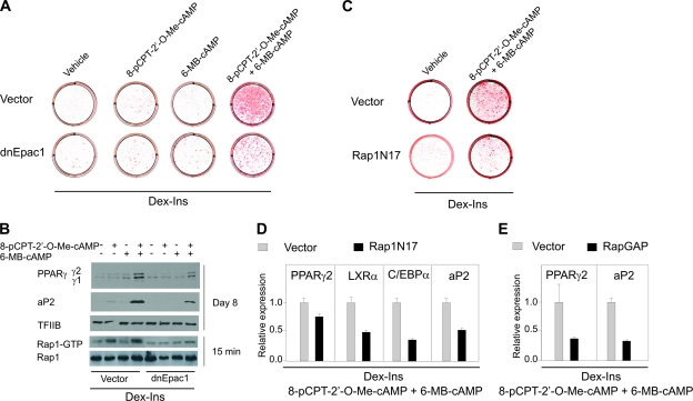
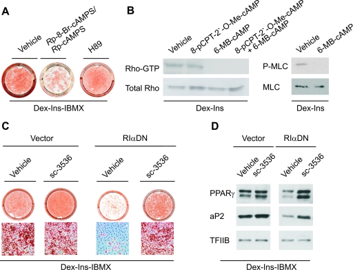
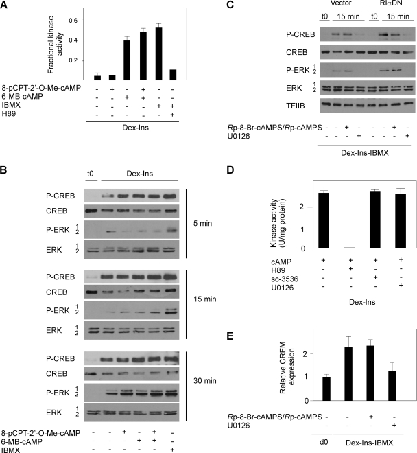
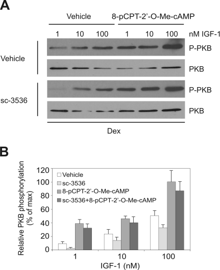
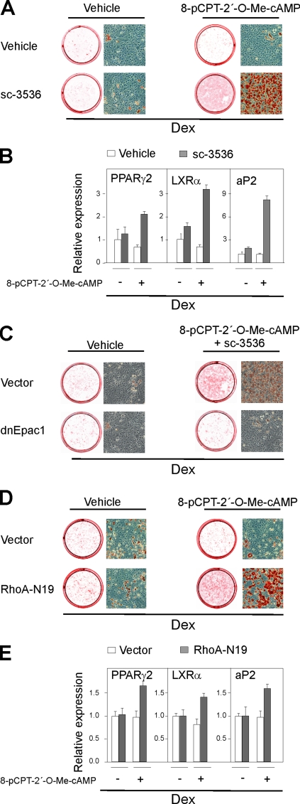
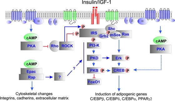
Cyclic AMP (cAMP)-dependent processes are pivotal during the early stages of adipocyte differentiation. We show that exchange protein directly activated by cAMP (Epac), which functions as a guanine nucleotide exchange factor for the Ras-like GTPases Rap1 and Rap2, was required for cAMP-dependent stimulation of adipocyte differentiation. Epac, working via Rap, acted synergistically with cAMP-dependent protein kinase (protein kinase A [PKA]) to promote adipogenesis. The major role of PKA was to down-regulate Rho and Rho-kinase activity, rather than to enhance CREB phosphorylation. Suppression of Rho-kinase impaired proadipogenic insulin/insulin-like growth factor 1 signaling, which was restored by activation of Epac. This interplay between PKA and Epac-mediated processes not only provides novel insight into the initiation and tuning of adipocyte differentiation, but also demonstrates a new mechanism of cAMP signaling whereby cAMP uses both PKA and Epac to achieve an appropriate cellular response.


Figures

FIG. 1.
FIG. 1.
Elevation of cAMP, but not selective activation of PKA, accelerates differentiation of 3T3-L1 preadipocytes. (A) Two-day-postconfluent 3T3-L1 cells were induced to differentiate by treatment with 1 μM Dex and 1 μg/ml Ins in the presence of 0.5 mM IBMX, 10 μM forskolin, 100 μM 8-CPT-cAMP, 100 μM 6-MB-cAMP, or 100 μM 6-Bnz-cAMP, as described in Materials and Methods. The cells were stained with oil red O and photographed on day 8. (B) RNA was isolated on day 8, and expression of PPARγ2, LXRα, C/EBPα, and aP2 was determined by RT-qPCR. The error bars represent standard deviations (n = 3). (C) Relative PKA activity in lysates of 3T3-L1 cells pretreated for 15 min in medium containing Dex plus Ins with vehicle, 0.5 mM IBMX, 50 μM forskolin, or 100 μM 6MB-cAMP. The error bars represent standard errors of the means (n = 3 to 5).
FIG. 2.
FIG. 2.
Expression of Epac in 3T3-L1 cells and mouse adipose tissues. (A) RNA was isolated on days 0, 1, 2, 3, 4, 6, and 10 from 3T3-L1 cells induced to differentiate by Dex, Ins, and IBMX and from mouse liver. (B) RNA was extracted from SVF or mature adipocytes (Ads) isolated from mouse eWAT, iBAT, and liver as described in Materials and Methods. The expression of Epac1 and Epac2 was determined using RT-qPCR and normalized to TBP expression. The error bars represent standard deviations (n = 3).
FIG. 3.
FIG. 3.
Activation of Epac and PKA synergistically induces differentiation of 3T3-L1 cells and MEFs into adipocytes. Two-day-postconfluent 3T3-L1 cells (A) or MEFs (C) were induced to differentiate by treatment with Dex and Ins in the presence of combinations of 200 μM 8-pCPT-2′-O-Me-cAMP and 100 μM 6-MB-cAMP or 0.5 mM IBMX as indicated. The cells were stained with oil red O on day 8. RNA was isolated on day 8, and expression of PPARγ2, LXRα, C/EBPα, and aP2 in 3T3-L1 cells (B) and MEFs (D) was determined by RT-qPCR. The error bars represent standard deviations (n = 3).
FIG. 4.
FIG. 4.
Activation of Epac is required for differentiation of 3T3-L1 cells into adipocytes. 3T3-L1 cells were retrovirally transduced with an empty vector, a vector expressing dnEpac1 (A and B), a vector expressing a dominant-negative form of Rap1A (Rap1N17) (C and D), or a vector expressing Rap GTPase activating protein (RapGAP) (E). The cells were grown to confluence, and at 2 days postconfluence they were induced to differentiate by Dex and Ins in the presence of combinations of 200 μM 8-pCPT-2′-O-Me-cAMP and 100 μM 6-MB-cAMP as indicated in the figure. (A and C) The cells were stained with oil red O and photographed on day 8. (B) GTP-bound Rap1 was measured by a Rap1 activation pull-down assay as described in Materials and Methods. Expression of aP2 and PPARγ was determined by Western blotting on day 8. TFIIB was used for control of equal protein loading on the gel. One representative experiment out of three independent experiments is shown. (D and E) RNA was isolated on day 8, and expression of LXRα, C/EBPα (D), PPARγ2, and aP2 (D and E) was determined by RT-qPCR. The error bars represent standard deviations (n = 3).
FIG. 5.
FIG. 5.
Activation of PKA is required for differentiation of 3T3-L1 cells, but dispensable when Rho-kinase is inhibited. (A) 3T3-L1 cells were grown to 2 days postconfluence and induced to differentiate by a standard differentiation cocktail consisting of Dex, Ins, and IBMX. Additionally, 100 μM Rp-8-Br-cAMPS/Rp-cAMPS or 10 μM H89 was present from day 0 to day 2. On day 8, the cells were stained with oil red O and photographed. (B) GTP-bound Rho was measured by a Rho activation pull-down assay after 15 min of treatment with 200 μM 8-pCPT-2′-O-Me-cAMP and 100 μM 6-MB-cAMP alone or in combination. Phosphorylated MLC and MLC were determined by Western blotting after 15 min of treatment with 100 μM 6-MB-cAMP or vehicle. One representative experiment out of three independent experiments is shown. (C) 3T3-L1 cells were retrovirally transduced with an empty vector or a vector expressing a dominant-negative form of the RIα PKA subunit (RIαDN). At 2 days postconfluence, the cells were induced to differentiate by Dex, IBMX, and Ins in the absence and presence of 10 μM sc-3536. On day 8, the cells were stained with oil red O and photographed or whole-cell extracts were prepared and the expression of PPARγ and aP2 was determined by Western blotting using TFIIB as a control for equal protein loading. One representative experiment out of three independent experiments is shown (D).
FIG. 6.
FIG. 6.
Induction of CREB phosphorylation during initiation of adipocyte differentiation is dependent on ERK1/2 activity. (A) 3T3-L1 cells were treated for 15 min in Dex-Ins medium with various combinations of 200 μM 8-pCPT-2′-O-Me-cAMP, 200 μM 6MB-cAMP, 0.5 mM IBMX, and 10 μM H89 as indicated. Subsequently the PKA activity in lysates of the cells was determined. The error bars represent standard errors of the means (n = 3). (B) 3T3-L1 preadipocytes at 2 days postconfluence were treated with Dex and Ins with combinations of 200 μM 8-pCPT-2′-O-Me-cAMP, 200 μM 6MB-cAMP, and 0.5 mM IBMX. Whole-cell extracts were prepared after 5, 15, and 30 min and analyzed for phosphorylation of CREB and ERK1/2 by Western blotting. One representative experiment out of three independent experiments is shown. (C) 3T3-L1 preadipocytes at 2 days postconfluence were treated with Dex, Ins, and IBMX with or without 100 μM Rp-8-Br-cAMPS/Rp-cAMPS or 10 μM U0126 as indicated. Whole-cell extracts were prepared after 15 min and analyzed for phosphorylation of CREB and ERK1/2 by Western blotting. One representative experiment out of three independent experiments is shown. (D) Effects of protein kinase inhibitors on PKA activity in 3T3-L1 lysates. The lysates were incubated with 10 μM H89, 10 μM sc-3536, or 10 μM U0126 in the presence of a maximally PKA-stimulating concentration of cAMP (1 μM). The error bars represent standard errors of the means (n = 3). (E) Two-day-postconfluent 3T3-L1 cells were induced to differentiate with Dex, Ins, and IBMX with or without 100 μM Rp-8-Br-cAMPS/Rp-cAMPS or 10 μM U0126. RNA was isolated after 6 h, and expression of CREB was determined by RT-qPCR. d0, day 0. The error bars represent standard deviations (n = 3).
FIG. 7.
FIG. 7.
Activation of Epac enhances Ins/IGF-1 signaling in 3T3-L1 cells (A) 3T3-L1 cells were grown to 2 days postconfluence and then treated with Dex and increasing concentrations of IGF-1 in the absence or presence of 10 μM sc-3536 and 200 μM 8-pCPT-2′-O-Me-cAMP, as indicated in the figure. After 15 min, whole-cell extracts were prepared and the levels of phosphorylated PKB and total PKB were determined by Western blotting. Shown is one representative experiment out of three independent experiments. (B) Quantification of the relative levels of PKB phosphorylation. Autoradiographs were analyzed by densitometric scanning, and the levels of phosphorylated PKB relative to total PKB were determined. The error bars represent standard deviations (n = 3).
FIG. 8.
FIG. 8.
Activation of Epac is sufficient to induce differentiation of 3T3-L1 cells when Rho-kinase is inhibited. 3T3-L1 cells were grown to 2 days postconfluence and induced to differentiate by Dex in the absence or presence of 10 μM sc-3536. Additionally, 200 μM 8-pCPT-2′-O-Me-cAMP was present from day 0 to day 2 as indicated. On day 8, the cells were stained with oil red O and photographed (A), total RNA was isolated, and the expression of PPARγ, LXRα, and aP2 was determined by RT-qPCR. The error bars represent standard deviations (n = 3) (B). 3T3-L1 cells were retrovirally transduced with an empty vector or a vector expressing a dominant negative form of Epac1 (dnEpac1). At 2-days post confluence they were induced to differentiate by Dex in the absence or presence of 10 μM sc-3536. Additionally, 200 μM 8-pCPT-2′-O-Me-cAMP was present from day 0 to day 2 as indicated. On day 8, the cells were stained with oil red O and photographed (C). 3T3-L1 cells were retrovirally transduced with an empty vector or a vector expressing a dominant-negative form of RhoA (RhoA-N19). At 2 days postconfluence, they were induced to differentiate by Dex in the absence or presence of 200 μM 8-pCPT-2′-O-Me-cAMP. On day 8, the cells were stained with oil red O and photographed. (D) Total RNA was isolated, and the expression of PPARγ, LXRα, and aP2 was determined by RT-qPCR. The error bars represent standard deviations (n = 3) (E).
FIG. 9.
FIG. 9.
Model for the role of cAMP-stimulated adipogenesis via PKA- end Epac1-dependent processes. Increased levels of cAMP activate both PKA- and Epac-dependent pathways. Activation of PKA leads to repression of Rho-kinase activity by targeting either the Rho-kinase or the upstream regulator Rho. High levels of Rho kinase activity inhibit Ins/IGF-1-dependent signaling, and attenuation of Rho-kinase activity is crucial for adipogenesis. However, low levels of Rho-kinase activity also enhance Ins/IGF-1-dependent signaling, and PKA-mediated inhibition of the Rho-kinase impairs Ins/IGF-1-dependent signaling. This is counteracted by the simultaneous activation of an Epac1/Rap1-dependent pathway, which also induces important changes in cytoskeletal organization, adhesion, and extracellular matrix. Activation of CREB is not dependent on PKA activity, but rather requires ERK activity during the initial stages of adipogenesis.

Similar articles

Cited by

References

    1. Baisamy, L., N. Jurisch, and D. Diviani. 2005. Leucine zipper-mediated homo-oligomerization regulates the Rho-GEF activity of AKAP-Lbc. J. Biol. Chem. 28015405-15412. - PubMed
    1. Begum, N., O. A. Sandu, and N. Duddy. 2002. Negative regulation of rho signaling by insulin and its impact on actin on cytoskeleton organization in vascular smooth muscle cells: role of nitric oxide and cyclic guanosine monophosphate signaling pathways. Diabetes 512256-2263. - PubMed
    1. Begum, N., O. A. Sandu, M. Ito, S. M. Lohmann, and A. Smolenski. 2002. Active Rho kinase (ROK-alpha) associates with insulin receptor substrate-1 and inhibits insulin signaling in vascular smooth muscle cells. J. Biol. Chem. 2776214-6222. - PubMed
    1. Belmonte, N., B. W. Phillips, F. Massiera, P. Villageois, B. Wdziekonski, P. Saint-Marc, J. Nichols, J. Aubert, K. Saeki, A. Yuo, S. Narumiya, G. Ailhaud, and C. Dani. 2001. Activation of extracellular signal-regulated kinases and CREB/ATF-1 mediate the expression of CCAAT/enhancer binding proteins β and -δ in preadipocytes. Mol. Endocrinol. 152037-2049. - PubMed
    1. Bennett, C. N., S. E. Ross, K. A. Longo, L. Bajnok, N. Hemati, K. W. Johnson, S. D. Harrison, and O. A. MacDougald. 2002. Regulation of Wnt signaling during adipogenesis. J. Biol. Chem. 27730998-31004. - PubMed

Publication types

MeSH terms