

Multivalent presentation of antihantavirus peptides on nanoparticles enhances infection blockade
- PMID: 18391034
- PMCID: PMC2415754
- DOI: 10.1128/AAC.01415-07
Multivalent presentation of antihantavirus peptides on nanoparticles enhances infection blockade
Abstract
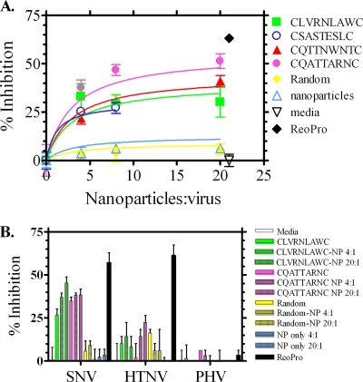
Viral entry into susceptible host cells typically results from multivalent interactions between viral surface proteins and host entry receptors. In the case of Sin Nombre virus (SNV), a New World hantavirus that causes hantavirus cardiopulmonary syndrome, infection involves the interaction between viral membrane surface glycoproteins and the human integrin alpha(v)beta(3). Currently, there are no therapeutic agents available which specifically target SNV. To address this problem, we used phage display selection of cyclic nonapeptides to identify peptides that bound SNV and specifically prevented SNV infection in vitro. We synthesized cyclic nonapeptides based on peptide sequences of phage demonstrating the strongest inhibition of infection, and in all cases, the isolated peptides were less effective at blocking infection (9.0% to 27.6% inhibition) than were the same peptides presented by phage (74.0% to 82.6% inhibition). Since peptides presented by the phage were pentavalent, we determined whether the identified peptides would show greater inhibition if presented in a multivalent format. We used carboxyl linkages to conjugate selected cyclic peptides to multivalent nanoparticles and tested infection inhibition. Two of the peptides, CLVRNLAWC and CQATTARNC, showed inhibition that was improved over that of the free format when presented on nanoparticles at a 4:1 nanoparticle-to-virus ratio (9.0% to 32.5% and 27.6% to 37.6%, respectively), with CQATTARNC inhibition surpassing 50% when nanoparticles were used at a 20:1 ratio versus virus. These data illustrate that multivalent inhibitors may disrupt polyvalent protein-protein interactions, such as those utilized for viral infection of host cells, and may represent a useful therapeutic approach.
Figures
Similar articles
-
Peptide antagonists that inhibit Sin Nombre virus and hantaan virus entry through the beta3-integrin receptor.J Virol. 2005 Jun;79(12):7319-26. doi: 10.1128/JVI.79.12.7319-7326.2005. J Virol. 2005. PMID: 15919886 Free PMC article.
-
Phage display selection of cyclic peptides that inhibit Andes virus infection.J Virol. 2009 Sep;83(17):8965-9. doi: 10.1128/JVI.00606-09. Epub 2009 Jun 10. J Virol. 2009. PMID: 19515773 Free PMC article.
-
Recognition of decay accelerating factor and alpha(v)beta(3) by inactivated hantaviruses: Toward the development of high-throughput screening flow cytometry assays.Anal Biochem. 2010 Jul 15;402(2):151-60. doi: 10.1016/j.ab.2010.03.016. Epub 2010 Apr 2. Anal Biochem. 2010. PMID: 20363206 Free PMC article.
-
Glycomimetic Peptides as Therapeutic Tools.Pharmaceutics. 2023 Feb 17;15(2):688. doi: 10.3390/pharmaceutics15020688. Pharmaceutics. 2023. PMID: 36840010 Free PMC article. Review.
-
Phage capsid nanoparticles as multivalent inhibitors of viral infections.Sci Bull (Beijing). 2020 Dec 30;65(24):2050-2052. doi: 10.1016/j.scib.2020.09.019. Epub 2020 Sep 15. Sci Bull (Beijing). 2020. PMID: 32953198 Free PMC article. Review. No abstract available.
Cited by
-
Leveraging the therapeutic, biological, and self-assembling potential of peptides for the treatment of viral infections.J Control Release. 2022 Aug;348:1028-1049. doi: 10.1016/j.jconrel.2022.06.037. Epub 2022 Jul 6. J Control Release. 2022. PMID: 35752254 Free PMC article. Review.
-
Ultracentrifugation Purification of Cache Valley Virus Using Iodixanol.Methods Mol Biol. 2025;2893:51-56. doi: 10.1007/978-1-0716-4338-9_5. Methods Mol Biol. 2025. PMID: 39671029
-
Effects of macromolecular crowding on the inhibition of virus assembly and virus-cell receptor recognition.Biophys J. 2011 Feb 2;100(3):738-746. doi: 10.1016/j.bpj.2010.12.3714. Biophys J. 2011. PMID: 21281589 Free PMC article.
-
Development of anti-infectives using phage display: biological agents against bacteria, viruses, and parasites.Antimicrob Agents Chemother. 2012 Sep;56(9):4569-82. doi: 10.1128/AAC.00567-12. Epub 2012 Jun 4. Antimicrob Agents Chemother. 2012. PMID: 22664969 Free PMC article. Review.
-
Vaccines and Therapeutics Against Hantaviruses.Front Microbiol. 2020 Jan 30;10:2989. doi: 10.3389/fmicb.2019.02989. eCollection 2019. Front Microbiol. 2020. PMID: 32082263 Free PMC article. Review.
References
-
- Ahmad, Z., S. Sharma, and G. K. Khuller. 2007. Chemotherapeutic evaluation of alginate nanoparticle-encapsulated azole antifungal and antitubercular drugs against murine tuberculosis. Nanomedicine 3:239-243. - PubMed
-
- Baudouin, V., A. Crusiaux, E. Haddad, L. Schandene, M. Goldman, C. Loirat, and D. Abramowicz. 2003. Anaphylactic shock caused by immunoglobulin E sensitization after retreatment with the chimeric anti-interleukin-2 receptor monoclonal antibody basiliximab. Transplantation 76:459-463. - PubMed
-
- Bharadwaj, M., C. R. Lyons, I. A. Wortman, and B. Hjelle. 1999. Intramuscular inoculation of Sin Nombre hantavirus cDNAs induces cellular and humoral immune responses in BALB/c mice. Vaccine 17:2836-2843. - PubMed
Publication types
MeSH terms
Substances
Grants and funding
LinkOut - more resources
Full Text Sources
Other Literature Sources
