. 2008 Apr 14;205(4):967-79.
doi: 10.1084/jem.20072051. Epub 2008 Apr 7.

Arthritis induced by posttranslationally modified (citrullinated) fibrinogen in DR4-IE transgenic mice

Affiliations

Arthritis induced by posttranslationally modified (citrullinated) fibrinogen in DR4-IE transgenic mice

Jonathan A Hill et al. J Exp Med. .

Abstract

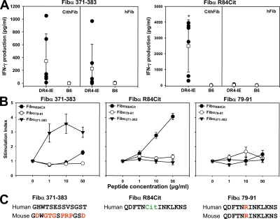
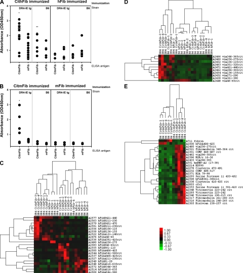
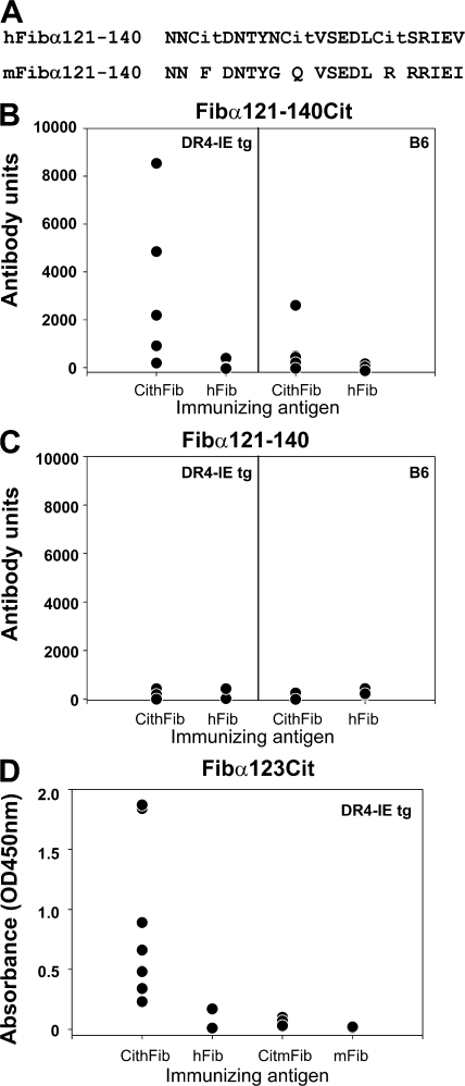
Rheumatoid arthritis (RA) is a common autoimmune disease that afflicts the synovium of diarthrodial joints. The pathogenic mechanisms inciting this disease are not fully characterized, but may involve the loss of tolerance to posttranslationally modified (citrullinated) antigens. We have demonstrated that this modification leads to a selective increase in antigenic peptide affinity for major histocompatibility complex (MHC) class II molecules that carry the RA-associated shared epitope, such as HLA-DRB1*0401 (DR4). We describe the induction of arthritis in DR4-IE transgenic (tg) mice with citrullinated fibrinogen, a protein commonly found in inflamed synovial tissue and a frequent target of autoantibodies in RA patients. The disease induced in these mice was characterized by synovial hyperplasia followed by ankylosis, but lacked a conspicuous polymorphonuclear cell infiltrate. Immunological analysis of these mice through T cell epitope scanning and antibody microarray analysis identified a unique profile of citrulline-specific reactivity that was not found in DR4-IE tg mice immunized with unmodified fibrinogen or in wild-type C57BL/6 mice immunized with citrullinated fibrinogen, two conditions where arthritis was not observed. These observations directly implicate citrullinated fibrinogen as arthritogenic in the context of RA-associated MHC class II molecules.


Figures

Figure 1.
Figure 1.
Clinical and histological evaluation of arthritis induced by CithFib in DR4-IE tg mice. (A) Normal ankle joint of a DR4-IE tg mouse immunized with hFib (day 70). (B) Maximal swelling in an arthritic DR4-IE tg mouse immunized with CithFib (day 70). (C) Progression of arthritis showing reduced erythema in an arthritic DR4-IE tg mouse immunized with CithFib (day 150). Corresponding HE-stained histological sections of tibiotalar joints from nonarthritic and arthritic DR4-IE tg mice (D–F from mice shown in A–C, respectively). (D) Normal histological appearance of synovial membrane and cartilage surfaces. (E) Hyperplastic synovium showing some areas of cartilage damage, but lacking conspicuous inflammatory cell infiltration. (F) Ankylosed joint showing fibrotic changes in the tissues spanning the talus and navicular bones. (G) High-powered magnification of normal synovial lining from a nonarthritic DR4-IE tg mouse immunized with hFib (day 70). (H) Synovial hyperplasia of the lining layer with subtle lymphocyte infiltration (arrows). This is representative of the earliest histological abnormality seen in arthritic DR4-IE tg mice immunized with CithFib. (I) Pannus tissue in an arthritic DR4-IE tg mouse highlighting areas of cartilage degradation. Bar, 100 μm.
Figure 2.
Figure 2.
Joint swelling and histological analysis of inflammatory infiltrate in arthritic DR4-IE tg mice. (A) Caliper measurements of DR4-IE tg mice immunized with CithFib were assessed every 7 d over 13 wk. Data are presented as mean Δ ankle width (± the SEM), which was calculated by caliper measurements at the indicated time point, minus the preimmunization ankle width, from 12 individual arthritic mice (filled circles) and 12 nonarthritic mice (open circles). P values were calculated by Student's t test. (B) High-powered view (hpv) histological analysis of inflammatory infiltrate in the joints of arthritic DR4-IE tg mice (day 70 postimmunization) and K/BxN mice (8 wk old). Lymphocyte and polymorphonuclear cell content was determined by nuclear morphology in HE-stained sections from hyperplastic synovial tissue. Points represent the number of cells identified in each high power view (63x objective), with 54 individual views from 5 arthritic DR4-IE tg mice and 15 views from 2 arthritic K/BxN mice. P values were obtained by Mann-Whitney test.
Figure 3.
Figure 3.
Immunohistochemical localization of citrullinated proteins and fibrinogen deposition in arthritic DR4-IE tg mice. Citrullinated protein was identified in arthritic mice 70 d after immunization with CithFib by staining with AMC antibodies in areas of synovial hyperplasia (A), with the most intense staining identifying intracellular proteins in synovial fibroblast cells (B). (C) Nonarthritic mice immunized with hFib did not stain positive for AMC in these regions. Although fibrin deposition was evident in perivascular regions of synovial tissue in arthritic mice (D), colocalization with citrullinated protein was minimal and virtually absent in areas of intense AMC staining (E). (F) No fibrin deposition was detected in joints of nonarthritic mice immunized with hFib. Control staining without the primary antibody for each section is shown in the insets. Bar, 100 μm.
Figure 4.
Figure 4.
Antigen-specific recall proliferation and cytokine production in DR4-IE tg and B6 mice immunized with CithFib or hFib (day 70). (A)Splenocytes were cultured in the presence of 50 μg/ml of the indicated in vitro antigens and proliferation (shown as stimulation index) was determined by [3H]thymidine incorporation. (B–E) Supernatant from these cultures were removed at 72 h and tested for the presence of IFN-γ by ELISA. Individual (filled circles) and mean (open boxes) responses ± the SEM (proliferation) or the SD (IFN-γ) are shown (n = 6 mice/strain/immunizing antigen). * indicates significant difference in cytokine production between DR4-IE tg and B6 mice. P < 0.05, paired Student's t test.
Figure 5.
Figure 5.
T cell responses to fibrinogen derived peptides in DR4-IE tg and B6 mice. (A) Splenocytes from DR4-IE tg and B6 mice immunized with CithFib or hFib (day 70) were cultured in the presence of 50 μg/ml of either Fibα 371–383 (left) or Fibα R84Cit (right). Supernatants from these cultures were removed at 72 h and tested for the production of IFN-γ by ELISA. Individual (filled circles) and mean (open boxes) responses ± the SD are shown (n = 6 mice/strain/immunizing antigen). Labels above boxes indicate the antigen used for in vitro challenge, and labels within boxes indicate the immunizing antigen. * indicates significant difference in cytokine production between DR4-IE tg immunized with CithFib and hFib (P < 0.05, paired Student's t test). (B) Confirmation of peptide immunogenicity by antigen-specific T cell proliferation. DR4-IE tg mice were immunized with either Fibα 371–383 (left), Fibα R84Cit (middle), or Fibα 79–91 (right); 10 d later, draining lymph node cells were cultured with various concentrations of peptide (Fibα R84Cit, closed circle; Fibα 79–91, open circle; Fibα 371–383, inverted closed triangle). Data represent the mean response ± the SEM of six mice per immunizing antigen. (C) Peptides corresponding to human α chain sequences used in A and B for determining T cell recall responses. Amino acids differing between mouse Fibα 371–383 are indicated in red. P4 amino acid from Fibα R84Cit and Fibα 79–81 predicted to be positioned at the SE are indicated in green and red, respectively, with the corresponding sequence from the α chain of mFib.
Figure 6.
Figure 6.
IgG antibody responses detected by ELISA and synovial proteome microarray. (A) IgG reactivity to either CitmFib or mFib in DR4-IE tg or B6 mice 70 d after immunization with either CithFib or hFib. Mean OD of duplicates for individual serum are shown (n = 12 for DR4-IE tg mice immunized with CithFib and n = 6 for all other groups). (B) Total IgG reactivity to either CitmFib or mFib in DR4-IE tg or B6 mice 100 d after immunization with either CitmFib or mFib (n = 7 for DR4-IE tg mice immunized with CitmFib and n = 5 for all other groups). * indicates significant difference in antibody reactivity (P < 0.05, paired Student's t test) between all groups, whereas ** indicates significant difference in antibody reactivity to CitmFib in B6 mice immunized with CithFib versus reactivity to CitmFib in B6 or DR4-IE tg mice immunized with hFib (P < 0.05, paired Student's t test). Analysis of serum IgG reactivity to fibrinogen- (C) or vimentin-derived (D) peptides identified by SAM and grouped by Cluster based on relationships. (E) Pairwise analysis of significantly different IgG antibody reactivity between DR4-IE tg and B6 mice immunized with CithFib (P < 0.05, Student's t test). Antigens shown were stratified based on having greater than fourfold increase in reactivity compared with immunization with hFib. Values shown are median subtracted and row normalized with relative expression shown in relation to scale bar. Raw data and peptide sequences can be found in Table S1–S3, available at http://www.jem.org/cgi/content/full/jem.20072051/DC1.
Figure 7.
Figure 7.
IgG antibody responses to heterologous regions of CithFib and CitmFib. (A) Amino acid substitutions between human and mFib are found in the α chain peptide 121–140 at arginine residues. Previous mass spectrometry analysis on in vitro CithFib identified αR123 and αR129 as sites of citrullination (25). (B) Increased antibody reactivity is evident in DR4-IE tg mice immunized with CithFib compared with B6 mice or either strain after immunization with hFib. Values represent normalized arbitrary antibody units from microarray analysis with n = 5 mice/strain/immunization. (C) Antibody reactivity to Fibα121-140Cit is citrulline specific because no reactivity is found to Fibα121-140. Values represent normalized arbitrary antibody units from microarray analysis with n = 5 mice/strain/immunization. (D) Antibody reactivity to Fibα123Cit in DR4-IE tg mice immunized with CithFib (n = 9), hFib (n = 5), CitmFib (n = 4), and mFib (n = 5) detected by ELISA. Mean OD of duplicates for individual serum are shown.

References

    1. Blass, S., J.M. Engel, and G.R. Burmester. 1999. The immunologic homunculus in rheumatoid arthritis. Arthritis Rheum. 42:2499–2506. - PubMed
    1. Gregersen, P.K., J. Silver, and R.J. Winchester. 1987. The shared epitope hypothesis. An approach to understanding the molecular genetics of susceptibility to rheumatoid arthritis. Arthritis Rheum. 30:1205–1213. - PubMed
    1. Hammer, J., F. Gallazzi, E. Bono, R.W. Karr, J. Guenot, P. Valsasnini, Z.A. Nagy, and F. Sinigaglia. 1995. Peptide binding specificity of HLA-DR4 molecules: correlation with rheumatoid arthritis association. J. Exp. Med. 181:1847–1855. - PMC - PubMed
    1. Schellekens, G.A., B. A.de Jong, F.H. van den Hoogen, L.B. van de Putte, and W.J. van Venrooij. 1998. Citrulline is an essential constituent of antigenic determinants recognized by rheumatoid arthritis-specific autoantibodies. J. Clin. Invest. 101:273–281. - PMC - PubMed
    1. Girbal-Neuhauser, E., J.J. Durieux, M. Arnaud, P. Dalbon, M. Sebbag, C. Vincent, M. Simon, T. Senshu, C. Masson-Bessiere, C. Jolivet-Reynaud, et al. 1999. The epitopes targeted by the rheumatoid arthritis-associated antifilaggrin autoantibodies are posttranslationally generated on various sites of (pro)filaggrin by deimination of arginine residues. J. Immunol. 162:585–594. - PubMed

Publication types

MeSH terms