. 2008 Apr 7;181(1):43-50.
doi: 10.1083/jcb.200710038.

Compensatory role for Pyk2 during angiogenesis in adult mice lacking endothelial cell FAK

Affiliations

Compensatory role for Pyk2 during angiogenesis in adult mice lacking endothelial cell FAK

Sara M Weis et al. J Cell Biol. .

Abstract

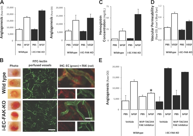
Focal adhesion kinase (FAK) plays a critical role during vascular development because knockout of FAK in endothelial cells (ECs) is embryonic lethal. Surprisingly, tamoxifen-inducible conditional knockout of FAK in adult blood vessels (inducible EC-specific FAK knockout [i-EC-FAK-KO]) produces no vascular phenotype, and these animals are capable of developing a robust growth factor-induced angiogenic response. Although angiogenesis in wild-type mice is suppressed by pharmacological inhibition of FAK, i-EC-FAK-KO mice are refractory to this treatment, which suggests that adult i-EC-FAK-KO mice develop a compensatory mechanism to bypass the requirement for FAK. Indeed, expression of the FAK-related proline-rich tyrosine kinase 2 (Pyk2) is elevated and phosphorylated in i-EC-FAK-KO blood vessels. In cultured ECs, FAK knockdown leads to increased Pyk2 expression and, surprisingly, FAK kinase inhibition leads to increased Pyk2 phosphorylation. Pyk2 can functionally compensate for the loss of FAK because knockdown or pharmacological inhibition of Pyk2 disrupts angiogenesis in i-EC-FAK-KO mice. These studies reveal the adaptive capacity of ECs to switch to Pyk2-dependent signaling after deletion or kinase inhibition of FAK.


Figures

Figure 1.
Figure 1.
Robust growth factor–induced angiogenesis in i-EC-FAK-KO mice. Matrigel containing PBS, bFGF, or VEGF was injected subcutaneously to assess angiogenesis in vivo. (A) Angiogenic responses to bFGF or VEGF were equal or more robust in i-EC-FAK-KO compared with the WT, quantified by FITC-lectin content. n = 7–15 each. (B, left) FITC-lectin–perfused blood vessels within Matrigel from the WT and i-EC-FAK-KO appear similar. Bar, 50 μm. (right) Staining for FAK (red) and EC markers (green) confirms the lack of FAK on i-EC-FAK-KO vessels. Bar, 5 μm. (C) Hemoglobin concentration is increased in i-EC-FAK-KO plugs, which is consistent with their bloodier appearance. n = 4 each. (D) i-EC-FAK-KO mice showed 30% less VEGF-induced leakage in the skin compared with the WT, which suggests that their robust angiogenic response was not caused by increased leakage. n = 11 each. (E) Treatment with the NVP-TAC544 FAK inhibitor blocked the angiogenic response induced by bFGF in WT but not i-EC-FAK-KO. n = 4 each; *, P < 0.05 versus vehicle. All graphs show mean ± SEM.
Figure 2.
Figure 2.
Integrin requirement for angiogenesis despite loss of EC FAK expression. (A) β3 integrin (blue) is expressed on vessels (green) within Matrigel plugs from WT or i-EC-FAK-KO mice. Bar, 5 μm. (B) The selective αvβ3 integrin antagonist cRGD-fK blocked the angiogenic response equivalently for both genotypes. n = 4 each group; *, P < 0.05 versus bFGF. Graph shows mean ± SEM.
Figure 3.
Figure 3.
Pyk2 is up-regulated in the absence of FAK. (A and B) FAK pY861 staining (a marker for activated FAK) appears on blood vessels in the WT but not i-EC-FAK-KO heart. Although minimally detected on blood vessels in the WT heart, Pyk2 expression on ECs is elevated in i-EC-FAK-KO. Graph represents mean ± SEM. Bar, 10 μm. (C) Expression of Pyk2, phosphorylation of Pyk2 on its autophosphorylation site Y402, and phosphorylation of FAK/Pyk2 substrates was increased 3- to 10-fold in heart lysates from i-EC-FAK-KO mice. (D) Pyk2 and pY402 were elevated in angiogenic Matrigel plugs from i-EC-FAK-KO mice. (E) Primary human ECs (HUVECs) were treated with shRNA for FAK or a nonsilencing scramble shRNA control “Scr.” Pyk2 expression was elevated after FAK deletion. (F) BAECs treated with the NVP-TAC544 FAK inhibitor for 1 h showed dose-dependent FAK blockade (pY397) and a surprising increase in Pyk2 (pY402) phosphorylation.
Figure 4.
Figure 4.
Ex vivo sprouting of aortic explants is normal in i-EC-FAK-KO mice. (A and B) Aortic ring explants from WT and i-EC-FAK-KO mice were grown as ex vivo cultures to assess sprouting. The mean sprout length at several time points was similar between genotypes, suggesting a normal angiogenic response to serum. n = 6 each; Graph shows mean ± SEM. (C) Immunohistochemical staining for FAK (red) and EC (green) confirms lack of FAK protein expression on vessels sprouting from rings isolated from i-EC-FAK-KO mice. Bars: (A) 0.5 mm; (C) 80 μm.
Figure 5.
Figure 5.
Pyk2 can functionally compensate for loss of FAK during angiogenesis. (A and B) Knockdown of FAK (but not Pyk2) slowed the rate of sprouting in WT aortic explants, indicating FAK plays the primary role during angiogenesis in normal blood vessels. For i-EC-FAK-KO, knockdown of Pyk2 (but not FAK) inhibited the angiogenic response. Broken lines indicate sprout length. Bar, 0.25 mm. (C) Sprouting of WT aortic explants was dose-dependently blocked by either a pharmacological inhibitor selective for FAK (PF-228) or a dual FAK/Pyk2 inhibitor (PF-271). Only the dual FAK/Pyk2 inhibitor (PF-271) blocked sprouting in i-EC-FAK-KO explants. n = 6 each; *, P < 0.05 versus control shRNA or vehicle. Graphs show mean ± SEM. (D) Our results suggest a necessity for FAK and/or Pyk2 expression in ECs that can preserve the angiogenic response in i-EC-FAK-KO mice.

References

    1. Allingham, M.J., J.D. van Buul, and K. Burridge. 2007. ICAM-1-mediated, Src- and Pyk2-dependent vascular endothelial cadherin tyrosine phosphorylation is required for leukocyte transendothelial migration. J. Immunol. 179:4053–4064. - PubMed
    1. Avraham, H.K., T.H. Lee, Y. Koh, T.A. Kim, S. Jiang, M. Sussman, A.M. Samarel, and S. Avraham. 2003. Vascular endothelial growth factor regulates focal adhesion assembly in human brain microvascular endothelial cells through activation of the focal adhesion kinase and related adhesion focal tyrosine kinase. J. Biol. Chem. 278:36661–36668. - PubMed
    1. Braren, R., H. Hu, Y.H. Kim, H.E. Beggs, L.F. Reichardt, and R. Wang. 2006. Endothelial FAK is essential for vascular network stability, cell survival, and lamellipodial formation. J. Cell Biol. 172:151–162. - PMC - PubMed
    1. Brooks, P.C., R.A. Clark, and D.A. Cheresh. 1994. Requirement of vascular integrin alpha v beta 3 for angiogenesis. Science. 264:569–571. - PubMed
    1. Brooks, P.C., S. Stromblad, R. Klemke, D. Visscher, F.H. Sarkar, and D.A. Cheresh. 1995. Anti-integrin alpha v beta 3 blocks human breast cancer growth and angiogenesis in human skin. J. Clin. Invest. 96:1815–1822. - PMC - PubMed

Publication types