Review
. 2008 Apr;88(2):581-609.
doi: 10.1152/physrev.00024.2007.

Mechanisms underlying acute protection from cardiac ischemia-reperfusion injury

Affiliations
Review

Mechanisms underlying acute protection from cardiac ischemia-reperfusion injury

Elizabeth Murphy et al. Physiol Rev. 2008 Apr.

Abstract

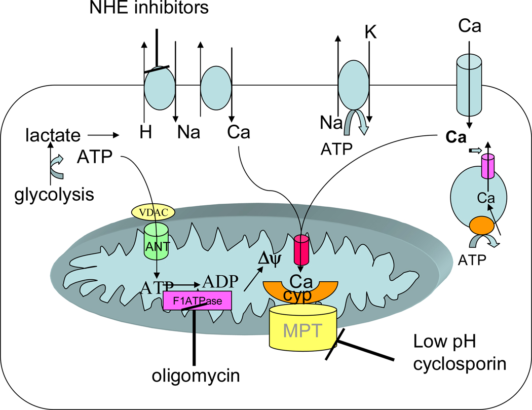
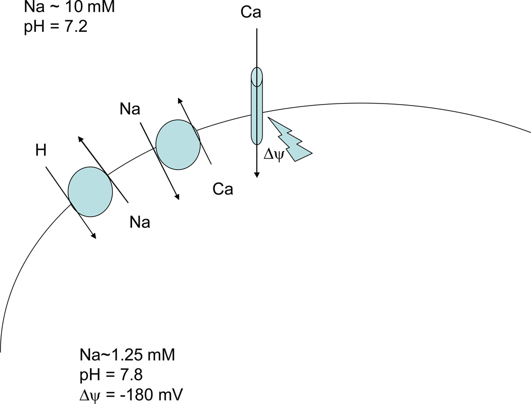
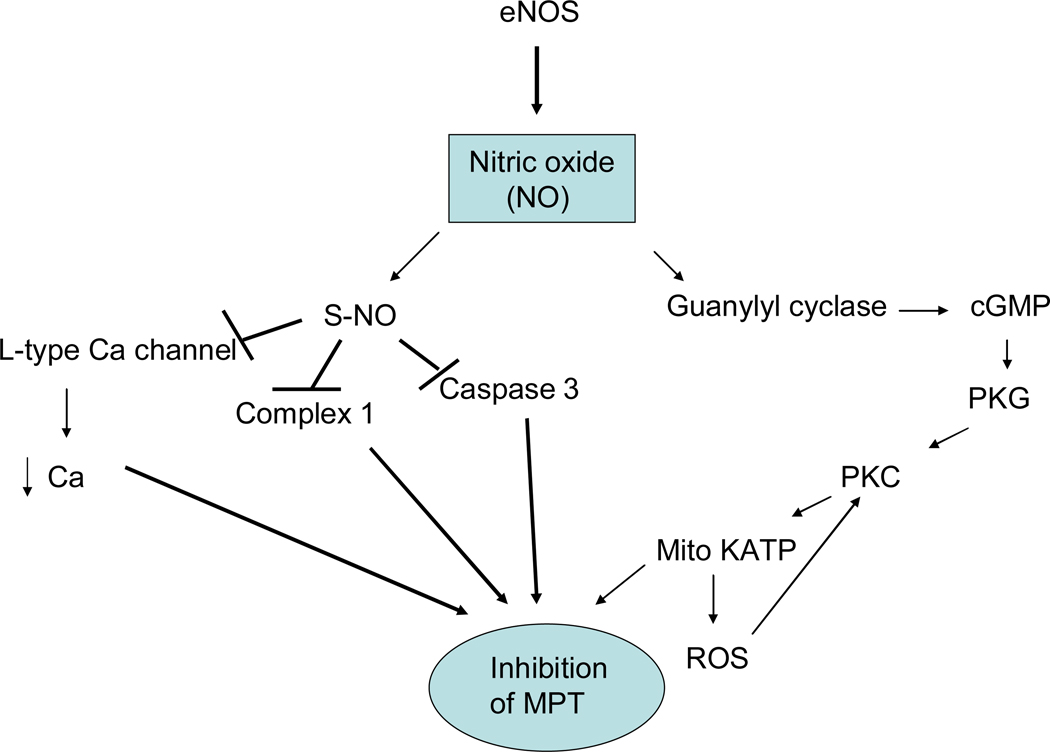
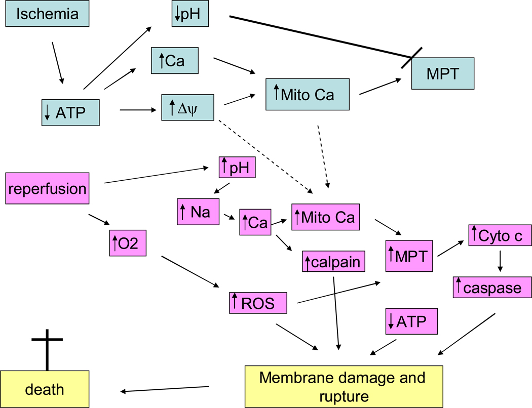
Mitochondria play an important role in cell death and cardioprotection. During ischemia, when ATP is progressively depleted, ion pumps cannot function resulting in a rise in calcium (Ca(2+)), which further accelerates ATP depletion. The rise in Ca(2+) during ischemia and reperfusion leads to mitochondrial Ca(2+) accumulation, particularly during reperfusion when oxygen is reintroduced. Reintroduction of oxygen allows generation of ATP; however, damage to the electron transport chain results in increased mitochondrial generation of reactive oxygen species (ROS). Mitochondrial Ca(2+) overload and increased ROS can result in opening of the mitochondrial permeability transition pore, which further compromises cellular energetics. The resultant low ATP and altered ion homeostasis result in rupture of the plasma membrane and cell death. Mitochondria have long been proposed as central players in cell death, since the mitochondria are central to synthesis of both ATP and ROS and since mitochondrial and cytosolic Ca(2+) overload are key components of cell death. Many cardioprotective mechanisms converge on the mitochondria to reduce cell death. Reducing Ca(2+) overload and reducing ROS have both been reported to reduce ischemic injury. Preconditioning activates a number of signaling pathways that reduce Ca(2+) overload and reduce activation of the mitochondrial permeability transition pore. The mitochondrial targets of cardioprotective signals are discussed in detail.


Figures

Figure 1
Figure 1
Changes in ions and metabolites during ischemia. During ischemia ATP declines resulting in a decrease in pH due to anaerobic glycolysis. The increase in proton stimulates Na-H exchange and Na-Ca exchange resulting in an increase in cytosolic Ca. Much of the ATP generated by glycolysis is consumed by the reverse mode of the mitochondrial F1F0ATPase which uses the energy to generate Δψ. The Δψ can then be used to take up Ca into the mitochondria. This increase in mitochondrial Ca can activate the MPT, but the low pH due ischemia inhibits MPT so that it is not activated until reperfusion when the pH is restored to normal.
Figure 2
Figure 2
Regulation of mitochondrial Ca2+. Ca2+ enters mitochondria on the uniporter, driven by the negative mitochondrial membrane potential. Ca2+ efflux from the mitochondrial matrix is via the mitochondrial Na-Ca exchanger.
Figure 3
Figure 3
Panel 3A illustrates the signaling pathways activated by PC during ischemia and at reperfusion. Panel 3B show the signaling pathways activated by postconditioning.
Figure 4
Figure 4
Details of the signaling pathways activated by PC. PC leads to release of agonist that activate GPCR, leading to activation of PI3K, which in turn activates AKT and P70S6K. Internalization of GPCRs involves sequential binding of β-arrestin, the clathrin adaptor (adaptor protein 2; AP-2), and clathrin. PI3K and its phosphoinositide products play a critical role in recruitment of β-arrestin GPCR complexes to endosomes. Many of these pathways converge on the mitochondria where the result in activation of the mitoKATP channel and inhibition of the MPT.
Figure 5
Figure 5
PI3K signaling. PI3K generates PIP3 which recruits AKT to the membrane where it can be phosphorylated by PDK. Newly synthesized PKC is attached to the plasma membrane where it is phosphorylated by PDK1. PDK1 phoshorylation of PKCs in the activation loop is required for PKC activation and stability. However, for most PKC isoforms, the rate of PKC processing by PDK1 is not dependent on phosphoinositides generated by PI3K and thus the rate of PKC processing is not inhibited by wortmannin (188). PDK can also phosphorylate and activate PKA, RSK and p70S6K.
Figure 6
Figure 6
AKT signaling. Akt is activated by phosphorylation of thr 308 by PDK1 and by phosphorylation of ser 473 by a poorly defined kinase. Phosphorylation of Akt at thr 308 is mediated by binding of PI3K generated lipid (PIP3) to the pleckstrin homology domain of Akt thereby facilitating translocation and association of Akt with PDK1. PIP3 binding to Akt also exposes the PDK1 phosphorylation site thus facilitating phosphorylation of Akt (188). A large number of kinases have been proposed to phosphorylate ser 473, including Akt autophosphorylation, PDK1, ILK1, MAPKAPK2, PKCbII, PIKK, ATM, DNA-PK, and TORC2 (see (68)). Increasing data seem to suggest a role for TORC2 in phosphorylating ser 473 on Akt (116). Akt can also be regulated by dephosphorylation, and PP2a (202) and PHLPP (33) have been reported to be important phosphatases regulating Akt phosphorylation.
Figure 7
Figure 7
Regulation of p70S6K phosphorylation and signaling. P70S6K is phosphorylated by PDK1 on thr 229 and by mTOR on thr 389. Prior phosphorylation of thr 389 is reported to be necessary before PDK1 can phosphorylate thr 229. Phosphorylation of thr 389 appears to be rate-limiting and correlates with activity; however activation of mTOR depends on Akt activation. P70S6K is also phosphorylated on the pseudosubstrate or auto-inhibitory domain at ser 411, ser 418, thr 421 and ser 424, but the kinases responsible for phosphorylating these sites are debated. Phosphorylation of these sites appears to be necessary but not sufficient for activation. The activation of mTOR appears to be mediated by activation of Akt. Akt phosphorylates and inhibits tuberin (TSC2), a GTPase, which results in activation of RHEB, which results in phosphorylation and activation of mTOR. Akt is also reported to directly phosphorylate mTOR on serine 2448, but phosphorylation of this site does not appear to regulate activity of mTOR. mTOR activity is also inhibited by AMP-kinase; thus under conditions of increased AMP (decreased ATP) activation of AMP-kinase will lead to inhibition of mTOR
Figure 8
Figure 8
GSK-β signaling in cardioprotection. In unstimulated cells, GSK-3β is unphosphorylated and active and it phosphorylates and usually inactivates its downstream targets. GSK-3β is phosphorylated and inactivated by a large number of kinases. Inactive GSK-3β has been suggested to be cardioprotective.
Figure 9
Figure 9
GSK-3β has a preference for primed substrates.
Figure 10
Figure 10
Nitric oxide signaling in cardioprotection. Nitric oxide (NO) is generated by nitric oxide synthases (NOS). NO can activate guanyly cyclase resulting in activation of protein kinase G. Alternatively NO can result in s-nitrosation of a number of proteins.
Figure 11
Figure 11
Pathways Leading to Cell Death in Ischemia-Reperfusion.

References

    1. MRC/BHF Heart Protection Study of antioxidant vitamin supplementation in 20,536 high-risk individuals: a randomised placebo-controlled trial. Lancet. 2002;360:23–33. - PubMed
    1. Adlam VJ, Harrison JC, Porteous CM, James AM, Smith RA, Murphy MP, Sammut IA. Targeting an antioxidant to mitochondria decreases cardiac ischemia-reperfusion injury. Faseb J. 2005;19:1088–1095. - PubMed
    1. Ahmad N, Wang Y, Haider KH, Wang B, Pasha Z, Uzun O, Ashraf M. Cardiac protection by mitoKATP channels is dependent on Akt translocation from cytosol to mitochondria during late preconditioning. Am J Physiol Heart Circ Physiol. 2006;290:H2402–H2408. - PubMed
    1. Ala-Rami A, Ylitalo KV, Hassinen IE. Ischaemic preconditioning and a mitochondrial KATP channel opener both produce cardioprotection accompanied by F1F0-ATPase inhibition in early ischaemia. Basic Res Cardiol. 2003;98:250–258. - PubMed
    1. Ali SM, Sabatini DM. Structure of S6 kinase 1 determines whether raptor-mTOR or rictor-mTOR phosphorylates its hydrophobic motif site. J Biol Chem. 2005;280:19445–19448. - PubMed

Publication types

LinkOut - more resources