

Differential roles for hippocampal areas CA1 and CA3 in the contextual encoding and retrieval of extinguished fear
- PMID: 18391185
- PMCID: PMC2327266
- DOI: 10.1101/lm.794808
Differential roles for hippocampal areas CA1 and CA3 in the contextual encoding and retrieval of extinguished fear
Abstract
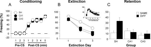
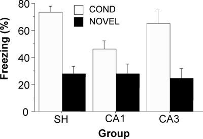
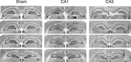
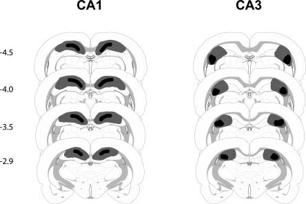
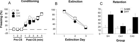
Recent studies demonstrate that context-specific memory retrieval after extinction requires the hippocampus. However, the contribution of hippocampal subfields to the context-dependent expression of extinction is not known. In the present experiments, we examined the roles of areas CA1 and CA3 of the dorsal hippocampus in the context specificity of extinction. After pairing an auditory conditional stimulus (CS) with an aversive footshock (unconditional stimulus or US), rats received extinction sessions in which the CS was presented without the US. In Experiment 1, pretraining neurotoxic lesions in either CA1 or CA3 eliminated the context dependence of extinguished fear. In Experiment 2, lesions of CA1 or CA3 were made after extinction training. In this case, only CA1 lesions impaired the context dependence of extinction. Collectively, these results reveal that both hippocampal areas CA1 and CA3 contribute to the acquisition of context-dependent extinction, but that only area CA1 is required for contextual memory retrieval.
Figures
Similar articles
-
Hippocampal inactivation disrupts the acquisition and contextual encoding of fear extinction.J Neurosci. 2005 Sep 28;25(39):8978-87. doi: 10.1523/JNEUROSCI.2246-05.2005. J Neurosci. 2005. PMID: 16192388 Free PMC article.
-
Lesions of the entorhinal cortex or fornix disrupt the context-dependence of fear extinction in rats.Behav Brain Res. 2008 Dec 12;194(2):201-6. doi: 10.1016/j.bbr.2008.07.011. Epub 2008 Jul 18. Behav Brain Res. 2008. PMID: 18692093 Free PMC article.
-
Dissociations across the dorsal-ventral axis of CA3 and CA1 for encoding and retrieval of contextual and auditory-cued fear.Neurobiol Learn Mem. 2008 Jan;89(1):61-9. doi: 10.1016/j.nlm.2007.08.016. Epub 2007 Oct 10. Neurobiol Learn Mem. 2008. PMID: 17931914 Free PMC article.
-
Electrolytic lesions of the dorsal hippocampus disrupt renewal of conditional fear after extinction.Learn Mem. 2005 May-Jun;12(3):270-6. doi: 10.1101/lm.91705. Learn Mem. 2005. PMID: 15930505 Free PMC article.
-
Hippocampal involvement in contextual modulation of fear extinction.Hippocampus. 2007;17(9):749-58. doi: 10.1002/hipo.20331. Hippocampus. 2007. PMID: 17604353 Review.
Cited by
-
Vitamin D deficiency is associated with reduced hippocampal volume and disrupted structural connectivity in patients with mild cognitive impairment.Hum Brain Mapp. 2019 Feb 1;40(2):394-406. doi: 10.1002/hbm.24380. Epub 2018 Sep 25. Hum Brain Mapp. 2019. PMID: 30251770 Free PMC article.
-
Adverse maternal environment alters Oprl1 variant expression in mouse hippocampus.Anat Rec (Hoboken). 2023 Jan;306(1):162-175. doi: 10.1002/ar.25056. Epub 2022 Aug 19. Anat Rec (Hoboken). 2023. PMID: 35983908 Free PMC article.
-
PTSD recovery, spatial processing, and the val66met polymorphism.Front Hum Neurosci. 2014 Feb 26;8:100. doi: 10.3389/fnhum.2014.00100. eCollection 2014. Front Hum Neurosci. 2014. PMID: 24616687 Free PMC article. No abstract available.
-
The metabotropic glutamate receptor, mGlu5, is required for extinction learning that occurs in the absence of a context change.Hippocampus. 2015 Feb;25(2):149-58. doi: 10.1002/hipo.22359. Epub 2014 Sep 30. Hippocampus. 2015. PMID: 25160592 Free PMC article.
-
Altered synaptic structure in the hippocampus in a mouse model of Alzheimer's disease with soluble amyloid-β oligomers and no plaque pathology.Mol Neurodegener. 2014 Oct 13;9:41. doi: 10.1186/1750-1326-9-41. Mol Neurodegener. 2014. PMID: 25312309 Free PMC article.
References
-
- Akbari E., Naghdi N., Motamedi F. Functional inactivation of orexin 1 receptors in CA1 region impairs acquisition, consolidation and retrieval in Morris water maze task. Behav. Brain Res. 2006;173:47–52. - PubMed
-
- Akbari E., Naghdi N., Motamedi F. The selective orexin 1 receptor antagonist SB-334867-A impairs acquisition and consolidation but not retrieval of spatial memory in Morris water maze. Peptides. 2007;28:650–656. - PubMed
-
- Amaral D.G., Witter M.P. The three-dimensional organization of the hippocampal formation: A review of anatomical data. Neuroscience. 1989;31:571–591. - PubMed
-
- Bardgett M.E., Jacobs P.S., Jackson J.L., Csernansky J.G. Kainic acid lesions enhance locomotor responses to novelty, saline, amphetamine, and MK-801. Behav. Brain Res. 1997;84:47–55. - PubMed
-
- Bardgett M.E., Boeckman R., Krochmal D., Fernando H., Ahrens R., Csernansky J.G. NMDA receptor blockade and hippocampal neuronal loss impair fear conditioning and position habit reversal in C57Bl/6 mice. Brain Res. Bull. 2003;60:131–142. - PubMed
Publication types
MeSH terms
Grants and funding
LinkOut - more resources
Full Text Sources
Miscellaneous