

doi: 10.1073/pnas.0802190105.
Epub 2008 Apr 7.
Quality control of overexpressed membrane proteins
Affiliations
- PMID: 18391190
- PMCID: PMC2311375
- DOI: 10.1073/pnas.0802190105
Item in Clipboard
Quality control of overexpressed membrane proteins
Proc Natl Acad Sci U S A.
.
Abstract
Overexpression of membrane proteins in Escherichia coli frequently leads to the formation of aggregates or inclusion bodies, which is undesirable for most studies. Ideally, one would like to optimize the expression conditions by monitoring simultaneously and rapidly both the amounts of properly folded and aggregated membrane protein, a requirement not met by any of the currently available methods. Here, we describe a simple gel-based approach with green fluorescent protein as folding indicator to detect well folded and aggregated proteins simultaneously. The method allows for rapid screening and, importantly, pinpointing the most likely bottlenecks in protein production.
Conflict of interest statement
The authors declare no conflict of interest.
Figures
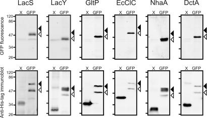
Electrophoretic mobility of membrane proteins fused or not fused to GFP. All proteins and GFP fusion proteins were expressed in E. coli MC1061, except for GltP and DctA, which were expressed in E. coli TOP10. Whole-cell samples of cultures induced with 1 × 10⁻²% (wt/vol) l -arabinose were disrupted and analyzed as described in Materials and Methods. (Upper) In gel GFP fluorescence. (Lower) Immunoblots of the same gels decorated with anti-His tag antibody. X and GFP indicate the absence or presence of a GFP moiety at the C terminus, respectively. Black and white arrows indicate the positions of nonfluorescent and fluorescent species of the GFP fusion proteins, respectively. Molecular masses (in kilodaltons) of the marker proteins are indicated on the left of each panel. Molecular mass of the nonmodified proteins are mentioned in Table S1 . The increase in molecular mass caused by the GFP fusion is ≈27 kDa. The faint doublet bands at ≈40 kDa (Upper) represent (an) endogenous fluorescent protein(s) from E. coli.
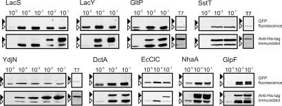
Electrophoretic mobility of GFP fusion proteins expressed to different levels. Expression of all GFP fusion proteins was controlled by the AraC/PBAD system, except for panels labeled T7, for which the T7 expression system was used. All proteins are polytopic membrane proteins (Table S1 ). Cultures were induced with 1 × 10⁻⁵%, 1 × 10⁻⁴%, 1 × 10⁻³%, 1 × 10⁻²%, or 1 × 10⁻¹% (wt/vol) l -arabinose as indicated above the lanes. For the T7 expression system, 0.4 mM IPTG was used. (Upper) In gel GFP fluorescence. (Lower) Immunoblots of the same gels decorated with anti-His tag antibody. Black and white arrows indicate the positions of nonfluorescent and fluorescent species of the GFP fusion proteins, respectively.
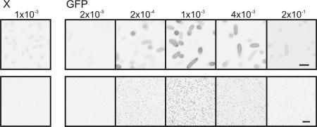
Confocal microscopy images of cells expressing LacS-GFP to different levels. E. coli MC1061 cells were induced with the percentages of l -arabinose indicated above the panels. X and GFP indicate the absence or presence of a GFP moiety at the C terminus of LacS, respectively. The fluorescence shown is inverted. For each sample, similar numbers of cells were imaged. (Upper) Close-up of cells to indicate the distribution of the fluorescence. (Scale bar, 2 μm.) (Lower) Overview of the culture. (Scale bar, 10 μm.)
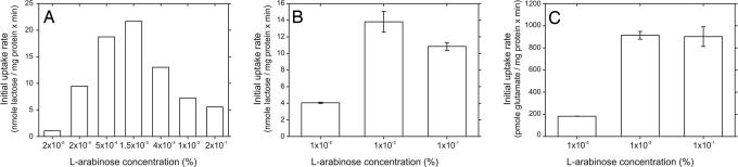
Transport activities of membrane proteins expressed at different levels. Initial rates of transport were determined in whole E. coli MC1061 cells for LacS (A) and LacY-GFP (B), or membrane vesicles for GltP-GFP (C). Arabinose concentrations during induction were varied from 1 × 10⁻⁵% to 1 × 10⁻¹% (wt/vol). E. coli MC1061 is devoid of the endogenous E. coli lactose transporter LacY, hence background lactose transport activity was absent. The GltP-GFP transport rates were corrected for background glutamate transport activity.
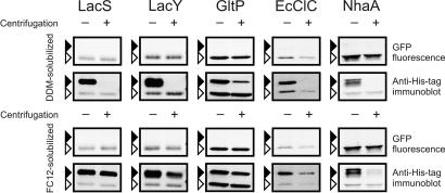
Differential sedimentation of the two species of GFP fusion proteins. E. coli MC1061 cells were induced with 1 × 10⁻¹% (wt/vol) l -arabinose. The GFP fusion proteins expressed are indicated above the panels. After disruption by bead beating, cells were solubilized by the addition of 1% DDM (Upper) or 1% FC-12 (Lower). Samples were taken before (−) and after (+) ultracentrifugation and analyzed by in gel fluorescence (indicated by GFP fluorescence) and immunodetection with an anti-His tag antibody (indicated by anti-His tag immunoblot). Black and white arrows indicate the positions of nonfluorescent and fluorescent species of the GFP fusion proteins, respectively.
References
-
- Drew D, Froderberg L, Baars L, de Gier JW. Assembly and overexpression of membrane proteins in Escherichia coli. Biochim Biophys Acta. 2003;1610:3–10. - PubMed
-
- Dalbey RE, Chen M. Sec-translocase-mediated membrane protein biogenesis. Biochim Biophys Acta. 2004;1694:37–53. - PubMed
-
- Wang DN, et al. Practical aspects of overexpressing bacterial secondary membrane transporters for structural studies. Biochim Biophys Acta. 2003;1610:23–36. - PubMed
-
- Waldo GS, Standish BM, Berendzen J, Terwilliger TC. Rapid protein-folding assay using green fluorescent protein. Nat Biotechnol. 1999;17:691–695. - PubMed
Publication types
MeSH terms
Substances
LinkOut - more resources
Full Text Sources
Other Literature Sources
Miscellaneous
