. 2008 Apr 15;105(15):5856-61.
doi: 10.1073/pnas.0801677105. Epub 2008 Apr 7.

Neurons derived from reprogrammed fibroblasts functionally integrate into the fetal brain and improve symptoms of rats with Parkinson's disease

Affiliations

Neurons derived from reprogrammed fibroblasts functionally integrate into the fetal brain and improve symptoms of rats with Parkinson's disease

Marius Wernig et al. Proc Natl Acad Sci U S A. .

Abstract

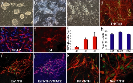
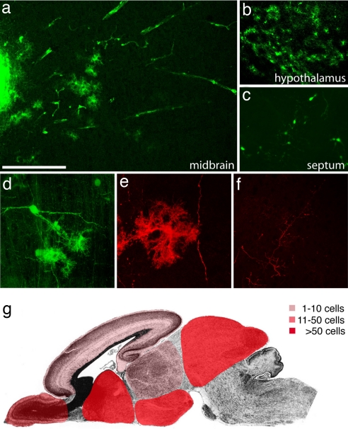
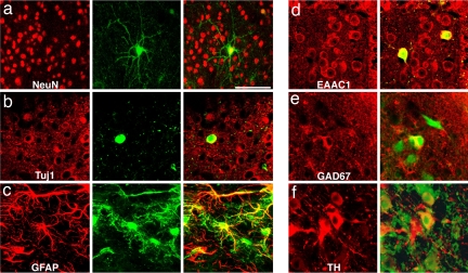
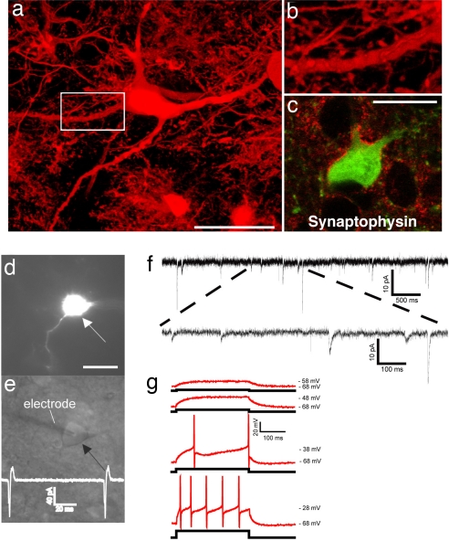
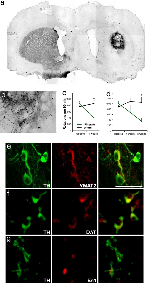
The long-term goal of nuclear transfer or alternative reprogramming approaches is to create patient-specific donor cells for transplantation therapy, avoiding immunorejection, a major complication in current transplantation medicine. It was recently shown that the four transcription factors Oct4, Sox2, Klf4, and c-Myc induce pluripotency in mouse fibroblasts. However, the therapeutic potential of induced pluripotent stem (iPS) cells for neural cell replacement strategies remained unexplored. Here, we show that iPS cells can be efficiently differentiated into neural precursor cells, giving rise to neuronal and glial cell types in culture. Upon transplantation into the fetal mouse brain, the cells migrate into various brain regions and differentiate into glia and neurons, including glutamatergic, GABAergic, and catecholaminergic subtypes. Electrophysiological recordings and morphological analysis demonstrated that the grafted neurons had mature neuronal activity and were functionally integrated in the host brain. Furthermore, iPS cells were induced to differentiate into dopamine neurons of midbrain character and were able to improve behavior in a rat model of Parkinson's disease upon transplantation into the adult brain. We minimized the risk of tumor formation from the grafted cells by separating contaminating pluripotent cells and committed neural cells using fluorescence-activated cell sorting. Our results demonstrate the therapeutic potential of directly reprogrammed fibroblasts for neuronal cell replacement in the animal model.


Conflict of interest statement

The authors declare no conflict of interest.

Figures

Fig. 1.
Fig. 1.
In vitro differentiation of induced pluripotent stem cells. (a) The undifferentiated Oct4-selected iPS cell line O9. (b) FGF2-responsive neural precursor cells. (c) Differentiated neural morphologies 7 days after growth factor withdrawal. (d) A fraction of β-III-tubulin positive neurons (red) are double labeled with antibodies against TH (green, yellow in merged image), 7 days after growth factor withdrawal. (e and f) At this stage, also, many GFAP-positive astrocytes (red) (e) and rare 04-positive oligodendrocytes (f) are found. (g) The fraction of TH-positive cells over β-III-tubulin positive cells increases along neuronal maturation (mean and standard deviation, three independent experiments). (h) The vast majority of TH-immunoreactive cells coexpress En1, Pitx3, and Nurr1 (mean, standard deviation). (i) Coexpression of En1 (green) and TH (red). (j) VMAT2 and TH [purple in the merged image indicates colocalization of TH (red) and VMAT2 (blue)]. (k and l) Most TH-positive cells (red) are also positive for Pitx3 (green) (k), and Nurr1 (green) (l) after 7 days of neuronal differentiation. [Scale bar in a: 200 μm (a and b), 100 μm (c, d, i, and j), 50 μm (e and k), and 20 μm (f and l).]
Fig. 2.
Fig. 2.
Extensive migration and differentiation of iPS cell-derived neural precursor cells in the embryonic brain. (a) Transplanted cells form an intraventricular cluster (left part of the image) and migrate extensively into the tectum 4 weeks after transplantation into the lateral brain ventricles of E13.5 mouse embryos. (b) A high density of integrated astrocyte-like cells in the hypothalamus. (c) Complex neuronal morphologies of GFP-positive cells in the septum. (d) Confocal reconstruction of grafted GFP-fluorescent cells in the tectum with neuronal and glial morphologies. (e) GFP immunofluorescence and confocal reconstruction identifies an astrocytic cell and a long neuronal process. (f) Similarly, GFP-immunoreactive, fine neuronal processes are crisply delineated. (g) Schematic representation of the main integration sites of iPS cell-derived neurons and glia. Color codes represent different ranges of cell numbers. Shown are the highest incorporation density across all analyzed brains. See Table 1 for more details. [Scale bar in a: 200 μm (a–c), 100 μm (d and f), and 50μ (e).]
Fig. 3.
Fig. 3.
Transplanted cells express neuronal and glial markers. (a) Confocal reconstruction of a GFP-positive cell in the midbrain (green) expressing NeuN (red) 4 weeks after intrauterine transplantation. (b) Another transplanted neuron (green) expresses cytoplasmatic β-III-tubulin (red) as shown in this confocal section. (c) Other cells can be colabeled with GFAP antibodies (red). (d) Both host neurons (red only) and transplanted cells (yellow) express the glutamate transporter EAAC1. (e) Soma of grafted cells (green) can be labeled with antibodies against GAD67 (red). (f) TH-immunoreactivity (red) can be found in both host and grafted neurons (green). [Scale bar in a: 100 μm (a–c) and 50 μm (d–f).]
Fig. 4.
Fig. 4.
Synaptic integration of functional iPS cell-derived neurons into the host brain. (a) GFP-immunofluorescence allows the high resolution characterization of dendritic morphologies of transplanted neurons. (b) Higher magnification of the indicated part in a suggests the presence of synaptic spines along this dendrite. (c) Integrated GFP-positive neurons are adjacent to many synaptophysin-positive patches (red), indicating synaptic contacts from host axon terminals. (d) A GFP-expressing neuron (arrow) in the dorsal midbrain was detected in acute brain slices of a P20 mouse brain after in utero transplantation. (e) GFP-positive neurons were identified by infrared differential interference contrast (arrow) and approached by a recording electrode (left). The trace indicates spontaneous generation of action potentials. (f) Voltage-clamp recording at −70 mV in extracellular solution containing 3 mM Mg2+. Traces show spontaneous slow and fast currents that indicate that this transplanted neuron receives synaptic contacts from host cells. All six recorded GFP-positive neurons from two mice (age P20 and P22) exhibited similar spontaneous currents. (g) Current-clamp recordings during current injection. Shown are superimposed membrane potential changes (Upper traces, red), which demonstrates the capability of the grafted neurons to fire action potentials in response to a series of current injection (Lower traces, black) from a holding potential of −68 mV. All six analyzed GFP-neurons showed these active membrane characteristic. (Scale bars: 20 μm.)
Fig. 5.
Fig. 5.
iPS cell-derived neurons integrate into the striatum of hemi-parkinsonian rats and improve behavioral deficits. (a) Overview of an iPS cell graft 4 weeks after transplantation stained with antibodies against TH (dark brown). (b) Higher magnification of another graft showing TH-positive soma and the dense innervation of the surrounding host striatum by donor-derived neurites (arrowheads). The dashed line indicates the edges of the graft. (c) Amphetamine-induced rotations are significantly reduced in animals grafted with unsorted iPS cell populations (green, n = 5) compared with the sham control animals (black, n = 10) (P = 0.0185, unpaired Student's t test). Shown are the number of rotations in 90 min after amphetamine injection as means and standard deviations. (d) Amphetamine-induced rotations in animals transplanted with iPS cell cultures after elimination of SSEA1-positive cells by FACS. Grafted animals (green, n = 4) show significant recovery compared with control animals (black, n = 10) (P = 0.006). (e–g) The grafted TH-positive cells (green) can be colabeled (red) with antibodies against VMAT2 (e), DAT (f), and En1 (g). (Scale bar: 50 μm.)

References

    1. Lerou PH, Daley GQ. Therapeutic potential of embryonic stem cells. Blood Rev. 2005;19:321–331. - PubMed
    1. Hochedlinger K, Jaenisch R. Nuclear transplantation, embryonic stem cells, and the potential for cell therapy. N Engl J Med. 2003;349:275–286. - PubMed
    1. Jaenisch R. Human cloning: The science and ethics of nuclear transplantation. N Engl J Med. 2004;351:2787–2791. - PubMed
    1. Weissman IL. Medicine: Politic stem cells. Nature. 2006;439:145–147. - PubMed
    1. Rideout WM, III, Hochedlinger K, Kyba M, Daley GQ, Jaenisch R. Correction of a genetic defect by nuclear transplantation and combined cell and gene therapy. Cell. 2002;109:17–27. - PubMed

Publication types