. 2008 Apr 8;105(14):5367-72.
doi: 10.1073/pnas.0801637105. Epub 2008 Apr 7.

TFIID component TAF7 functionally interacts with both TFIIH and P-TEFb

Affiliations

TFIID component TAF7 functionally interacts with both TFIIH and P-TEFb

Anne Gegonne et al. Proc Natl Acad Sci U S A. .

Abstract

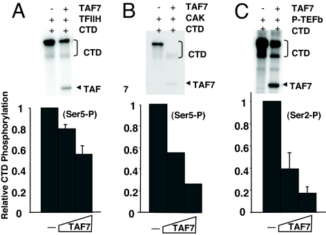
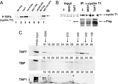
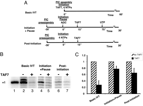
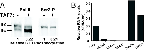
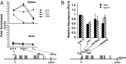
Transcription consists of a series of highly regulated steps: assembly of the preinitiation complex (PIC) at the promoter, initiation, elongation, and termination. PIC assembly is nucleated by TFIID, a complex composed of the TATA-binding protein (TBP) and a series of TBP-associated factors (TAFs). One component, TAF7, is incorporated in the PIC through its interaction with TFIID but is released from TFIID upon transcription initiation. We now report that TAF7 interacts with the transcription factors, TFIIH and P-TEFb, resulting in the inhibition of their Pol II CTD kinase activities. Importantly, in in vitro transcription reactions, TAF7 inhibits steps after PIC assembly and formation of the first phosphodiester bonds. Further, in vivo TAF7 coelongates with P-TEFb and Pol II downstream of the promoter. We propose a model in which TAF7 contributes to the regulation of the transition from PIC assembly to initiation and elongation.


Conflict of interest statement

The authors declare no conflict of interest.

Figures

Fig. 1.
Fig. 1.
TAF7 inhibits the kinase activities of TFIIH and P-TEFb. (A) TAF7 inhibits TFIIH-mediated phosphorylation of the Pol II CTD Ser-5. (Upper) Purified TFIIH and 100 ng of GST-tagged Pol II CTD were incubated in the presence or absence of recombinant Flag-tagged TAF7 (F-TAF7) in an in vitro kinase assay using [32P]ATP. Phosphorylation was determined by autoradiography after gel electrophoresis; positions of TAF7 and CTD are indicated based on mobility markers (data not shown). (Lower) Purified TFIIH and 100 ng of GST-tagged Pol II CTD were incubated in the presence of increasing amount of recombinant F-TAF7 (125 and 250 ng), in an in vitro kinase assay. Ser-5 CTD phosphorylation was determined by Western blot analysis, using H14 anti-phosphoSer5 antibody, and quantified by densitometry. (B) TAF7 inhibits the CAK-mediated phosphorylation of the Pol II CTD. (Upper) In vitro kinase assay with purified CAK, 100 ng of GST-tagged Pol II CTD, and F-TAF7 as in A Upper. (Lower) In vitro kinase assay with purified CAK, 100 ng of GST-tagged Pol II CTD, and increasing F-TAF7 as in A Lower. (C) TAF7 inhibits P-TEFb-mediated phosphorylation of the Pol II CTD Ser-2. (Upper) In vitro kinase assay with 50 ng of purified P-TEFb, 100 ng of GST-tagged Pol II CTD, and F-TAF7 as in A Upper. (Lower) In vitro kinase assay with 50 ng of P-TEFb, 100 ng of GST-tagged Pol II CTD, and increasing F-TAF7 as in A Lower. CTD Ser-2 phosphorylation was determined by Western blot analysis, using H5 anti-phosphoSer2 antibody.
Fig. 2.
Fig. 2.
TAF7 interacts with P-TEFb in vitro and in vivo and exists in a TFIID-independent complex. (A) TAF7 binds to P-TEFb through its central domain in in vitro pull-down assays. Purified recombinant P-TEFb (200 ng) was incubated with F-TAF7 or each of the F-TAF7 mutants immobilized on Sepharose beads. TAF7-bound fractions were assayed for retention of P-TEFb by immunoblotting, using anti-cyclin T1 antibody. (B) TAF7 binds to P-TEFb in vivo. Extracts from cells transiently transfected with either F-TAF7, pcDNA3, or Flag-SKD1, an irrelevant control protein, were immunoprecipitated with anti-cyclin T1 antibody. Immunoprecipitates were analyzed for cyclin T1 (Upper) or F-TAF7 (Lower) by Western blot. (Upper) Anti-cyclin T1. Total extract from cells transfected with vector (lane 1), Flag-SKD1 (lane 2), or F-TAF7 (lane 3) before immunoprecipitation and immunoprecipitated cyclin T1 (lanes 4–6). (Lower) Anti-Flag M2 antibody, detection of F-TAF7 coimmunoprecipitated from extracts of cells transfected with vector control (lane 4), Flag-SKD1 (lane 5), or F-TAF7 expression vector (lane 6). (C) TAF7 exists in a TFIID-independent form. C8166 whole cell extracts were fractionated on an FPLC column and fractions analyzed by Western blotting as indicated. Arrows refer to the indicated molecular sizes. Purified TFIID was included as a marker. TFIID consistently appears in the excluded volume in column fractionation of whole cell extracts. Fractions 16–28 and 30–36 were run in parallel but on two separate SDS/PAGEs.
Fig. 3.
Fig. 3.
TAF7 regulates multiple distinct steps in transcription initiation. (A) Scheme of experimental protocol. Basic IVT, TAF7, and the four NTPs are present during PIC assembly and initiation; Initiation+Pause, PIC preassembly occurs in the absence of NTPs or TAF7. TAF7 is added after transcription is initiated with AGC; UTP is added at 75 sec to permit clearance and elongation. Post-Initiation, PIC preassembly occurs in the absence of NTPs or TAF7, which is added after transcription is initiated with all four NTPs. (B) TAF7 regulates in vitro transcription. Lane 1, marker; lanes 2–3, HeLa nuclear extract in the presence or absence of purified TAF7 preincubated for 15 min in presence of rNTPs. Transcription was initiated by the addition of the MHC class I −313CAT plasmid. In lanes 4–7, −313CAT DNA and extracts were preincubated for 30 min in presence of 40 μM ATP to permit PIC assembly. Transcription was initiated by addition of ATP/CTP/GTP mix (lanes 4 and 5) or rNTP (lanes 6 and 7). Control TKEG buffer (−) or purified F-TAF7 (+) were added 15 seconds after initiation of transcription and incubated for 30min. Analysis of −313CAT transcripts was by primer extension. [In previous studies (27), addition of TAF7 was after promoter clearance so did not affect transcription.] (C) Quantitation of results of three experiments, showing averages and SEM. Addition of TAF7 in each of these conditions results in significant inhibition of transcription: IVT, P < 0.003; pause, P < 0.03; initiation, P < 0.04.
Fig. 4.
Fig. 4.
TAF7 regulates promoter activity in vivo. (A) TAF7 inhibits Pol II CTD phosphorylation in vivo. Nuclear extracts from either 293/F-TAF7 (+) or control 293T (−) cells were analyzed by Western blotting with either an antibody specific for the Ser-2 CTD phosphorylated form of pol II (Right) or an antibody reactive with all forms of Pol II CTD (Left). (Quantitation) (Left) Phosphorylated Pol II (II-0) is expressed relative to nonphosphorylated Pol II (II-a). (Right) The amount of Ser-2-P is expressed relative to the total amount of Pol II in the same sample (Left). (B) Depletion of TAF7 results in reduced levels of endogenous MHC class I RNA. Human 293T cells were transiently transfected with TAF7 siRNA or scrambled siRNA, as described; levels of TAF7, HLA class I RNAs, actin, and GAPDH RNAs were determined by real time PCR. Results are presented relative to control siRNA.
Fig. 5.
Fig. 5.
TAF7 remains associated with the transcription elongation complex in vivo. (A) TAF7 is associated with MHC class I coding sequences in spleen but not brain. A representative ChIP assay out of four performed by using chromatin from spleen cells and brain of mice transgenic for the pig MHC class I gene, PD1 (B10.PD1). The fold enrichments of TAF7, Pol II, AcHistone H4, and CREB were calculated for different regions of the PD1 gene as described in Methods. The PD1 gene segments analyzed and primer sets in the ChIP assays are shown diagrammatically. Boxes indicate exons. PCR fragments are nonoverlapping. (B) TAF7 and Cyclin T1 remain associated with Pol II through exon 2 of the class I gene. ChIP assays were performed on B10.PD1 spleen cells. The amounts of TAF7 or cyclin T1 were determined relative to Pol II for different regions of the PD1 gene and are expressed as “relative abundance”: the fold enrichment (V+/V−) of TAF7 and cyclin T1 relative to the fold enrichment of Pol II, to normalize for variations in efficiency of PCR amplification of primer sets across the gene. The PD1 gene segments analyzed and primer sets in the ChIP assays are shown diagrammatically. Gray boxes indicate exons. PCR fragments are nonoverlapping. The graph represents the composite of three independent experiments.

References

    1. Woychik NA, Hampsey M. The RNA polymerase II machinery: Structure illuminates function. Cell. 2002;108:453–463. - PubMed
    1. Zawel L, Kumar KP, Reinberg D. Recycling of the general transcription factors during RNA polymerase II transcription. Genes Dev. 1995;9:1479–1490. - PubMed
    1. Yudkovsky N, Ranish JA, Hahn S. A transcription reinitiation intermediate that is stabilized by activator. Nature. 2000;408:225–229. - PubMed
    1. Schwartz BE, Larochelle S, Suter B, Lis JT. Cdk7 is required for full activation of Drosophila heat shock genes and RNA polymerase II phosphorylation in vivo. Mol Cell Biol. 2003;23:6876–6886. - PMC - PubMed
    1. Boehm AK, Saunders A, Werner J, Lis JT. Transcription factor and polymerase recruitment, modification, and movement on dhsp70 in vivo in the minutes following heat shock. Mol Cell Biol. 2003;23:7628–7637. - PMC - PubMed

MeSH terms

Substances