. 2008 Apr 15;105(15):5739-44.
doi: 10.1073/pnas.0710626105. Epub 2008 Apr 7.

Structure of human monoamine oxidase A at 2.2-A resolution: the control of opening the entry for substrates/inhibitors

Affiliations

Structure of human monoamine oxidase A at 2.2-A resolution: the control of opening the entry for substrates/inhibitors

Se-Young Son et al. Proc Natl Acad Sci U S A. .

Abstract

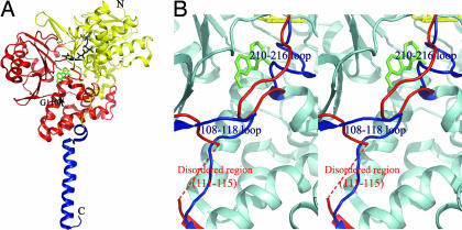
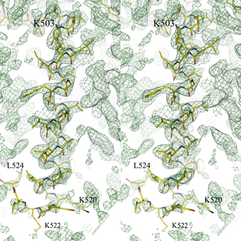
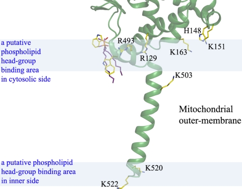
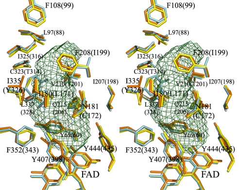
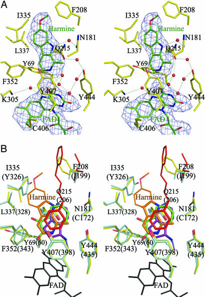
The mitochondrial outer membrane-anchored monoamine oxidase (MAO) is a biochemically important flavoenzyme that catalyzes the deamination of biogenic and xenobiotic amines. Its two subtypes, MAOA and MAOB, are linked to several psychiatric disorders and therefore are interesting targets for drug design. To understand the relationship between structure and function of this enzyme, we extended our previous low-resolution rat MAOA structure to the high-resolution wild-type and G110A mutant human MAOA structures at 2.2 and 2.17 A, respectively. The high-resolution MAOA structures are similar to those of rat MAOA and human MAOB, but different from the known structure of human MAOA [De Colibus L, et al. (2005) Proc Natl Acad Sci USA 102:12684-12689], specifically regarding residues 108-118 and 210-216, which surround the substrate/inhibitor cavity. The results confirm that the inhibitor selectivity of MAOA and MAOB is caused by the structural differences arising from Ile-335 in MAOA vs. Tyr-326 in MAOB. The structures exhibit a C-terminal transmembrane helix with clear electron density, as is also seen in rat MAOA. Mutations on one residue of loop 108-118, G110, which is far from the active center but close to the membrane surface, cause the solubilized enzyme to undergo a dramatic drop in activity, but have less effect when the enzyme is anchored in the membrane. These results suggest that the flexibility of loop 108-118, facilitated by anchoring the enzyme into the membrane, is essential for controlling substrate access to the active site. We report on the observation of the structure-function relationship between a transmembrane helical anchor and an extra-membrane domain.


Conflict of interest statement

The authors declare no conflict of interest.

Figures

Fig. 1.
Fig. 1.
The structure of human MAOA and the comparison with the early known structure. (A) The overall structure drawn in ribbon mode. N, N terminus; C, C terminus. The structure can be divided into two domains, extra-membrane domain (shown in yellow and red) and membrane binding domain (shown in blue). The extra-membrane domain was further divided as two regions, FAD binding region (yellow) and substrate/inhibitor binding region (red). FAD (black) and harmine (green) molecules are shown as stick models. The black arrow indicates the position of G110, a residue at which we introduced mutations. (B) Stereoview of the superposed structures of human MAOA and the early published human MAOA. The identical parts between the two structures are shown in cyan. The different folds at loops 108–118 and 210–216 in the two structures are shown in blue (our structure) and red (early known structure). The fragment of A111-V115 in the early structure is disordered and not visible. Pictures were generated by PyMOL (26).
Fig. 2.
Fig. 2.
Stereoview of the C-terminal transmembrane helical structure. The composite-omit map for the C-terminal domain is contoured at the 1.0-σ level at 2.2-Å resolution. A structural model of the transmembrane helix from residues 498 to 524 is superposed on the composite-omit map. The other parts of the protein structure are not shown for clarity.
Fig. 3.
Fig. 3.
The detergent molecules bound at the one-turn helix next to the transmembrane helix. The composite-omit map (1.0-σ level) was generated by CNSsolve (22). The pocket is surrounded by hydrophobic amino acid residues: W116, P118, Y121, L122, and W491. These models were generated by PyMOL (26).
Fig. 4.
Fig. 4.
Binding model of MAOA into the mitochondrial outer membrane. The positively charged residues Arg-129, His-148, Lys-151, Lys-163, Arg-493, Lys-503, Lys-520, and Lys-522 are shown. These residues are presumed to interact with the phospholipid hydrophilic head group at the membrane surface shown as blue semitransparent areas. The upper area represents the cytosolic side.
Fig. 5.
Fig. 5.
Stereoview of the substrate/inhibitor binding sites of human MAOA, human MAOB, and rat MAOA. Residues of human MAOA are shown in yellow, rat MAOA in orange, and human MAOB in cyan. The residues that are important in forming the substrate/inhibitor cavity are labeled. The residue numbering is according to the residue positions in human MAOA, which are the same as in rat MAOA. The residue numbers of human MAOB are shown in parentheses. Two residues, I199 of human MAOB and I335 of human or rat MAOA, are present as different rotamers in different complexes. The cavity was calculated by VOIDOO (13) with a 1.57-Å radius probe. These models were generated in PyMOL (26) (rmsd was 0.545 Å for human MAOB and 0.612 Å for rat MAOA).
Fig. 6.
Fig. 6.
Stereoviews of the substrate/inhibitor binding site. (A) The FoFc difference Fourier map contoured at 2.0 σ was generated at 2.2-Å resolution for the inhibitor (harmine) and FAD. Amino acid residues are shown in yellow, and FAD and harmine are shown in green. Dotted lines indicate hydrogen bonds. (B) The structure of the substrate/inhibitor binding sites in human MAOA and MAOB complexed with specific inhibitors. The residues are numbered according to human MAOA, and the numbers in parentheses are for human MAOB. MAOA and MAOB residues are shown in yellow and light blue, respectively. Inhibitors are colored as follows: orange, harmine; green, isatin (PDB ID code 1OJA); purple, rasagiline analogue (PDB ID code 2C67); and red, 1,4-diphenyl-2-butene (PDB ID code 1OJ9). FAD is shown in black. Nitrogen and oxygen atoms are shown in blue and red, respectively. Residue I199 of MAOB is present as different rotamers in different complexes. The rotamer of this residue, in MAOB with 1,4-diphenyl-2-butene, is shown in red. The residues Q215 and Y407 that form important hydrophobic interactions to the inhibitors are shown as thick stick models.

References

    1. Walker WH, Kearney EB, Seng RL, Singer TP. The covalently bound flavin of hepatic monoamine oxidase. 2. Identification and properties of cysteinyl riboflavin. Eur J Biochem. 1971;24:328–331. - PubMed
    1. Bach AW, et al. cDNA cloning of human liver monoamine oxidase A and B: Molecular basis of differences in enzymatic properties. Proc Natl Acad Sci USA. 1988;85:4934–4938. - PMC - PubMed
    1. Youdim MBH, Riederer P. The relevance of glial monoamine oxidase B and polyamines to the action of selegiline in Parkinson's disease. Movement Disorders. 1993;8:S8–S13. - PubMed
    1. Saura J, et al. Localization of monoamine oxidases in human peripheral tissues. Life Sci. 1996;59:1341–1349. - PubMed
    1. Binda C, Newton-Vinson P, Hubalek F, Edmondson DE, Mattevi A. Structure of human monoamine oxidase B, a drug target for the treatment of neurological disorders. Nat Struct Biol. 2002;9:22–26. - PubMed

Substances