Expression, purification, crystallization and preliminary X-ray diffraction analysis of thioredoxin Trx1 from Saccharomyces cerevisiae
- PMID: 18391437
- PMCID: PMC2374243
- DOI: 10.1107/S1744309108004612
Expression, purification, crystallization and preliminary X-ray diffraction analysis of thioredoxin Trx1 from Saccharomyces cerevisiae
Abstract
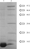
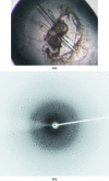
Thioredoxins play key roles in the cellular response to oxidative stress. Three isoforms of thioredoxin have been identified in Saccharomyces cerevisiae: two that are cytosolic (Trx1 and Trx2) and one that is mitochondrial (Trx3). In the present work, the cytosolic form Trx1 was cloned, expressed, purified and crystallized. Crystals were obtained by the hanging-drop vapour-diffusion method. A data set was collected from a single crystal to 1.7 A resolution. The crystal belongs to space group P2(1)2(1)2(1), with unit-cell parameters a = 32.29, b = 46.59, c = 64.20 A, alpha = beta = gamma = 90 degrees .
Figures


Similar articles
-
Purification, crystallization and preliminary X-ray diffraction analysis of glutathionylated Trx1 C33S mutant from yeast.Acta Crystallogr Sect F Struct Biol Cryst Commun. 2009 Jan 1;65(Pt 1):39-41. doi: 10.1107/S1744309108039316. Epub 2008 Dec 25. Acta Crystallogr Sect F Struct Biol Cryst Commun. 2009. PMID: 19153453 Free PMC article.
-
Expression, purification, crystallization and preliminary X-ray diffraction analysis of mitochondrial thioredoxin Trx3 from Saccharomyces cerevisiae.Acta Crystallogr Sect F Struct Biol Cryst Commun. 2006 Nov 1;62(Pt 11):1161-3. doi: 10.1107/S1744309106041467. Epub 2006 Oct 25. Acta Crystallogr Sect F Struct Biol Cryst Commun. 2006. PMID: 17077505 Free PMC article.
-
Purification, crystallization and preliminary X-ray diffraction analysis of yeast regulatory particle non-ATPase subunit 6 (Nas6p).Acta Crystallogr D Biol Crystallogr. 2002 May;58(Pt 5):859-60. doi: 10.1107/s0907444902004808. Epub 2002 Apr 26. Acta Crystallogr D Biol Crystallogr. 2002. PMID: 11976503
-
Purification, crystallization and preliminary X-ray analysis of Hsp33 from Saccharomyces cerevisiae.Acta Crystallogr Sect F Struct Biol Cryst Commun. 2007 Feb 1;63(Pt 2):114-6. doi: 10.1107/S1744309107000681. Epub 2007 Jan 17. Acta Crystallogr Sect F Struct Biol Cryst Commun. 2007. PMID: 17277453 Free PMC article.
-
Tricks of the trade used to accelerate high-resolution structure determination of membrane proteins.FEBS Lett. 2010 Jun 18;584(12):2539-47. doi: 10.1016/j.febslet.2010.04.015. Epub 2010 Apr 13. FEBS Lett. 2010. PMID: 20394746 Review.
Cited by
-
Purification, crystallization and preliminary X-ray diffraction analysis of glutathionylated Trx1 C33S mutant from yeast.Acta Crystallogr Sect F Struct Biol Cryst Commun. 2009 Jan 1;65(Pt 1):39-41. doi: 10.1107/S1744309108039316. Epub 2008 Dec 25. Acta Crystallogr Sect F Struct Biol Cryst Commun. 2009. PMID: 19153453 Free PMC article.
-
The N-terminal β-sheet of peroxiredoxin 4 in the large yellow croaker Pseudosciaena crocea is involved in its biological functions.PLoS One. 2013;8(2):e57061. doi: 10.1371/journal.pone.0057061. Epub 2013 Feb 25. PLoS One. 2013. PMID: 23451146 Free PMC article.
References
-
- Bao, R., Chen, Y., Tang, Y. J., Janin, J. & Zhou, C.-Z. (2007). Proteins, 66, 246–249. - PubMed
-
- Collaborative Computational Project, Number 4 (1994). Acta Cryst. D50, 760–763. - PubMed
-
- Gan, Z. R. (1991). J. Biol. Chem.266, 1692–1696. - PubMed
-
- Garrido, E. O. & Grant, C. M. (2002). Mol. Microbiol.43, 993–1003. - PubMed
Publication types
MeSH terms
Substances
LinkOut - more resources
Full Text Sources
Molecular Biology Databases

