. 2008 May;31(4):359-69.
doi: 10.1097/CJI.0b013e318165f5d2.

Using dendritic cell maturation and IL-12 producing capacity as markers of function: a cautionary tale

Affiliations

Using dendritic cell maturation and IL-12 producing capacity as markers of function: a cautionary tale

Anjum S Kaka et al. J Immunother. 2008 May.

Abstract

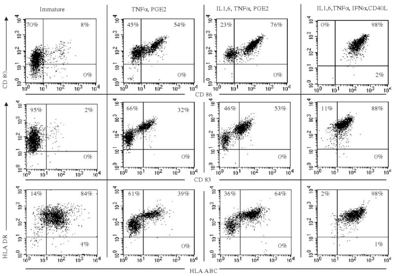
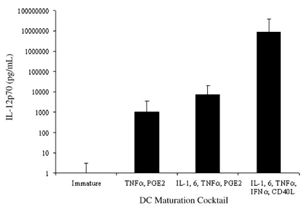
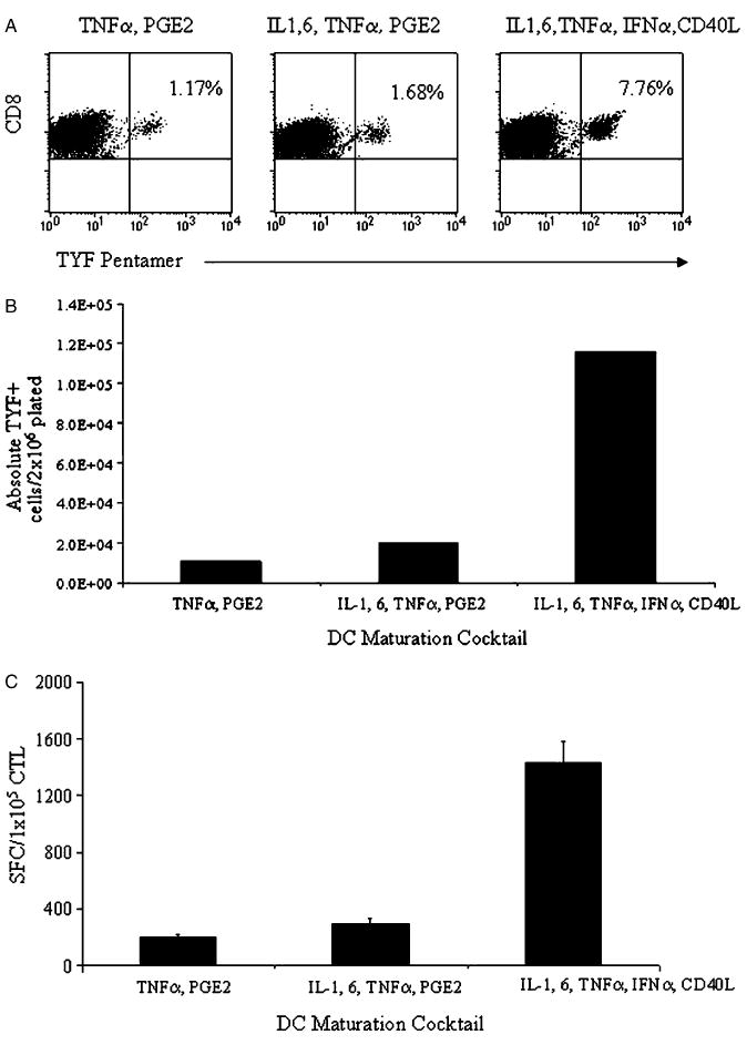
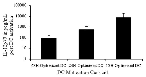
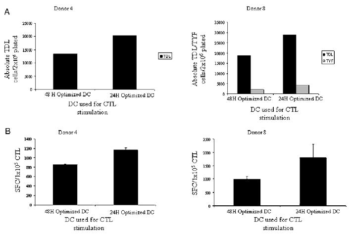
Effective dendritic cell (DC) function depends on sufficient expression of antigen and costimulatory molecules, and secretion of interleukin (IL)-12. We sought to augment DC stimulatory capacity by optimizing DC phenotype and IL-12 production. DCs, obtained by CD14-selection, were matured using 8 different cytokine cocktails, and expression of costimulatory/major histocompatibility complex molecules and IL-12 production at the end of maturation was assessed. DC stimulatory capacity was determined after pulsing with immunogenic adenoviral CD8 peptide epitopes or after transduction with an Ad5f35-null vector. Resultant T-cell cultures were analyzed using pentamer and interferon-gamma enzyme-linked immunosorbent spot assays. On the basis of DC expression of maturation markers and IL-12 production, we defined prototype "minimal" [tumor necrosis factor-alpha (TNF-alpha), prostaglandin E2], "standard" (IL-1, IL-6, TNF-alpha, prostaglandin E2), and "optimal" (IL-1, IL-6, TNF-alpha, interferon-alpha, CD40 ligand) DC cocktails. Optimal DCs were functionally superior when pulsed with CD8 peptides, but when transduced with Ad5f35, functioned poorly as antigen-presenting cells. We investigated the mechanisms underlying this discrepancy and suggest that prolonged stimulation with potent cytokines (optimal cocktail) in combination with adenoviral transduction alters the kinetics of DC maturation such that the DCs are functionally exhausted by the traditional 48-hour maturation time point. Shortening the DC maturation period posttransduction restored optimal DC stimulatory capacity. Thus, maturation stimuli and viral transduction affects DC phenotype, IL-12 producing capacity, and kinetics of maturation, and all must be considered before designing protocols to generate the optimal DC for cytotoxic T lymphocyte generation.


Figures

Figure 1
Figure 1
DC phenotype depends on maturation cocktail. DCs were incubated with granulocyte macrophage colony-stimulating factor (GM-CSF) and IL-4 for 5 days, and then matured with 1 of 3 different cytokine combinations: (i) TNF-α, PGE2 (minimal); (ii) IL-1, 6, TNF-α, and PGE2 (standard); and (iii) IL-1, IL-6, TNF-α, IFN-α, CD40L (optimal) for 2 days (A). At the end of 48 hours of maturation, DCs were analyzed by flow cytometry for expression of maturation markers, shown as dot plots. The results shown are for donor 1; similar results were obtained in 3/3 different donors.
Figure 2
Figure 2
DC maturation cocktail affects IL-12p70 production. DCs were incubated with GM-CSF and IL-4 for 5 days, and then matured with 3 different cytokine combinations: (i) TNF-α, PGE2; (ii) IL-1, IL-6, TNF-α, and PGE2; and (iii) IL-1, IL-6, TNF-α, IFN-α, and CD40L for 48 hours. At the end of 48 hours, DC supernatant was collected for the measurement of IL-12p70 by ELISA. The results are shown as mean IL-12p70 in pg/mL/1 × 106 DC ± SD averaged from results of all 8/8 different donors tested.
Figure 3
Figure 3
DCs matured using the optimal cocktail generate the most adenoviral peptide antigen-specific cells. DCs matured using the 3 prototype maturation cocktails: (i) TNF-α, PGE2; (ii) IL-1, IL-6, TNF-α, and PGE2; and (iii) IL-1, IL-6, TNF-α, IFN-α, and CD40L were loaded with peptide adenoviral antigen (TYF) and used to stimulate autologous peripheral blood T cells. At the end of 9 days, antigen-specific T-cell frequency and proliferation were measured. A, The frequency of antigen specific cells was determined using pentamer staining for the relevant peptide (TYF). The results are expressed as percent TYF-pentamer+ CD8+ cells. B, Absolute numbers of TYF-specific cells were quantitated using absolute CTL proliferation, CD8%, and pentamer %. C, IFN-γ production by the CTL in response to cognate TYF peptide was determined in an IFN-γ ELISPOT and is presented as SFC/1 × 105 CTL (subtracted from irrelevant control). The results shown are for donor 2.
Figure 4
Figure 4
DCs transduced with Ad5f35-null vector and matured using the standard cocktail generate the most adenovirus antigen-specific cells. DCs transduced with Ad5f35 were matured using the 3 prototype maturation cocktails: (i) TNF-α, PGE2; (ii) IL-1, IL-6, TNF-α, and PGE2; and (iii) IL-1, IL-6, TNF-α, IFN-α, and CD40L were used to stimulate autologous responder T cells at a stimulator to responder ratio of 1:20. Proliferation of antigen-specific T cells was determined after 9 days. A, The frequency of CD8+ TDL and TYF-specific cells was determined using pentamer staining. The results are expressed as percent CD8+ pentamer+ cells. B, Absolute numbers of TDL and TYF specific cells were determined using the absolute CTL proliferation and pentamer percentages. C, IFN-γ production in response to irradiated Ad5f35-transduced stimulators using an IFN-γ ELISPOT assay is presented as SFC/1 × 105 CTL (subtracted from control). The results shown are for donor 3.
Figure 5
Figure 5
CD4+ responder cells enhance the frequency of adenoviral antigen-specific response in all DC conditions tested. DCs transduced with Ad5f35 were matured using the 3 prototype maturation cocktails: (i) TNF-α, PGE2; (ii) IL-1, IL-6, TNF-α, and PGE2; and (iii) IL-1, IL-6, TNF-α, IFN-α, and CD40L, and used to stimulate autologous responder T cells at a stimulator to responder ratio of 1:20. Responder cells were unseparated CD4+/CD8+ or CD8+ T cells alone. A, Expansion of CD8+ antigen specific cells was determined using TDL and TYF pentamer. The results are expressed as percent pentamer + cells of all CD8+. B, Absolute numbers of TDL and TYF specific cells were determined using the absolute CTL proliferation, CD8+ percentage in CTL, and pentamer percentages. C, IFN-γ production by CTL in response to irradiated Ad5f35-transduced stimulators was determined in an IFN-γ ELISPOT assay and is presented as SFC/1 × 105 CTL (subtracted from control). The results shown are for donor 3. Similar results were obtained in 4/4 donors tested.
Figure 6
Figure 6
Ad5f35-null vector transduced optimal DCs are increasingly refractory to T-cell stimulation with increasing time postmaturation. DCs were incubated with GM-CSF and IL-4 for 5 days, and then Ad5f35 transduced and matured with the optimal cocktail cytokines- IL-1, IL-6, TNF-α, IFN-α, and CD40L for 12, 24, and 48 hours. At the end of each maturation time period, the DCs were harvested, washed, resuspended in CTL medium and stimulated with CD40L (1 μg/mL) and IFN-γ (1000 IU/mL) for 24 hours at the end of which supernatant was collected for IL-12 measurement. The results are shown as mean IL-12p70 produced from 3/3 donors tested and are represented as IL-12p70 in pg/mL/1 × 106 DCs ± SD.
Figure 7
Figure 7
DCs transduced with Ad5f35 and matured using optimal cocktail are exhausted at the end of 48 hours of maturation. DC matured using the prototype optimal cocktail (IL-1, IL-6, TNF-α, IFN-α, CD40L) for either 24 or 48 hours were used to stimulate autologous CD4+/CD8+ responder cells at a 1:20 stimulator to responder ratio. A, Expansion of TDL and TYF specific T cells were determined using the absolute T-cell numbers, CD8+ percent in CTL and pentamer percentages. B, IFN-γ production by CTL in response to irradiated Ad5f35-transduced stimulators was determined in an IFN-γ ELISPOT assay and is presented as SFC/1 × 10e5 CTL (subtracted from control). The results shown are for donors 4 and 8. Results with a similar trend were obtained for 3/3 donors tested.
Figure 8
Figure 8
DC phenotype achieved after Ad5f35 transduction and maturation with the optimal cocktail for 12, 24, and 48 hours. DCs were incubated with GM-CSF and IL-4 for 5 days, and then matured with the optimal; cytokine cocktail—IL-1, IL-6, TNF-α, IFN-α, CD40L for 12, 24, and 48 hours. At the end of each time period, the DCs were analyzed by flow cytometry for expression of maturation markers, shown as dot plots. The results, shown for donor 9, were similar in 3/3 different donors.

References

    1. Banchereau J, Steinman RM. Dendritic cells and the control of immunity. Nature. 1998;392:245–252. - PubMed
    1. Bollard CM, Straathof KC, Huls MH, et al. The generation and characterization of LMP2-specific CTLs for use as adoptive transfer from patients with relapsed EBV-positive Hodgkin disease. J Immunother (1997) 2004;27:317–327. - PubMed
    1. Mackensen A, Meidenbauer N, Vogl S, et al. Phase I study of adoptive T-cell therapy using antigen-specific CD8+ T cells for the treatment of patients with metastatic melanoma. J Clin Oncol. 2006;24:5060–5069. - PubMed
    1. Banchereau J, Briere F, Caux C, et al. Immunobiology of dendritic cells. Annu Rev Immunol. 2000;18:767–811. - PubMed
    1. Figdor CG, de Vries IJ, Lesterhuis WJ, et al. Dendritic cell immunotherapy: mapping the way. Nat Med. 2004;10:475–480. - PubMed

Publication types

MeSH terms