

doi: 10.1038/nm1752.
Epub 2008 Apr 6.
Dopamine neurons implanted into people with Parkinson's disease survive without pathology for 14 years
Affiliations
- PMID: 18391961
- PMCID: PMC2656682
- DOI: 10.1038/nm1752
Item in Clipboard
Dopamine neurons implanted into people with Parkinson's disease survive without pathology for 14 years
Nat Med.
2008 May.
Abstract
Postmortem analysis of five subjects with Parkinson's disease 9-14 years after transplantation of fetal midbrain cell suspensions revealed surviving grafts that included dopamine and serotonin neurons without pathology. These findings are important for the understanding of the etiopathogenesis of midbrain dopamine neuron degeneration and future use of cell replacement therapies.
Figures
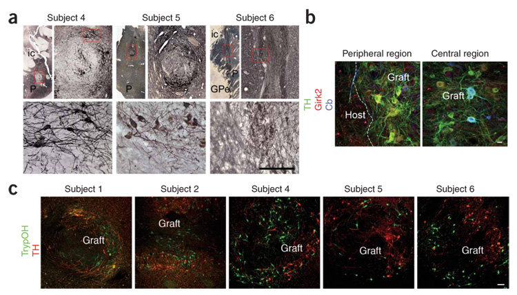
(a) Fetal ventral midbrain cell suspension grafts in the post-commissural putamina of subjects 4, 5 and 6 contained tyrosine hydroxylase–immunoreactive (TH+) neurons that were well integrated with the host and did not cause any tissue displacement. Scale bar: 5 mm (top, left-hand images), 1,000 µm (top, right-hand images) and 150 µm (bottom) for each subject. The top right-hand image for each subject is an enlargement of the boxed area in the top left-hand image, and the bottom image is an enlargement of the boxed area in the top right-hand image. P, putamen; ic, internal capsule; GPe, globus pallidus, pars externus. (b) Representative confocal images of triple immunofluorescence staining of TH (green), Girk2 (red) and calbindin (Cb, blue) within a putaminal graft of subject 6, showing peripheral and central regions of the graft. TH+Girk2+ neurons were preferentially located in the peripheral areas of the graft, whereas TH+Cb+ neurons were preferentially located in central areas. Scale bar, 20 µm. (c) Representative confocal images of double immunofluorescence studies of TH+ (red) and tryptophan hydroxylase–immunoreactive (TrypOH+, a marker for serotoninergic neurons, green) neurons within graft deposits of subjects 1, 2, 4, 5 and 6. Colocalization between TrypOH and tyrosine hydroxylase immunoreactivity was rare, and tyrosine hydroxylase did not cross-react with TrypOH. To avoid ambiguity, all neurons labeled with TH were counted as dopaminergic neurons, and all TrypOH+ neurons that did not colocalize with tyrosine hydroxylase were counted as serotoninergic neurons. Scale bar, 100 µm. All studies were conducted under the strict guidelines of a protocol approved by the Queen Elizabeth II Health Sciences Centre Ethics Review Board, Nova Scotia, Canada. Fetal ventral midbrain tissue was collected with maternal consent. Informed consent for the transplantation procedures was obtained from each subject. Permission was granted by the subjects’ next of kin to retrieve the brains for histological analysis.
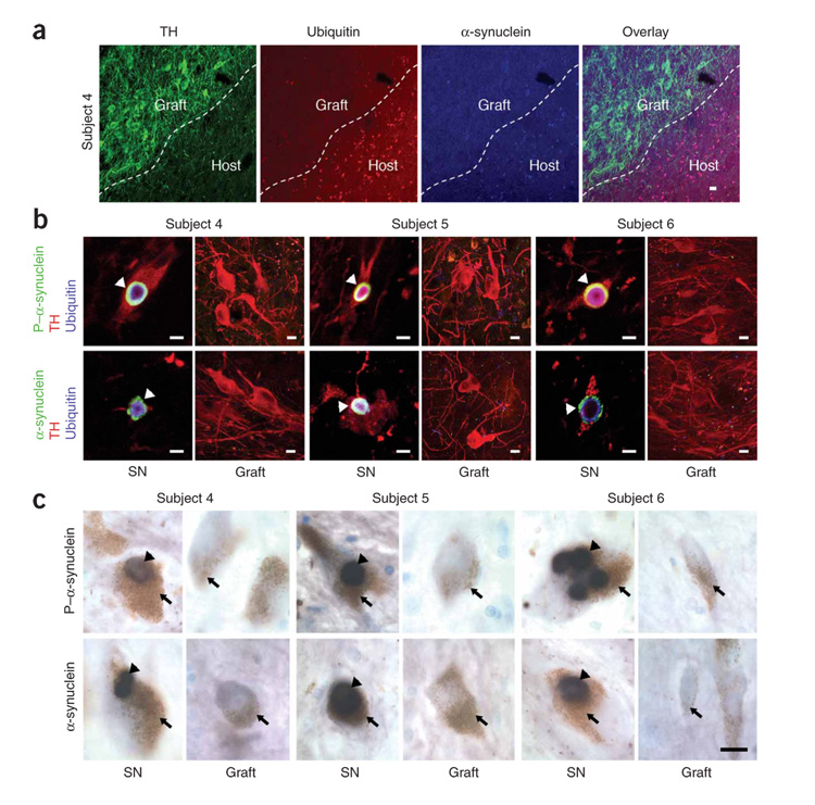
(a) There was no evidence of α-synuclein– or ubiquitin-positive inclusions within the grafts, as typified by confocal images of triple immunofluorescence labeling of TH (green), α-synuclein (blue) and ubiquitin (red) in the grafted and adjacent host putamen of subject 4. Scale bar, 10 µm. (b) TH+ host nigral neurons (red) of all subjects showed α-synuclein (green), phosphorylated α-synuclein (P–α-synuclein) (green) and ubiquitin-positive (blue) Lewy body inclusions (arrowheads), consistent with the diagnosis of Parkinson’s disease (top and bottom left-hand images for each subject). In contrast, no Lewy body pathology was found in grafted TH+ neurons, as shown in these representative images from the putamina of subjects 4, 5 and 6 (top and bottom right-hand images for each subject). SN, substantia nigra. Scale bars, 20 µm. (c) Heavily neuromelanized dopamine neurons (brown, arrows) in the substantia nigras of the subjects with Parkinson’s disease contained Lewy bodies (black, arrowheads), the pathological hallmark of Parkinson’s disease, as seen in these representative images of phosphorylated α-synuclein (top left-hand images for each subject) and α-synuclein (bottom left-hand images for each subject) immunostaining in the substantia nigras of subjects 4, 5 and 6. In the subjects’ putamina, no grafted dopamine neurons, which were lightly neuromelanized, contained Lewy bodies (top and bottom right-hand images for each subject). Scale bar, 15 µm.
Comment in
-
Assessing fetal nerve cell grafts in Parkinson's disease.Nat Med. 2008 May;14(5):483-5. doi: 10.1038/nm0508-483. Nat Med. 2008. PMID: 18463652 No abstract available.
References
Publication types
MeSH terms
Substances
Grants and funding
LinkOut - more resources
Full Text Sources
Other Literature Sources
Medical
Molecular Biology Databases
