. 2008 Apr 4:14:644-51.

Increased mitochondrial DNA damage and down-regulation of DNA repair enzymes in aged rodent retinal pigment epithelium and choroid

Affiliations

Increased mitochondrial DNA damage and down-regulation of DNA repair enzymes in aged rodent retinal pigment epithelium and choroid

Ai Ling Wang et al. Mol Vis. .

Abstract

Purpose: In the central nervous system (CNS), increased mitochondrial DNA (mtDNA) damage is associated with aging and may underlie, contribute to, or increase the susceptibility to neurodegenerative diseases. Because of the focus on the retinal pigment epithelium (RPE) and choroid as tissue relevant to age-related macular degeneration (AMD), we examined young and aged RPE and choroid, harvested from rodent eyes, for DNA damage and for changes in selected DNA repair enzymes.

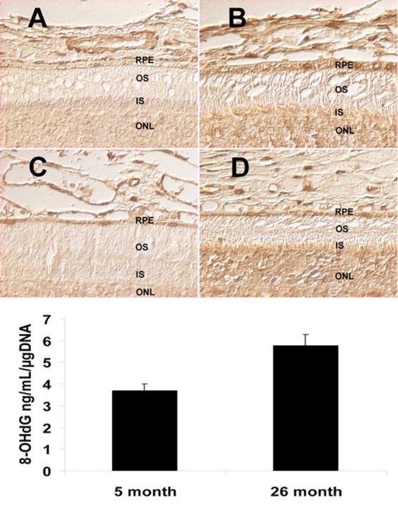
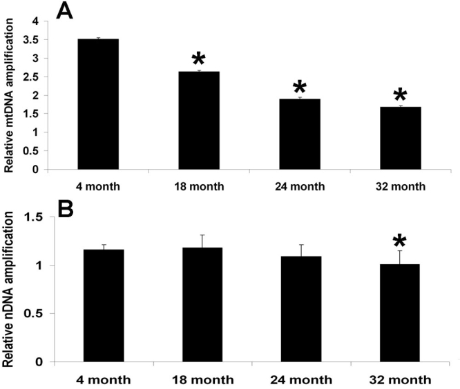
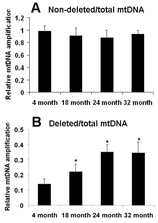
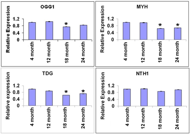
Methods: Immunohistochemical labeling and quantitative ELISA for the oxidative DNA damage marker, 8-hydroxy-2'-deoxy-guanosine (8-OHdG), were measured in young and aged rodent RPE and choroid. mtDNA and nuclear DNA (nDNA) damage was determined by quantitative polymerase chain reaction (PCR) by comparing the relative amplification of small and large DNA fragments. Expression of several DNA repair enzymes was measured using real-time quantitative reverse transcription -PCR (qRT-PCR) and immunoblot.

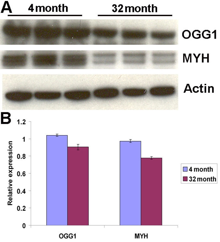
Results: Immunohistochemical labeling for 8-OHdG increased in aged rodent RPE and choroid. Quantitative ELISA confirmed increased levels of 8-OHdG. Measurements of nDNA and mtDNA lesions indicated that DNA damage is primarily in mtDNA in aged RPE and choroid. Using qRT-PCR, we found that gene expression of DNA repair enzymes, 8-oxoguanine-DNA glycosylase 1 (OGG1), mutY homolog (MYH), and thymine DNA glycosylase were decreased in an age-dependent pattern in RPE and choroid. However, endonuclease III homolog 1 was not significantly changed in aged RPE and choroid. Using immunoblots, we found that protein levels of OGG1 and MYH were decreased in aged RPE and choroid.

Conclusions: Our results show that there is increased mtDNA damage in aged RPE and choroid, which is likely due to decreased DNA repair capability. mtDNA damage in the RPE and choroid may be a susceptibility factor that underlies the development of AMD.


Figures

Figure 1
Figure 1
Increased 8-hydroxy-2’-deoxy-guanosine in the aged retinal pigment epithelium and choroid. Immunolabeling for 8-hydroxy-2’-deoxy-guanosine (8-OHdG) of retinal pigment epithelium (RPE) and choroid from from young mice (5 month, A), young rats (5 month, C) compared to old mice (22 month, B) and old rats (26 month, D). The labeling intensity of 8-OHdG was higher in the old RPE and choroid from mice (B) and rats (D), compared to the young RPE and choroid (A and C). Scale bar equals 20 µm. The oxidative DNA adduct, 8-OHdG, in rat RPE and choroid was quantitatively measured by 8-OHdG ELISA (E). There was a significant increase (p=0.007, n=6) in the amount of 8-OHdG in aged RPE and choroid.
Figure 2
Figure 2
Increased mitochondrial DNA damage compared to nucleic DNA in aged retinal pigment epithelium and choroid. DNA products from the polymerase (rTth) mediated amplification of long fragments of mitochondrial DNA (mtDNA) (13.4 kb) and nuclear DNA (nDNA) (12.5 kb) from retinal pigment epithelium (RPE) and choroid were measured. These data were normalized by the measured levels of the short fragment of mtDNA and nDNA obtained using the sample DNA template sample. As shown in Panel A, there was a significant decrease in mtDNA amplification beginning at 18 month (p=0.004, n=5), which decreased progressively at 24 month (p<0.0001, n=5) and at 32 month (p<0.0001, n=5), compared with the 4 month group. As shown in Panel B, there was no change in the nDNA amplification at 18 month (p=0.54, n=5) and 24 month (p=0.06, n=5), compared with the 4 month group. At 32 month (p=0.002, n=5), there was a significant decrease in nDNA amplification, compared with the 4 month group
Figure 3
Figure 3
Increased levels of deleted mitochondrial DNA in aged retinal pigment epithelium and choroid. Measurements of levels of the PCR products of non-deleted (A, not damaged) and deleted (B, damaged) mitochondrial DNA (mtDNA) normalized by total mtDNA normalized by total mtDNA were done using the PicoGreen reagent. Each PCR reaction started with 10 ng of genomic DNA (nuclear and mitochondrial) from rat retinal pigment epithelium (RPE) and choroid as a template. There was no difference in the non-deleted mtDNA at 18 month (p=0.02, n=5), 24 month (p=0.06, n=5), and 32 month (p=0.70, n=5), compared with the 4 month group. However, there was a significant increase in deleted mtDNA at 18 month (p=0.003, n=5), 24 month (p<0.0001, n=5), and 32 month (p<0.0001, n=5), as compared with the 4 month group. Values are the mean±SEM.
Figure 4
Figure 4
mRNA levels of DNA repair enzymes are decreased in aged retinal pigment epithelium and choroid. Comparisons of the mRNA levels of DNA repair enzymes: OGG1 (A), MYH (B), TDG (C) and NTH1 (D), in mouse retinal pigment epithelium (RPE) and choroid at different ages 18s mRNA was used as internal standard for normalization. The expression levels of OGG1 were decreased at 18 month (p=0.01, n=6), but showed no difference at 12 month (p=0.73, n=6) and at 24 month (p=0.07, n=6) as compared with the 4 month group (n=5) (A). The expression levels of MYH were decreased at 18 month (p=0.001, n=6) and at 24 month (p=0.007, n=6), but showed no difference at 12 month (p=0.87, n=6), compared with the 4 month group (B). The expression levels of TDG were decreased at 18 month (p<0.0001, n=6) and at 24 month (p=0.03, n=6), but showed no difference at 12 month (p=0.35, n=6), compared with the 4 months group (C). The expression levels of NTH1 were not different at all ages tested (p>0.05, n=6; Panel D). Values are the mean ±SEM.
Figure 5
Figure 5
Protein levels of DNA repair enzymes are decreased in aged retinal pigment epithelium and choroid. A: Immunoblots of the DNA repair enzymes, OGG1 and MYH, are shown. Actin was used as internal reference. Each group of three bands represents protein extracts from both retinal pigment epithelium (RPE) and choroid of three different animals. B: The differences in expression levels were determined by scanning gels and determining the integrated areas of the bands using Image-J software. Data are expressed as normalized ratios to actin. There were significant decreases in aged RPE and choroid of OGG1 (p=0.02, n=3) and MYH (p=0.001, n=3), compared to young RPE and choroid.

Similar articles

Cited by

References

    1. Drachman DA. Aging of the brain, entropy, and Alzheimer disease. Neurology. 2006;67:1340–52. - PubMed
    1. Burke D, Hickie I, Breakspear M, Gotz J. Possibilities for the prevention and treatment of cognitive impairment and dementia. Br J Psychiatry. 2007;190:371–2. - PubMed
    1. Rattan SI. Theories of biological aging: genes, proteins, and free radicals. Free Radic Res. 2006;40:1230–8. - PubMed
    1. Lu T, Pan Y, Kao SY, Li C, Kohane I, Chan J, Yankner BA. Gene regulation and DNA damage in the ageing human brain. Nature. 2004;429:883–91. - PubMed
    1. Martin LJ. Mitochondriopathy in Parkinson disease and amyotrophic lateral sclerosis. J Neuropathol Exp Neurol. 2006;65:1103–10. - PubMed

Publication types

MeSH terms