

Effect of trans-retinoic acid in the inhibition of cholesteatoma in guinea pigs
- PMID: 18392502
- PMCID: PMC9450672
- DOI: 10.1016/s1808-8694(15)30751-5
Effect of trans-retinoic acid in the inhibition of cholesteatoma in guinea pigs
Abstract
Middle ear cholesteatoma affected more than 5 million people until the 80;s. Many animal models were used, unsuccessfully, to study an alternative therapy to cholesteatoma.
Aim: observe the effect of the trans-retinoic acid in the inhibition of middle ear cholesteatomas induced by propylene glycol.
Study design: Clinical and Experimental.
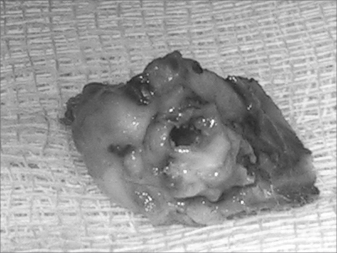
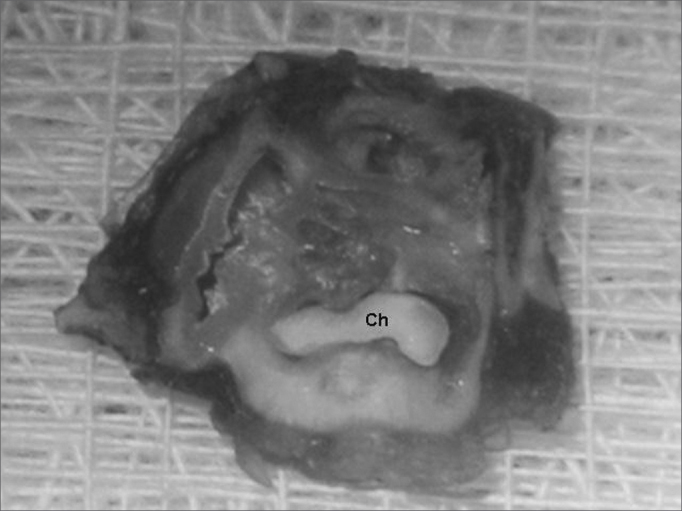
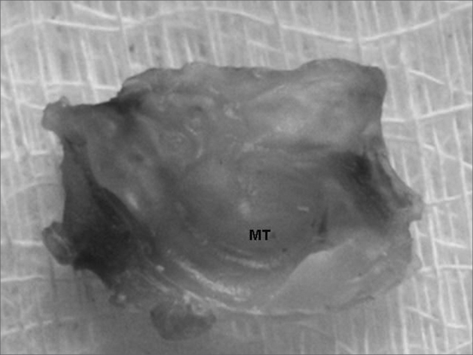
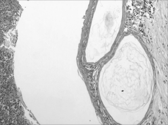
Methods: 25 guinea pigs were submitted to the application of a 100% propylene glycol solution in their bulla bilaterally and a solution of trans-retinoic acid was applied locally in the external right ear, while in the left ear saline solution was applied (control ear). The guinea pigs were slaughtered and their temporal bones were prepared for macroscopic and histological analysis.
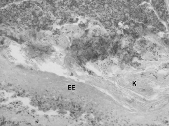
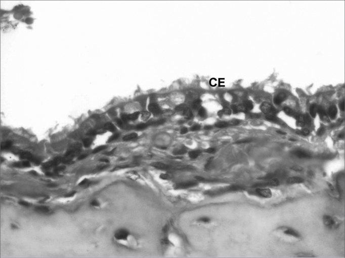
Results: The macroscopic findings had evidenced the presence of cholesteatoma in 25% of the right ears and 85% of the left ears (P=0.0003 *). The histological study had evidenced the presence of cholesteatoma in 30% of right ears and 75% of the left ears (P=0.0104*).
Conclusion: The local use of the trans-retinoic acid is effective in inhibiting the induced formation of cholesteatomas in guinea pigs.
Figures
Similar articles
-
Animal models of middle ear cholesteatoma.J Biomed Biotechnol. 2011;2011:394241. doi: 10.1155/2011/394241. Epub 2011 Apr 6. J Biomed Biotechnol. 2011. PMID: 21541229 Free PMC article. Review.
-
Single dose intratympanic mesna application inhibits propylene glycol induced cholesteatoma formation.J Laryngol Otol. 2017 Mar;131(3):215-220. doi: 10.1017/S002221511600983X. Epub 2016 Dec 20. J Laryngol Otol. 2017. PMID: 27995828
-
Effect of topical hyaluronic acid on experimental cholesteatoma.Am J Otolaryngol. 1995 Sep-Oct;16(5):312-8. doi: 10.1016/0196-0709(95)90059-4. Am J Otolaryngol. 1995. PMID: 7503374
-
Effect of Tacrolimus on Development of Experimental Cholesteatoma.Int J Pediatr Otorhinolaryngol. 2025 Jun;193:112357. doi: 10.1016/j.ijporl.2025.112357. Epub 2025 Apr 24. Int J Pediatr Otorhinolaryngol. 2025. PMID: 40300248
-
[Congenital cholesteatomas in Japanese--forty from our experience and fifty-five from a survey of the Japanese literature].Nihon Jibiinkoka Gakkai Kaiho. 1996 Sep;99(9):1200-7. Nihon Jibiinkoka Gakkai Kaiho. 1996. PMID: 8914417 Review. Japanese.
Cited by
-
Animal models of middle ear cholesteatoma.J Biomed Biotechnol. 2011;2011:394241. doi: 10.1155/2011/394241. Epub 2011 Apr 6. J Biomed Biotechnol. 2011. PMID: 21541229 Free PMC article. Review.
-
Inhibitory effect of mesna and 5-fluorouracil on propylene glycol-induced cholesteatoma in rats.Acta Otorhinolaryngol Ital. 2021 Oct;41(5):481-486. doi: 10.14639/0392-100X-N1392. Acta Otorhinolaryngol Ital. 2021. PMID: 34734585 Free PMC article.
References
-
- Sadé J. Prologue cholesteatoma and mastoid surgery. Proceedings of the Second International Conference. Kugler Publications; Sadé. Amsterdam: 1982.
-
- Rüedi L. Cholesteatoma formation in the middle ear in animal experiments. Acta Otolaryngol (Stockh) 1959;50:233–242. - PubMed
-
- Masaki M, Wright CG, Lee DH, Meyerhoff WL. Effects of Otic Drops on Chinchilla Tympanic Membrane. Arch Otolaryngol Head Neck Surg. 1988;114:1007–1011. - PubMed
-
- Vassali L, Harris DM, Gradini R, Applebaum EL. Propylene Glycol-Induced Cholesteatoma in Chinchilla Middle Ears. Am J Otolaryngol. 1988;9:180–188. - PubMed
-
- Huang CC, Shi GS, Yi ZX. Experimental Induction of middle ear cholesteatoma in rats. Am J Otolaryngol. 1988;9:165–172. - PubMed
Uncited Reference
-
- Nageris BI, Grushko I, Feinmesser R. Cholesteatoma prevention by local treatment with Vitamin A. Otol Neurotol. 2001;22(5):576–578. - PubMed
MeSH terms
Substances
LinkOut - more resources
Full Text Sources
