. 2008 May;47(5):1754-63.
doi: 10.1002/hep.22203.

Vascular biology and pathobiology of the liver: Report of a single-topic symposium

Affiliations

Vascular biology and pathobiology of the liver: Report of a single-topic symposium

Yasuko Iwakiri et al. Hepatology. 2008 May.

Abstract

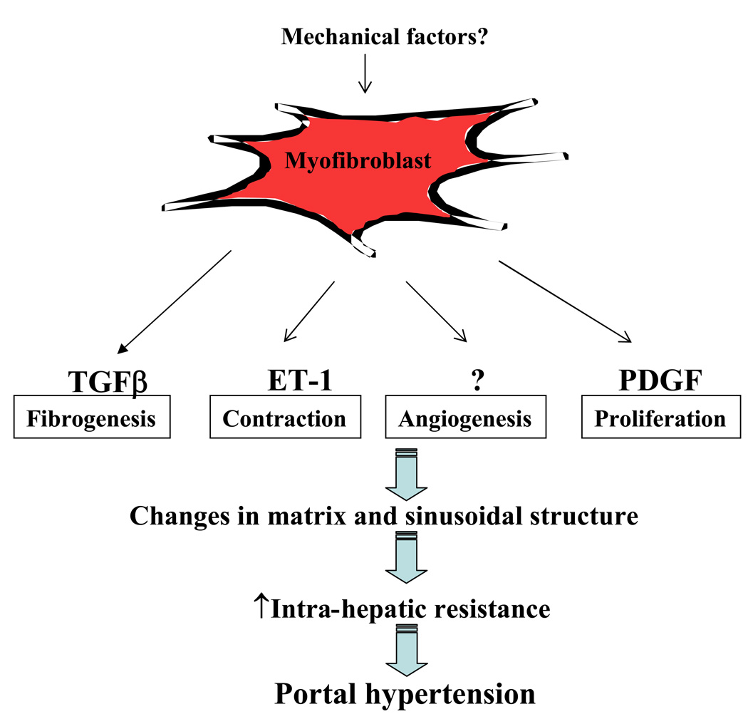
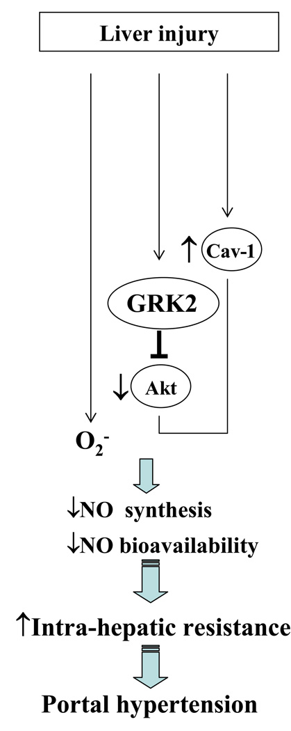
Portal hypertension and its complications account for the majority of morbidity and mortality that occurs in patients with cirrhosis. In addition to portal hypertension, a number of other vascular syndromes are also of great importance, especially the ischemia-reperfusion (IR) injury. With the identification of major vascular defects that could account for many of the clinical sequelae of these syndromes, the liver vasculature field has now integrated very closely with the broader vascular biology discipline. In that spirit, the Henry and Lillian Stratton Basic Research Single Topic Conference was held on the topic of Vascular Biology and Pathobiology of the Liver. The course took place approximately 10 years after the first American Association for the Study of Liver Disease (AASLD)-sponsored conference on this topic that occurred in Reston, Virginia. The conference initiated with an introduction to basic vascular cell signaling and then explored vascular biology specifically as it relates to liver cells. Subsequently, specific disease syndromes were discussed in more detail including portal hypertension and IR injury. Finally, clinical and translational sessions focused on emerging therapies and technologies to treat vascular diseases of the liver.


Figures

Figure 1
Figure 1. eNOS regulation by AKT and caveolin
One of the key pathways that transduce vascular cell signals is NO. Endothelial cells (EC) produce NO through the enzyme endothelial NO synthase (eNOS). eNOS is regulated in a multi-protein complex, which includes the activating kinase, Akt and putative negative regulator caveolin-1 (Cav-1). Vascular endothelial growth factor (VEGF) is one of the most potent activators of eNOS and especially relevant in the context of splanchnic vasodilation and portal hypertension.
Figure 2
Figure 2. NO signaling mechanisms
NO acts in part by stimulating soluble guanylate cyclase (sGC) to produce intracellular second messenger cyclic guanosine monophosphate (cGMP), which in turn mediates vasodilatation. Direct activation of sGC through drugs such as BAY41-2272 and BAY58-2667 has been shown to have a remarkable ability to dilate blood vessels, and thus may provide an alternate therapeutic target downstream from NO in vascular diseases that benefit from enhanced NO signaling. Besides the conventional NO signaling via cGMP, NO induced S-nitrosylation, the coupling of an NO moiety to a reactive thiol side chain of cysteine to form an S-nitrosothiol, has been proposed as a specific post-translational regulatory mechanism for many proteins and may mediate diverse biological functions.
Figure 3
Figure 3. Interactions among hepatic cells and myofibroblast activation
Hepatic circulation is unique from most other vascular beds owing to complex interactions between several liver specific cell-types, including specialized sinusoidal endothelial cells (SEC), pericyte-like hepatic stellate cells (HSC), macrophage-like - Kupffer cells (Mϕ) and additional blood-derived cells. Signaling between these cells maintains sinusoidal homeostasis and conversely, alterations in signaling lead to sinusoidal pathobiology. In particular, SEC can maintain HSC in a quiescent state and limit HSC mass through NO generation, conversely HSC (and other cells) produce VEGF that maintains SEC phenotype and specialized function. This cross-talk between SEC and HSC is disrupted in chronic liver disease and contributes to alterations in the hepatic sinusoid including HSC activation.
Figure 4
Figure 4. Myofibroblast phenotypes in portal hypertension
Differentiation of myofibroblast cells in liver requires a complex cytokine profile that includes transforming growth factor-β (TGF-β), platelet-derived growth factor (PDGF), chemokines, adipokines, components of the renin-angiotensin system, lectins, and others. Mechanical factors such as stretch and other dynamic mechanical changes are also being increasingly recognized to contribute to HSC and portal fibroblast phenotype and activation. This activation phenotype is multifaceted and includes components of proliferation, contractility, fibrogenesis, and also perhaps angiogenesis. The role of HSC in liver angiogenesis is an area of active investigation especially as it may relate to cirrhotic vascular changes and portal hypertension.
Figure 5
Figure 5. Regulation of intrahepatic NO generation in liver cirrhosis and portal hypertension
One of the most well established defects in SEC-HSC crosstalk in liver injury is the decreased bioavailability of the vasodilator NO. which occurs independent of changes in eNOS expression and through defects in post-translational regulation of the enzyme within endothelial cells. During the process of liver injury, reduction of NO bioavailability occurs due to decreased synthesis of NO, enhanced inactivation of NO by the overproduction of superoxide (O2) or both. An inactivation of eNOS occurs through an increase in cav-1 expression as well as one recently recognized protein, the G-protein-coupled receptor kinase 2 (GRK2), which directly interacts with and inhibits Akt, thereby decreasing eNOS activity.

Similar articles

Cited by

References

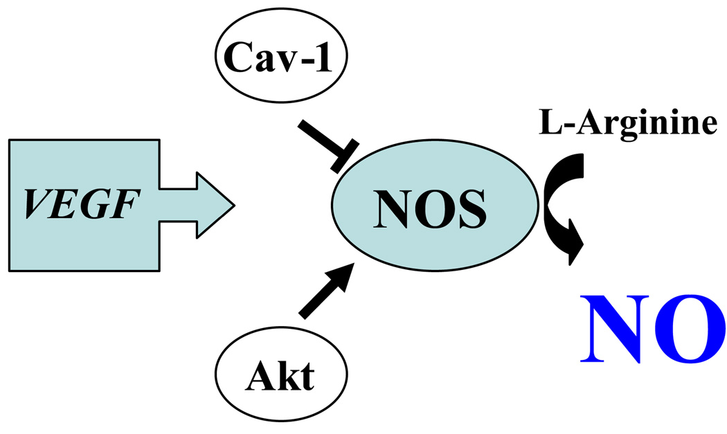
    1. Sessa WC. eNOS at a glance. J Cell Sci. 2004;117:2427–2429. - PubMed
    1. Yu J, deMuinck ED, Zhuang Z, Drinane M, Kauser K, Rubanyi GM, Qian HS, et al. Endothelial nitric oxide synthase is critical for ischemic remodeling, mural cell recruitment, and blood flow reserve. Proc Natl Acad Sci U S A. 2005;102:10999–11004. - PMC - PubMed
    1. Ackah E, Yu J, Zoellner S, Iwakiri Y, Skurk C, Shibata R, Ouchi N, et al. Akt1/protein kinase Balpha is critical for ischemic and VEGF-mediated angiogenesis. J Clin Invest. 2005;115:2119–2127. - PMC - PubMed
    1. Yu J, Bergaya S, Murata T, Alp IF, Bauer MP, Lin MI, Drab M, et al. Direct evidence for the role of caveolin-1 and caveolae in mechanotransduction and remodeling of blood vessels. J Clin Invest. 2006;116:1284–1291. - PMC - PubMed
    1. Marks DS, Vita JA, Folts JD, Keaney JF, Jr, Welch GN, Loscalzo J. Inhibition of neointimal proliferation in rabbits after vascular injury by a single treatment with a protein adduct of nitric oxide. J Clin Invest. 1995;96:2630–2638. - PMC - PubMed

Publication types

Substances