. 2008 May;47(5):1655-66.
doi: 10.1002/hep.22231.

S-adenosylmethionine inhibits lipopolysaccharide-induced gene expression via modulation of histone methylation

Affiliations

S-adenosylmethionine inhibits lipopolysaccharide-induced gene expression via modulation of histone methylation

Ainhoa Iglesias Ara et al. Hepatology. 2008 May.

Abstract

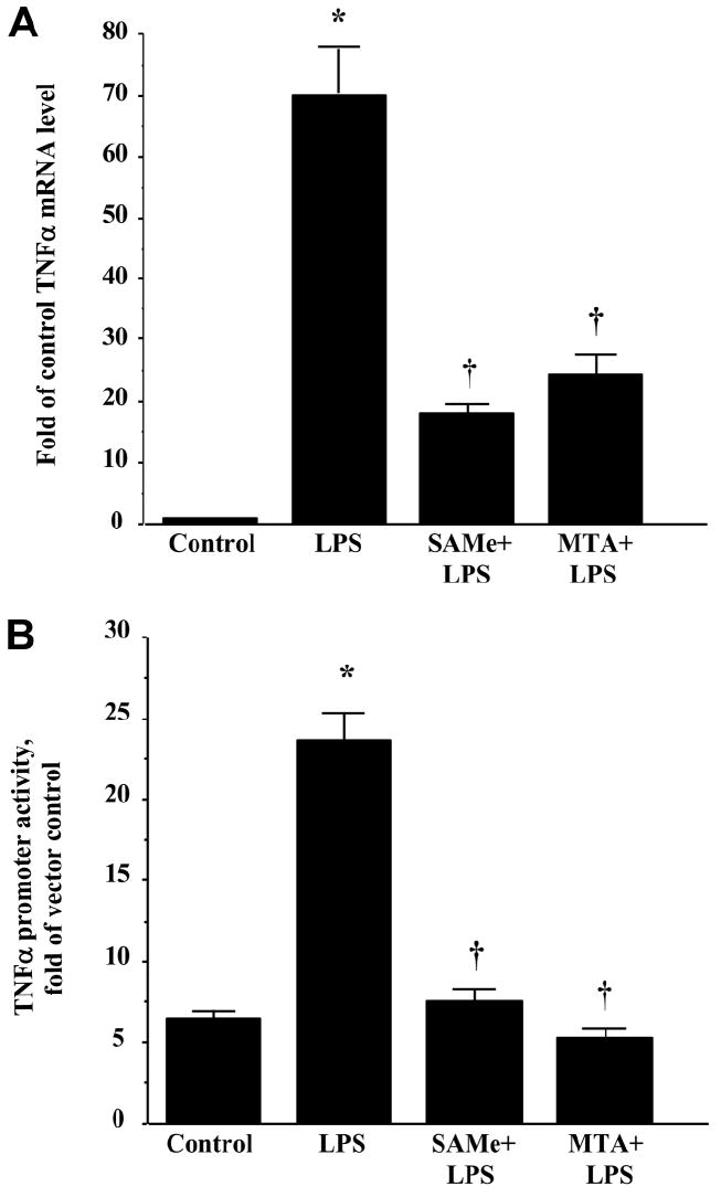
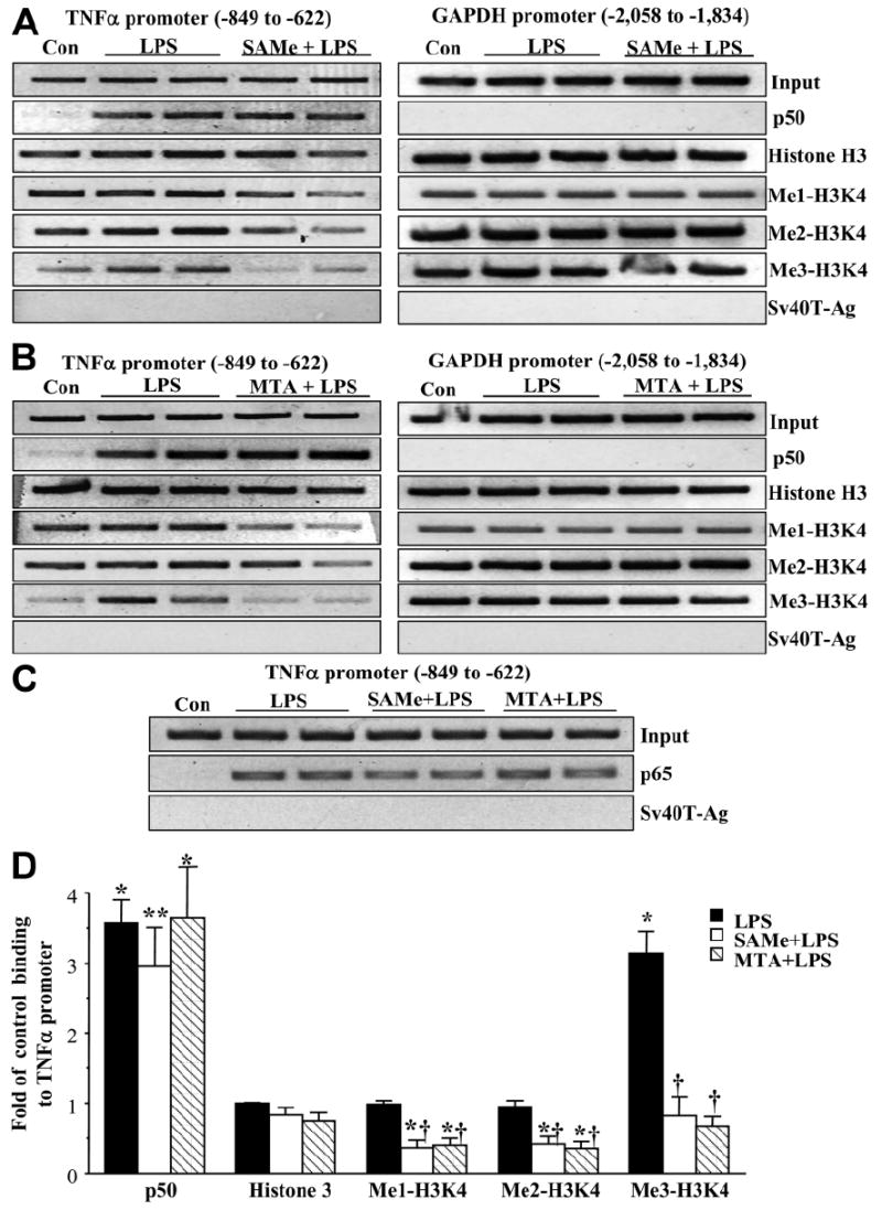
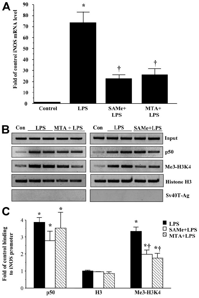
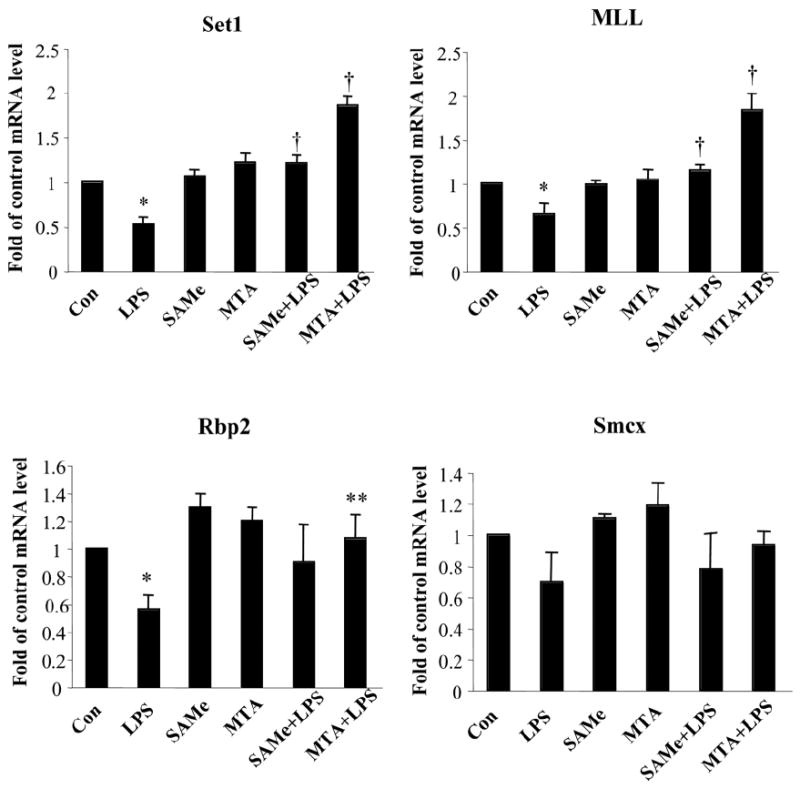
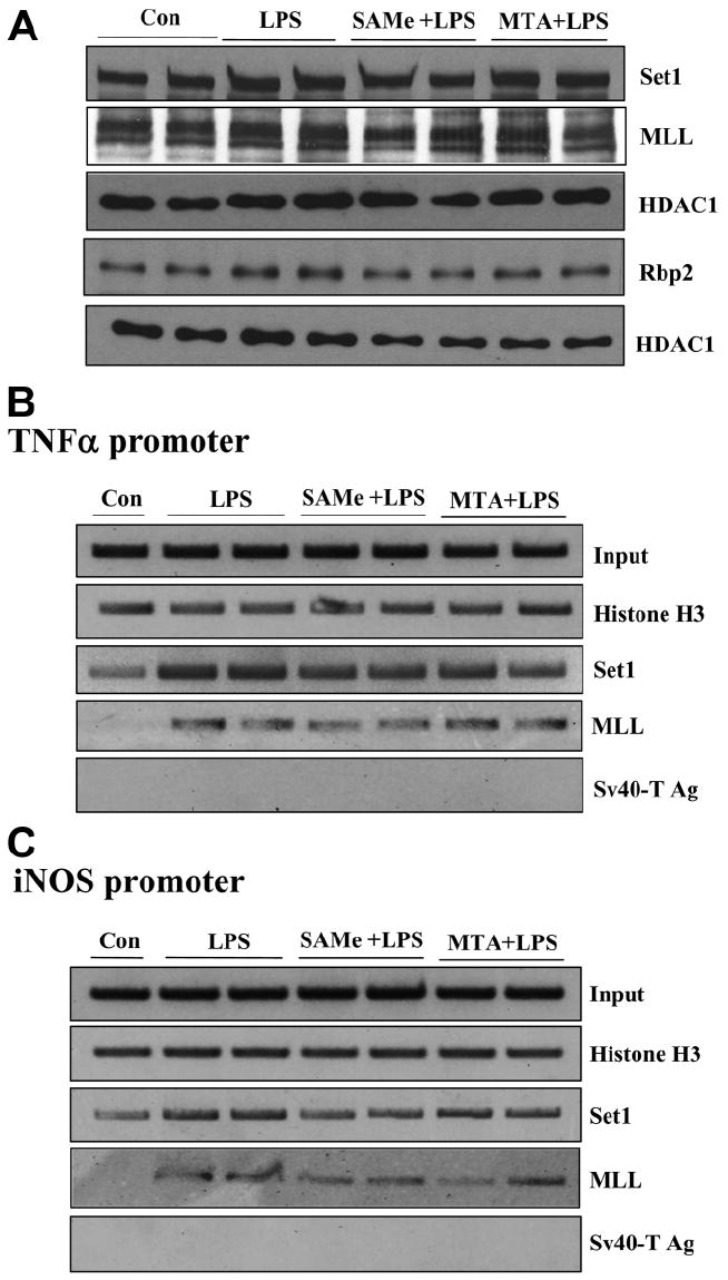
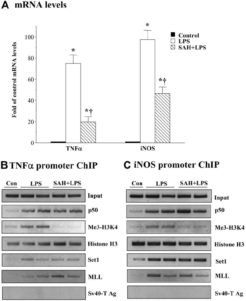
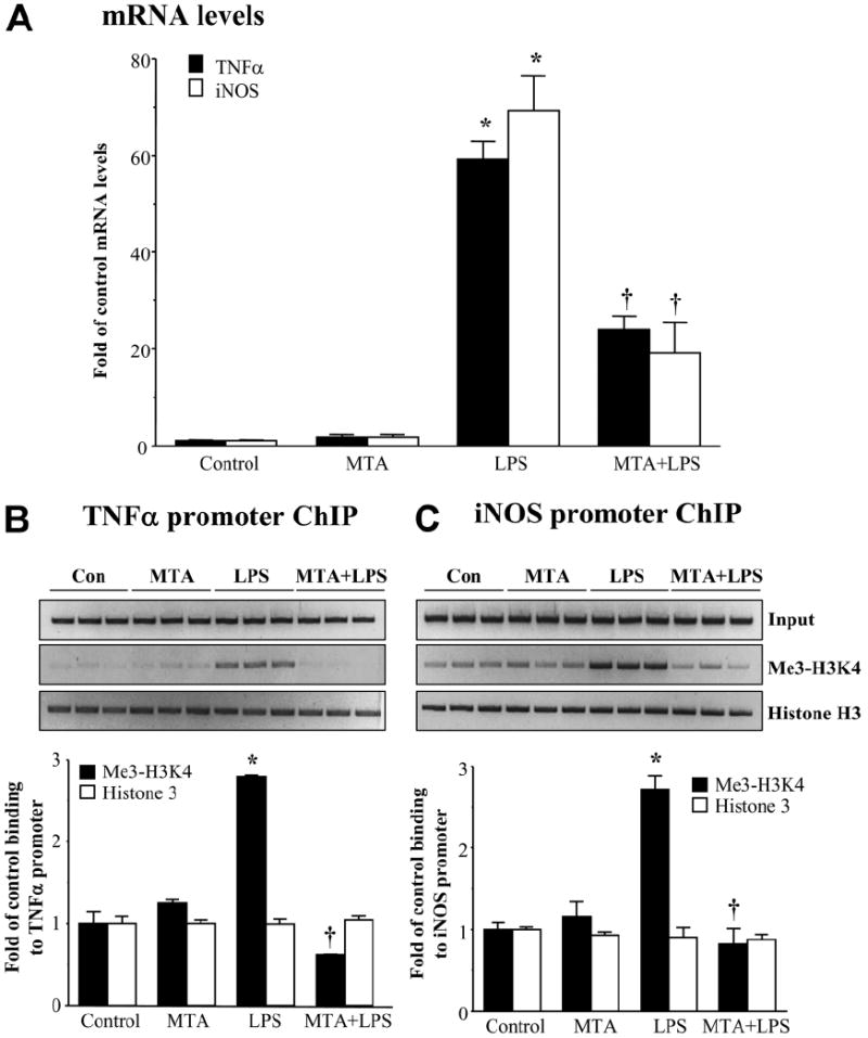
We previously showed that S-adenosylmethionine (SAMe) and its metabolite methylthioadenosine (MTA) blocked lipopolysaccharide (LPS)-induced tumor necrosis factor alpha (TNFalpha) expression in RAW (murine macrophage cell line) and Kupffer cells at the transcriptional level without affecting nuclear factor kappa B nuclear binding. However, the exact molecular mechanism or mechanisms of the inhibitory effect were unclear. While SAMe is a methyl donor, MTA is an inhibitor of methylation. SAMe can convert to MTA spontaneously, so the effect of exogenous SAMe may be mediated by MTA. The aim of our current work is to examine whether the mechanism of SAMe and MTA's inhibitory effect on proinflammatory mediators might involve modulation of histone methylation. In RAW cells, we found that LPS induced TNFalpha expression by both transcriptional and posttranscriptional mechanisms. SAMe and MTA treatment inhibited the LPS-induced increase in gene transcription. Using the chromatin immunoprecipitation assay, we found that LPS increased the binding of trimethylated histone 3 lysine 4 (H3K4) to the TNFalpha promoter, and this was completely blocked by either SAMe or MTA pretreatment. Similar effects were observed with LPS-mediated induction of inducible nitric oxide synthase (iNOS). LPS increased the binding of histone methyltransferases Set1 and myeloid/lymphoid leukemia to these promoters, which was unaffected by SAMe or MTA. The effects of MTA in RAW cells were confirmed in vivo in LPS-treated mice. Exogenous SAMe is unstable and converts spontaneously to MTA, which is stable and cell-permeant. Treatment with SAMe doubled intracellular MTA and S-adenosylhomocysteine (SAH) levels. SAH also inhibited H3K4 binding to TNFalpha and iNOS promoters.

Conclusion: The mechanism of SAMe's pharmacologic inhibitory effect on proinflammatory mediators is mainly mediated by MTA and SAH at the level of histone methylation.


Conflict of interest statement

Potential conflict of interest: Nothing to report.

Figures

Fig. 1
Fig. 1
Effects of SAMe and MTA on LPS-stimulated TNFα expression. (A) SAMe and MTA reduced an LPS-induced increase in TNFα mRNA levels. RAW cells were pretreated with 0.75 mM SAMe or 0.5 mM MTA for 16 hours and then stimulated with LPS (500 ng/mL) or vehicle (water) for 4 hours. RNA was extracted and subjected to quantitative real-time PCR analysis with a TNFα TaqMan probe with 18S rRNA for housekeeping. Results are expressed as fold over control cells (mean ± SEM) from three independent experiments performed in duplicate. *P < 0.001 versus control; †P < 0.05 versus control and LPS. (B) SAMe and MTA completely blocked LPS-induced TNFα promoter activity. RAW cells were transiently transfected with 2 μg of TNFα promoter-luciferase plasmid and 20 ng of Renilla phRL-TK with Targefect-Raw. SAMe (0.75 mM) or MTA (0.5 mM) pretreatment started 24 hours after the addition of the vectors for 16 hours. Then, LPS (500 ng/mL) or vehicle (water) was added for 4 hours. Cell lysates were collected for luciferase assay. Levels of TNFα-luc promoter were normalized by transfection efficiency as determined by Renilla luciferase activity. Results are expressed as fold over vector control (mean ± SEM) from three independent experiments performed in triplicate. *P < 0.001 versus control; †P < 0.001 versus control and LPS.
Fig. 2
Fig. 2
(A) SAMe and (B) MTA lower the binding of methylated H3K4 to the TNFα promoter. [A,B (left) and C] RAW cells were pretreated with 0.75 mM SAMe or 0.5 mM MTA for 16 hours and then stimulated with LPS (500 ng/mL) or vehicle (water) for 4 hours, and the ChIP assay was used to assess the binding of p50, p65, histone H3, and the three methylated forms of H3K4 (Me1-H3K4, Me2-H3K4, and Me3-H3K4) to the kB sites within the −849 to −622 region of the murine TNFα promoter in an endogenous chromatin configuration as described in the Materials and Methods section. Input genomic DNA (Input) was used as a loading control, and immunoprecipitation with an antibody against Sv40-Ag was used as a negative control. [A,B (right)] PCR products from the amplification of a κB site–free region within −2058 to −1834 of the murine GAPDH promoter were used as specificity controls. (D) Densitometry was performed. Results were normalized to the input and expressed as fold of control binding to TNFα promoter (mean ± SEM) from three to seven independent experiments. *P < 0.01 versus control; **P < 0.05 versus control; †P < 0.01 versus LPS.
Fig. 3
Fig. 3
(A) SAMe and MTA lower the binding of methylated H3K4 to the TNFα promoter region containing AP-1 and Egr-1 sites. RAW cells were pretreated with 0.75 mM SAMe or 0.5 mM MTA for 16 hours and then stimulated with LPS (500 ng/mL) or vehicle (water) for 4 hours, and the ChIP assay was used to assess the binding of c-Fos, c-Jun, Egr-1, and the trimethylated form of H3K4 (Me3-H3K4) to the AP-1 and Egr-1 sites within the −384 to −225 region of the murine TNFα promoter in an endogenous chromatin configuration as described in the Materials and Methods section. Input genomic DNA (Input) was used as a loading control, and immunoprecipitation with an antibody against Sv40-Ag was used as a negative control. (B,C) SAMe and MTA completely blocked LPS-induced AP-1 transcriptional activity. RAW cells were transiently transfected with 2 μg of (B) Jun2-luc or (C) TRE-luc plasmids and 20 ng of Renilla phRL-TK with Targefect-Raw. SAMe (0.75 mM) or MTA (0.5 mM) pretreatment was started 24 hours after the addition of the vectors for 16 hours. Then, LPS (500 ng/mL) or vehicle (water) was added for another 4 hours. Cell lysates were collected for luciferase assay. Levels of Jun2-luc or TRE-luc were normalized by transfection efficiency as determined by Renilla luciferase activity. Results are expressed as fold over control (mean ± SEM) from triplicates. *P < 0.005 versus control, SAMe + LPS, and MTA + LPS.
Fig. 4
Fig. 4
(A) SAMe and MTA reduced an LPS-induced increase in iNOS mRNA levels. RAW cells were pretreated with 0.75 mM SAMe or 0.5 mM MTA for 16 hours and then stimulated with LPS (500 ng/mL) or vehicle (water) for 4 hours. RNA was extracted and subjected to quantitative real-time PCR analysis with an iNOS TaqMan probe with 18S rRNA for housekeeping. Results are expressed as fold over control cells (mean ± SEM) from four independent experiments performed in duplicate. *P < 0.001 versus control; †P < 0.05 versus control and LPS. (B) MTA (left panel) and SAMe (right panel) lowered the binding of trimethylated H3K4 (Me3-H3K4) to the iNOS promoter. RAW cells were pretreated with 0.75 mM SAMe or 0.5 mM MTA for 16 hours and then stimulated with LPS (500 ng/mL) or vehicle (water) for 4 hours, and the ChIP assay was used to assess the binding of p50, histone H3, and Me3-H3K4 to the kB site within the −972 to −753 region of the murine iNOS promoter in an endogenous chromatin configuration as described in the Materials and Methods section. Input genomic DNA (Input) was used as a loading control, and immunoprecipitation with an antibody against Sv40-Ag was used as a negative control. (C) Densitometry was performed. Results were normalized to the input and expressed as fold of control binding to the iNOS promoter (mean ± SEM) from three to seven independent experiments. *P < 0.01 versus control; †P < 0.05 versus LPS.
Fig. 5
Fig. 5
Effect of LPS, SAMe, and MTA on expression of histone methyltransferases and demethylases. RAW cells were pretreated with 0.75 mM SAMe or 0.5 mM MTA for 16 hours and then stimulated with LPS (500 ng/mL) or vehicle (water) for 4 hours. RNA was extracted and subjected to quantitative real-time PCR analysis with Set1, MLL, Rbp2, and Smcx Taq-Man probes with 18S rRNA for housekeeping. Results are expressed as fold over control cells (mean ± SEM) from four to seven independent experiments performed in duplicate. *P < 0.01 versus control; †P < 0.05 versus control and LPS; **P < 0.05 versus LPS.
Fig. 6
Fig. 6
(A) Effect of the LPS, SAMe, and MTA treatments on the nuclear protein levels of Set1, MLL, and Rbp2. RAW cells were pretreated with 0.75 mM SAMe or 0.5 mM MTA for 16 hours and then stimulated with LPS (500 ng/mL) or vehicle (water) for 4 hours. Nuclear extracts (5-30 μg) were loaded onto 7%-10% sodium dodecyl sulfate–polyacrylamide gel electrophoresis followed by western blotting against Set1, MLL, Rbp2, or HDAC1. (B,C) Effect of LPS, SAMe, and MTA on the binding of Set1 and MLL to (B) TNFα and (C) iNOS promoters. RAW cells were pretreated with 0.75 mM SAMe or 0.5 mM MTA for 16 hours and then stimulated with LPS (500 ng/mL) or vehicle (water) for 4 hours, and the ChIP assay was used to assess the binding of histone H3, Set1, and MLL (B) to the kB sites within the −849 to −622 region of the murine TNFα promoter and (C) to the kB site within the −972 to −753 region of the murine iNOS promoter. Representative ChIP assays from three independent experiments are shown.
Fig. 7
Fig. 7
SAH recapitulates the inhibitory actions of SAMe and MTA. RAW cells were pretreated with 1 mM SAH for 16 hours and then stimulated with LPS (500 ng/mL) or vehicle (water) for 4 hours. (A) RNA was extracted and subjected to quantitative real-time PCR analysis with TNFα or iNOS TaqMan probes with 18S rRNA for housekeeping. Results are expressed as fold over control cells (mean ± SEM) from four independent experiments performed in duplicate. *P < 0.001 versus control; †P < 0.001 versus LPS. (B,C) ChIP assay was used to assess the binding of p50, trimethyl-H3K4 (Me3-H3K4), histone H3, Set1, and MLL (B) to the kB sites within the −849 to −622 region of the murine TNFα promoter and (C) to the kB site within the −972 to −753 region of the murine iNOS promoter. Representative ChIP assays from three independent experiments are shown.
Fig. 8
Fig. 8
MTA inhibits LPS-induced TNFα and iNOS expression and binding of trimethylated H3K4 in vivo. Mice were pretreated with MTA or vehicle, and this was followed by LPS treatment for 6 hours; hepatic TNFα and iNOS mRNA levels and the binding of trimethylated H3K4 to these promoters were examined as described in the Materials and Methods section. (A) RNA was extracted and subjected to quantitative real-time PCR analysis with TNFα or iNOS TaqMan probes with 18S rRNA for housekeeping. Results are expressed as fold over control animals (mean ± SEM) from three animals per group. *P < 0.005 versus control; †P < 0.05 versus control and LPS. (B,C) ChIP assay was used to assess the binding of trim-ethyl-H3K4 (Me3-H3K4) and histone H3 (B) to the kB sites within the −849 to −622 region of the murine TNFα promoter and (C) to the kB site within the −972 to −753 region of the murine iNOS promoter. Densitometry results are summarized below respective ChIPs. Results are expressed as fold of control binding to the TNFα or iNOS promoter (mean ± SEM) from three animals per group. *P < 0.0005 versus control; †P < 0.001 versus LPS.

References

    1. Mato JM, Lu SC. Role of S-adenosyl-L-methionine in liver health and injury. HEPATOLOGY. 2007;45:1306–1312. - PubMed
    1. Su GL. Lipopolysaccharides in liver injury: molecular mechanisms of Kupffer cell activation. Am J Physiol Gastrointest Liver Physiol. 2002;283:G256–G265. - PubMed
    1. Tracey KJ, Cerami A. Tumor necrosis factor: a pleiotropic cytokine and therapeutic target. Annu Rev Med. 1994;45:491–503. - PubMed
    1. Li J, Billiar TR. Nitric oxide. IV. Determinants of nitric oxide protection and toxicity in liver. Am J Physiol. 1999;276:G1069–G1073. - PubMed
    1. Veal N, Hsieh CL, Xiong S, Mato JM, Lu S, Tsukamoto H. Inhibition of lipopolysaccharide-stimulated TNF-alpha promoter activity by S-adenosylmethionine and 5′-methylthioadenosine. Am J Physiol Gastrointest Liver Physiol. 2004;287:G352–G362. - PubMed

Publication types