

Review
doi: 10.2174/156802608783790983.
Vascularization of engineered tissues: approaches to promote angio-genesis in biomaterials
Affiliations
- PMID: 18393893
- PMCID: PMC3929210
- DOI: 10.2174/156802608783790983
Item in Clipboard
Review
Vascularization of engineered tissues: approaches to promote angio-genesis in biomaterials
Curr Top Med Chem.
2008.
Abstract
Although there have been extensive research efforts to create functional tissues and organs, most successes in tissue engineering have been limited to avascular or thin tissues. The major hurdle in development of more complex tissues lies in the formation of vascular networks capable of delivering oxygen and nutrients throughout the engineered constructs. Sufficient neovascularization in scaffold materials can be achieved through coordinated application of angiogenic factors with proper cell types in biomaterials. This review present the current research developments in the design of biomaterials and their biochemical and biochemical modifications to produce vascularized tissue constructs.
Figures

Distribution of oxygen in tissue. Sufficient levels of oxygen and nutrient exchange occur within a few hundred micrometers away from capillaries. Limited oxygen and nutrient delivery throughout thick, implanted tissues contributes to implant failure.

Processes of neovascularization. Primitive endothelial tubes are generated through A) vasculogenesis and B) angiogenesis and eventually become stabilized by recruitment of pericytes to form C) capillary structures.
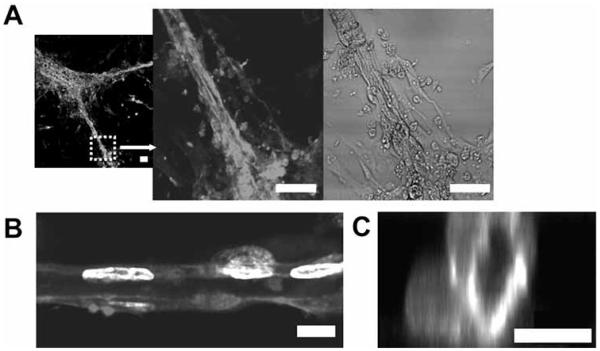
Angiogenic responses by ECs cultured on PEG hydrogels grafted with ephrin-A1. A) Ephrin-A1 immobilized on the hydrogels stimulated formation of extensive capillary-like network with lumens. Cells were stained with phalloidin-TRITC and DAPI and visualized with confocal microscopy to reveal B) longitudinal and C) vertical cross-sections. Scale bars = 50 μm in A), and 10 μm in B, C). Adapted and reprinted with permission from Moon J.J. et al. [39] @ 2007 American Chemical Society.

Schematic diagrams of various micropatterning techniques. Photolithography, microcontact printing, micromolding, and laser lithography have been used to regulate angiogenesis on biomaterials.

Directed cell migration in hydrogels with multi-photon laser lithography. Biodegradable PEG hydrogels were encapsulated with fibroblasts, and RGDS were later covalently conjugated in the hydrogels with multi-photon laser photolithography. Y-shaped RGDS channels were patterned with multi-photon laser lithography and FITC-conjugated RGDS reveals the patterned regions. Staining with phalloidin-TRITC shows that fibroblasts have migrated out of the cell clusters into selective regions of RGDS channels patterned with multi-photon laser lithography [68]. Scale bar = 100 μm.

Fibrin matrix containing heparin-binding growth factor delivery system. Linker peptides with a heparin-binding domain were conjugated in fibrin matrices. Addition of heparin and heparin-binding growth factors allows their incorporation into the delivery system. Adapted from Sakiyama-Elbert, S.E. et al. [77].

Neovascularization of fibrin matrices on chicken chorioallantoic membrane. Fibrin matrices were loaded with A, B) no drug, C, D) freely diffusible VEGF, and E, F) matrix-bound VEGF. Matrix-bound form of VEGF stimulated strong neovascularization with more normal hierarchical organizations compared to freely diffusible VEGF. Scale bars = 1 mm. Adapted and reprinted with permission from Ehrbar, M. et al. [93] @ 2004 American Heart Association.
References
-
- Jain RK, Au P, Tam J, Duda DG, Fukumura D. Engineering vascularized tissue. Nat. Biotechnol. 2005;23:821–823. - PubMed
-
- Langer R, Vacanti JP. Tissue engineering. Science. 1993;260:920–926. - PubMed
-
- Naughton GK. From lab bench to market: critical issues in tissue engineering. Ann. N. Y. Acad. Sci. 2002;961:372–385. - PubMed
-
- Oberpenning F, Meng J, Yoo JJ, Atala A. De novo reconstitution of a functional mammalian urinary bladder by tissue engineering. Nat. Biotechnol. 1999;17:149–155. - PubMed
-
- Atala A, Bauer SB, Soker S, Yoo JJ, Retik AB. Tissue-engineered autologous bladders for patients needing cystoplasty. Lancet. 2006;367:1241–1246. - PubMed
Publication types
MeSH terms
Substances
Grants and funding
LinkOut - more resources
Full Text Sources
Other Literature Sources
