

Three-layered structure shared between Lewy bodies and lewy neurites-three-dimensional reconstruction of triple-labeled sections
- PMID: 18394008
- PMCID: PMC8095600
- DOI: 10.1111/j.1750-3639.2008.00140.x
Three-layered structure shared between Lewy bodies and lewy neurites-three-dimensional reconstruction of triple-labeled sections
Abstract
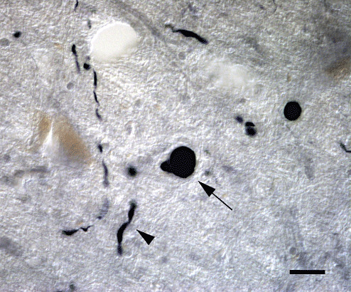
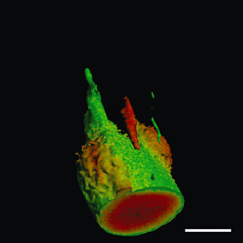
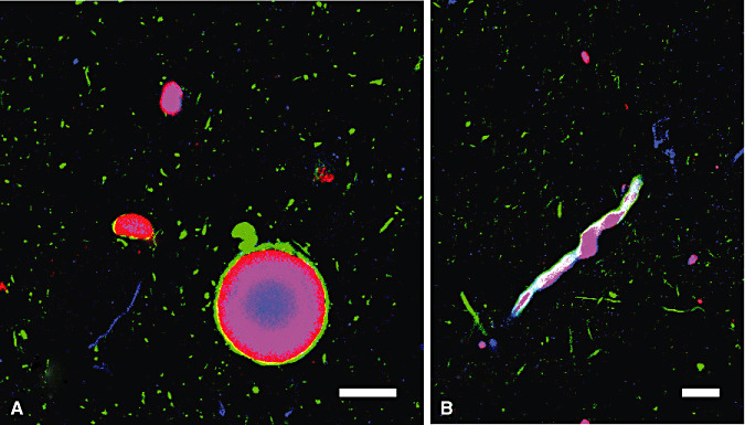
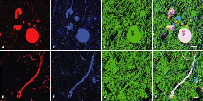
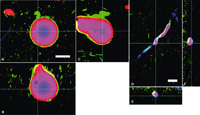
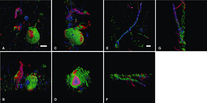
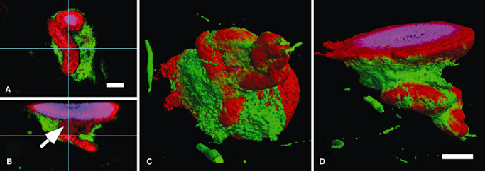
Lewy bodies (LBs) and Lewy neurites (LNs) are the hallmarks of Parkinson's disease (PD). Although LBs and LNs, frequently coexistent, share some histological properties, their appearances are quite different under conventional two-dimensional observation. In order to clarify how these apparently different structures (LBs and LNs) are related during their formation, we performed three-dimensional observation on post-mortem brainstem tissues with PD. Sixty-microm thick floating sections were multi-immunofluorolabeled for alpha-synuclein (alphaS), ubiquitin (Ub) and neurofilament (NF). Serial confocal images were reconstructed with software. External three-dimensional configuration of LBs, double-labeled for alphaS and NF, exhibited frequent continuity with LNs (70%). Internally, alphaS and Ub formed the three-dimensional concentric inner layers and NF rimmed these inner layers. This layered structure was shared among spherical LBs, rod-shaped LNs and even convoluted forms of LBs/LNs. Furthermore, each layer exhibited continuity without interruption even in the convoluted form and around its junction to spherical LBs. This three-layered structure shared among various Lewy pathologies and their layered continuity on three-dimensional basis favor the hypothesis that LNs evolve into LBs. Besides progression from pale bodies to LBs, structural evolution from LNs into LBs may provide an alternative explanation for the variability of alphaS deposits and their interrelation.
Figures
Similar articles
-
Pale neurites, premature α-synuclein aggregates with centripetal extension from axon collaterals.Brain Pathol. 2012 Jan;22(1):67-78. doi: 10.1111/j.1750-3639.2011.00509.x. Epub 2011 Aug 16. Brain Pathol. 2012. PMID: 21672073 Free PMC article.
-
Parkin localizes to the Lewy bodies of Parkinson disease and dementia with Lewy bodies.Am J Pathol. 2002 May;160(5):1655-67. doi: 10.1016/S0002-9440(10)61113-3. Am J Pathol. 2002. PMID: 12000718 Free PMC article.
-
Limbic neuropathology in idiopathic Parkinson's disease with concomitant dementia.Folia Neuropathol. 2004;42(3):141-50. Folia Neuropathol. 2004. PMID: 15535032
-
Modeling Lewy pathology propagation in Parkinson's disease.Parkinsonism Relat Disord. 2014 Jan;20 Suppl 1(0 1):S85-7. doi: 10.1016/S1353-8020(13)70022-1. Parkinsonism Relat Disord. 2014. PMID: 24262196 Free PMC article. Review.
-
[Lewy body formation in Parkinson's disease: neurodegeneration or neuroprotection?].Rinsho Shinkeigaku. 2008 Nov;48(11):981-3. doi: 10.5692/clinicalneurol.48.981. Rinsho Shinkeigaku. 2008. PMID: 19198138 Review. Japanese.
Cited by
-
Effects of local reduction of endogenous α-synuclein using antisense oligonucleotides on the fibril-induced propagation of pathology through the neural network in wild-type mice.Acta Neuropathol Commun. 2024 May 14;12(1):75. doi: 10.1186/s40478-024-01766-3. Acta Neuropathol Commun. 2024. PMID: 38745295 Free PMC article.
-
Propagation of alpha-synuclein pathology: hypotheses, discoveries, and yet unresolved questions from experimental and human brain studies.Acta Neuropathol. 2016 Jan;131(1):49-73. doi: 10.1007/s00401-015-1485-1. Epub 2015 Oct 7. Acta Neuropathol. 2016. PMID: 26446103 Free PMC article. Review.
-
Posttranscriptional regulation of neurofilament proteins and tau in health and disease.Brain Res Bull. 2023 Jan;192:115-127. doi: 10.1016/j.brainresbull.2022.10.017. Epub 2022 Oct 29. Brain Res Bull. 2023. PMID: 36441047 Free PMC article. Review.
-
Axonal degeneration as a therapeutic target in the CNS.Cell Tissue Res. 2012 Jul;349(1):289-311. doi: 10.1007/s00441-012-1362-3. Epub 2012 Mar 6. Cell Tissue Res. 2012. PMID: 22392734 Free PMC article. Review.
-
Bringing CLARITY to the human brain: visualization of Lewy pathology in three dimensions.Neuropathol Appl Neurobiol. 2016 Oct;42(6):573-87. doi: 10.1111/nan.12293. Epub 2015 Dec 7. Neuropathol Appl Neurobiol. 2016. PMID: 26526972 Free PMC article.
References
-
- Arima K, Uéda K, Sunohara N, Hirai S, Izumiyama Y, Tonozuka‐Uehara H, Kawai M (1998) Immunoelectron‐microscopic demonstration of NACP/alpha‐synuclein‐epitopes on the filamentous component of Lewy bodies in Parkinson's disease and in dementia with Lewy bodies. Brain Res 808:93–100. - PubMed
-
- Betarbet R, Sherer TB, MacKenzie G, Garcia‐Osuna M, Panov AV, Greenamyre JT (2000) Chronic systemic pesticide exposure reproduces features of Parkinson's disease. Nat Neurosci 3:1301–1306. - PubMed
-
- Braak E, Sandmann‐Keil D, Rüb U, Gai WP, De Vos RA, Jansen Steur ENH et al (2001) Alpha‐synuclein immunopositive Parkinson's disease‐related inclusion bodies in lower brain stem nuclei. Acta Neuropathol (Berl) 101:195–201. - PubMed
-
- Braak H, Sandmann‐Keil D, Gai WP, Braak E (1999) Extensive axonal Lewy neurites in Parkinson's disease: a novel pathological feature revealed by alpha‐synuclein immunocytochemistry. Neurosci Lett 265:67–69. - PubMed
Publication types
MeSH terms
Substances
LinkOut - more resources
Full Text Sources
Medical
