. 2008 Jul;18(3):378-86.
doi: 10.1111/j.1750-3639.2008.00137.x. Epub 2008 Apr 2.

Expression of integrin alphavbeta3 in gliomas correlates with tumor grade and is not restricted to tumor vasculature

Affiliations

Expression of integrin alphavbeta3 in gliomas correlates with tumor grade and is not restricted to tumor vasculature

Oliver Schnell et al. Brain Pathol. 2008 Jul.

Abstract

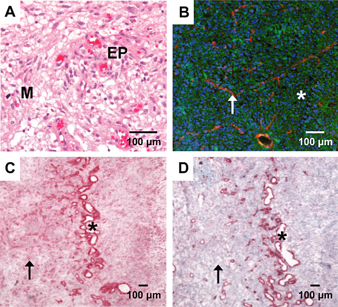
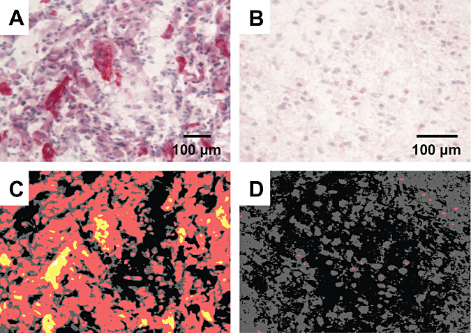
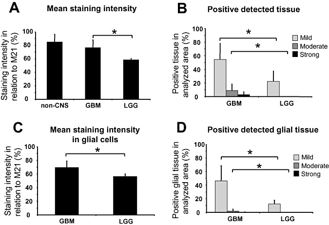
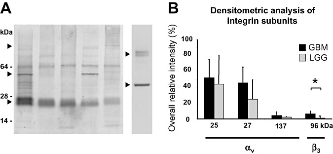
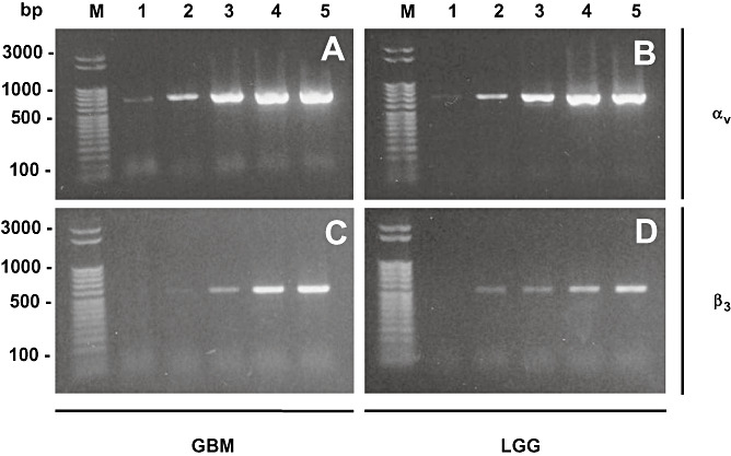
In malignant gliomas, the integrin adhesion receptors seem to play a key role for invasive growth and angiogenesis. However, there is still a controversy about the expression and the distribution of alpha(v)beta(3) integrin caused by malignancy. The aim of our study was to assess the extent and pattern of alpha(v)beta(3) integrin expression within primary glioblastomas (GBMs) compared with low-grade gliomas (LGGs). Tumor samples were immunostained for the detection of alpha(v)beta(3) integrin and quantified by an imaging software. The expression of alpha(v)beta(3) was found to be significantly higher in GBMs than in LGGs, whereby focal strong reactivity was restricted to GBMs only. Subsequent analysis revealed that not only endothelial cells but also, to a large extent, glial tumor cells contribute to the overall amount of alpha(v)beta(3) integrin in the tumors. To further analyze the integrin subunits, Western blots from histologic sections were performed, which demonstrated a significant difference in the expression of the beta(3) integrin subunit between GBMs and LGGs. The presented data lead to new insights in the pattern of alpha(v)beta(3) integrin in gliomas and are of relevance for the inhibition of alpha(v)beta(3) integrin with specific RGD peptides and interfering drugs to reduce angiogenesis and tumor growth.


Figures

Figure 1
Figure 1
Hematoxilin & Eosin (H&E) (A) and immunostain of integrin αvβ3 (C), CD31 (D) and fluorescent overlay (B) in samples of glioblastoma. Standard H&E staining (A) shows typical morphology of a malignant glioma. Immunohistochemical staining of αvβ3 (C) is intense in vascular structures (asterisk) as confirmed by a consecutive section stained for the endothelial cell marker CD31 (D). Rather, αvβ3 is clearly detectable throughout the whole section as expressed by the glial tumor cells (arrow). A fluorescent overlay picture of another area with dense tumor growth (B) demonstrates that the integrin αvβ3 (green) is not restricted to vascular structures (red, asterisk) but shows a ubiquitous distribution pattern (cell nuclei blue). Scale bars: 100 µm. Abbreviations: EP = endothelial proliferation; M = mitosis.
Figure 2
Figure 2
Detection of αvβ3 integrin in malignant (A) and a low‐grade (B) glioma and analysis by an imaging software (C,D). The malignant glioma (A) exhibit a much higher expression of αvβ3 integrin (red) than the low‐grade glioma (B), where only very few glial cells are immunopositive for the integrin. The detection of the staining intensity in the malignant glioma shows a large overall baseline (red) expression with certain strong (yellow) or even intense (white) expression in microvascular structures (C). In contrast, only some red dots can be detected in the low‐grade glioma (D). Scale bars: 100 µm
Figure 3
Figure 3
Statistical analysis of the αvβ3 integrin immunohistochemistry in patients with glioblastomas (GBMs) and low‐grade gliomas (LGGs) and non‐CNS tumors (non‐CNS). A. Mean staining intensity of non‐CNS tumors, GBMs and LGGs. B. Fraction of mild, moderate and strong stained tissue in GBMs and LGGs. C. Mean staining intensity of glial tissue only in GBMs and LGGs. D. Fraction of mild, moderate and strong stained tissue of glial tissue only in GBMs and LGGs. Significant differences (P < 0.05, Student's t‐test) are indicated with an asterisk.
Figure 4
Figure 4
Western blot analysis of subunits and degradation products of the αvβ3 integrin. The Western blot analysis of five different glioblastoma (GBM) tissue samples (A, left lanes) and densitometric analysis of integrin subunits (B). In all glial tumors, degradation products of the αv subunit (αv light chains) were detected as a double band at 25/27 kDa (▸). Expression was constantly present but heterogeneous between different GBMs. The β3 subunit was detected at about 96 kDa (▸). Degradation products of αv and β3 subunit were found at 55 kDa (▸). The detection of CD31 at about 80 kDa (right ▸) and β‐actin at about 42 kDa (right ▸) is additionally demonstrated in the lane on the right. B. Densitometric analysis revealed significant differences for the β3 subunit between malignant gliomas and low‐grade gliomas (LGGs) (P < 0.05; Student's t‐test; indicated with an asterisk). The αv subunit (about 137 kDa) and its degradation products (αv light chains, 25/27 kDa) showed no significant differences between high‐ and low‐grade gliomas.
Figure 5
Figure 5
PCR analysis of αvβ3 integrin subunits. PCR analysis of αvβ3 integrin subunits αv (A,B) and β3 (C,D) of a glioblastoma (GBM; A,C) and low‐grade glioma (LGG; B,D) after different recombination cycles. Lane 1: 24 cycles, lane 2: 27 cycles, lane 3: 30 cycles, lane 4: 33 cycles and lane 5: 36 recombination cycles. Standard PCR revealed that both subunits αv (690 bp) and β3 (510 bp) are synthesized in high as well as low‐grade gliomas indicating that both tumor entities are able to express a functionally active αvβ3 integrin receptor. Marker (M) was a 50 bp ladder (Novagen, Perfect DNA ladder).

Similar articles

Cited by

References

    1. Bader BL, Rayburn H, Crowley D, Hynes RO (1998) Extensive vasculogenesis, angiogenesis, and organogenesis precede lethality in mice lacking all alpha v integrins. Cell 95:507–519. - PubMed
    1. Beer AJ, Schwaiger M (2007) Molecular imaging with new PET tracers. Radiologe 47:8–17. - PubMed
    1. Beer AJ, Haubner R, Goebel M, Luderschmidt S, Spilker ME, Wester HJ et al (2005) Biodistribution and pharmacokinetics of the alphavbeta3‐selective tracer 18F‐galacto‐RGD in cancer patients. J Nucl Med 46:1333–1341. - PubMed
    1. Beer AJ, Haubner R, Sarbia M, Goebel M, Luderschmidt S, Grosu AL et al (2006) Positron emission tomography using [18F] Galacto‐RGD identifies the level of integrin alpha(v)beta3 expression in man. Clin Cancer Res 12:3942–3949. - PubMed
    1. Bello L, Francolini M, Marthyn P, Zhang J, Carroll RS, Nikas DC et al (2001) Alpha(v)beta3 and alpha(v)beta5 integrin expression in glioma periphery. Neurosurgery 49:380–389. - PubMed

Publication types

Substances