. 2008 Apr;14(4):592-9.
doi: 10.3201/eid1404.071044.

Retrospective analysis of monkeypox infection

Affiliations

Retrospective analysis of monkeypox infection

Melissa E Dubois et al. Emerg Infect Dis. 2008 Apr.

Abstract

Serologic cross-reactivity between orthopoxviruses is a substantial barrier to laboratory diagnosis of specific orthopoxvirus infections and epidemiologic characterization of disease outbreaks. Historically, time-consuming and labor-intensive strategies such as cross-adsorbed neutralization assays, immunofluorescence assays, and hemagglutination-inhibition assays have been used to identify orthopoxvirus infections. We used cross-adsorption to develop a simple and quantitative postadsorption ELISA for distinguishing between monkeypox and vaccinia infections. Despite the difficulty of diagnosing clinically inapparent monkeypox in previously vaccinated persons, this technique exhibited 100% sensitivity and 100% specificity for identifying clinically overt monkeypox infection irrespective of vaccination history. We also describe a Western blot technique in which up to 3 diagnostic bands may be used to distinguish between vaccinia and monkeypox infection. The techniques described provide independent diagnostic tests suitable for retrospective analysis of monkeypox outbreaks.


Figures

Figure 1
Figure 1
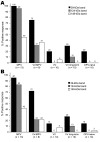
Diagnosis of monkeypox by postadsorption ELISA. Plasma samples were obtained from monkeypox-immune persons (2–30 months postinfection), vaccinia-immune persons (2–4 months postinfection), or uninfected orthopoxvirus-naive persons and tested on ELISA plates coated with inactivated monkeypox antigen. A) A representative monkeypox-specific ELISA with plasma samples from an unvaccinated monkeypox-infected person (MPV), a previously vaccinated (i.e., vaccinia-immune) monkeypox-infected person (VV-MPV), a vaccinia-immune person (VV), a vaccinia-immune person who was revaccinated with vaccinia (VV-VV), and an uninfected orthopoxvirus-naive person (OPV-naive). Plasma was not preadsorbed (∅, gray bars), preadsorbed with inactivated vaccinia antigen (black bars), or preadsorbed with inactivated monkeypox antigen (white bars) before ELISA on monkeypox-coated plates. Numbers above bars refer to differences in postadsorption MPV ELISA titers after adsorption with vaccinia antigen compared with adsorption with monkeypox antigen. Plasma from 1 orthopoxvirus-naive person (representative of n = 12) was not preadsorbed with viral antigen because it was seronegative (<100 ELISA units) and below our detection limit (dashed horizontal line). B) Plasma samples from monkeypox-infected persons (•, n = 13), vaccinia-immune monkeypox-infected persons (■, n = 8), vaccinia-immune persons (▲, n = 10), and revaccinated vaccinia-immune persons (▼, n = 10) were tested by postadsorption ELISA. Data show fold-differences of monkeypox antibody titers after adsorption with vaccinia antigen compared with adsorption with monkeypox antigen. Dashed horizontal line indicates a diagnostic cutoff indicative of a positive result, which was determined as a postadsorption difference score of >2.5. *Denotes results of plasma samples obtained from persons with clinically inapparent monkeypox infection.
Figure 2
Figure 2
Development of a monkeypox (MPV)–specific diagnostic assay using Western blot analysis. Adsorption of cross-reactive orthopoxvirus antibodies with vaccinia antigen before Western blot analysis provided easier identification of monkeypox-specific bands. A) Two micrograms of sucrose gradient–purified monkeypox virus or vaccinia virus (VV) were separated by sodium dodecyl sulfate–polyacrylamide gel electrophoresis (4%–20% gels) and stained with GelCode Blue (Pierce, Rockford, IL, USA) to compare banding patterns and confirm equivalent protein loading. Proteins were electrophoretically transferred to polyvinylidene difluoride membranes and probed with plasma from B) a monkeypox-immune person, C) a vaccinia-immune person, or D) an orthopoxvirus-naive person after adsorption of plasma with control antigen (uninfected H2O2-treated BSC40 cell lysate) or vaccinia antigen (H2O2-inactived vaccinia-infected BSC40 cell lysate). Immunoreactive bands were detected with peroxidase-conjugated antihuman immunoglobulin G plus chemiluminescent substrate and exposed to x-ray film. Arrows indicate location of diagnostic bands with apparent molecular masses of 148, 124, and 39 kDa. Rectangles indicate locations of diagnostic bands.
Figure 3
Figure 3
Diagnosis of monkeypox infection by Western blot analysis. Plasma samples from unvaccinated monkeypox-infected (2–30 months postinfection) (MPV), vaccinia-immune monkeypox-infected (2–6 months postinfection) (VV-MPV), primary vaccinia-immune (2–4 months postimmunization) (VV), long-term vaccinia-immune (>20 years postimmunization) (VV long-term), and orthopoxvirus-naive (OPV) persons were analyzed by Western blot after adsorption with vaccinia-infected BSC40 cell lysate to reduce cross-reactive antibodies as described in Figure 2. Immunoreactivity to diagnostic protein bands of ≈39 kDa, 124 kDa, and 148 kDa was assessed by A) unblinded analysts with knowledge of subject medical history (n = 2) and B) blinded analysts who did not have knowledge of subject medical history (n = 4). Findings of each analyst were averaged for each person and percentages shown represent a composite of all data points.

References

    1. Nalca A, Rimoin AW, Bavari S, Whitehouse CA. Reemergence of monkeypox: prevalence, diagnostics, and countermeasures. Clin Infect Dis. 2005;41:1765–71. 10.1086/498155 - DOI - PubMed
    1. Rimoin AW, Kisalu N, Kebela-Ilunga B, Mukaba T, Wright LL, Formenty P, et al. Endemic human monkeypox, Democratic Republic of Congo, 2001–2004. Emerg Infect Dis. 2007;13:934–7. - PMC - PubMed
    1. Jezek Z, Fenner F, eds. Human monkeypox. Basel: Karger; 1988.
    1. Reed KD, Melski JW, Graham MB, Regnery RL, Sotir MJ, Wegner MV, et al. The detection of monkeypox in humans in the Western Hemisphere. N Engl J Med. 2004;350:342–50. 10.1056/NEJMoa032299 - DOI - PubMed
    1. Sejvar JJ, Chowdary Y, Schomogyi M, Stevens J, Patel J, Karem K, et al. Human monkeypox infection: a family cluster in the midwestern United States. J Infect Dis. 2004;190:1833–40. 10.1086/425039 - DOI - PubMed

Publication types

Substances

LinkOut - more resources