. 2008 Apr;14(4):600-7.
doi: 10.3201/eid1404.071016.

Wild ducks as long-distance vectors of highly pathogenic avian influenza virus (H5N1)

Affiliations

Wild ducks as long-distance vectors of highly pathogenic avian influenza virus (H5N1)

Juthatip Keawcharoen et al. Emerg Infect Dis. 2008 Apr.

Abstract

Wild birds have been implicated in the expansion of highly pathogenic avian influenza virus (H5N1) outbreaks across Asia, the Middle East, Europe, and Africa (in addition to traditional transmission by infected poultry, contaminated equipment, and people). Such a role would require wild birds to excrete virus in the absence of debilitating disease. By experimentally infecting wild ducks, we found that tufted ducks, Eurasian pochards, and mallards excreted significantly more virus than common teals, Eurasian wigeons, and gadwalls; yet only tufted ducks and, to a lesser degree, pochards became ill or died. These findings suggest that some wild duck species, particularly mallards, can potentially be long-distance vectors of highly pathogenic avian influenza virus (H5N1) and that others, particularly tufted ducks, are more likely to act as sentinels.


Figures
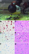
Figure 1
Figure 1
Central nervous system changes in wild ducks experimentally infected with highly pathogenic avian influenza virus (H5N1). A) Torticollis in a pochard. B) Severe multifocal encephalitis, characterized by abundant influenza virus antigen expression in neurons and glial cells and C) extensive necrosis and inflammation, in a tufted duck. D) Rare virus antigen expression in neurons and E) mild necrosis and inflammation in a gadwall that did not show neurologic signs and had only mild focal encephalitis. F) Lack of virus antigen expression and G) lack of necrosis and inflammation in brain tissue of a mallard that did not show neurologic signs. Tissues were stained either by immunohistochemistry that used a monoclonal antibody against the nucleoprotein of influenza A virus as a primary antibody (B, D, F) or with hematoxylin and eosin (C, E, G); original magnification ×100.
Figure 2
Figure 2
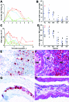
Mean pharyngeal excretion of highly pathogenic avian influenza virus (H5N1) of wild ducks by A) virus isolation and C) reverse transcription–PCR (RT-PCR). Pochard (red, closed circle), tufted duck (orange, open circle), mallard (dark blue, closed triangle), teal (light blue, open triangle), wigeon (dark green, closed square), gadwall (light green, open square); TCID50, median tissue culture infectious dose; Ct, cycle threshold. Area under the curve in the first 4 days postinoculation (mean ± 95% confidence interval) for B) virus isolation and D) RT-PCR. TU, tufted duck; PO, pochard; MA, mallard; TE, teal; WI, wigeon; GA, gadwall; red triangles, birds with severe clinical signs; black triangles, birds with mild or no clinical signs. E) Influenza virus antigen expression in epithelial cells in bronchus, parabronchus, atrium, and air capillaries of a tufted duck. F) Bronchointerstitial pneumonia, characterized by flooding of parabronchi (PB), atria (AT), and air capillaries (AC) with proteinaceous fluid and inflammatory cells in a tufted duck. G) Influenza virus antigen expression in epithelial cells lining the air sac wall and H) epithelial necrosis and lymphocytic infiltration in a gadwall. E–H original magnification ×100. Tissues were stained either by immunohistochemistry that used a monoclonal antibody against the nucleoprotein of influenza A virus as a primary antibody (E, G) or with hematoxylin and eosin (F, H).
Figure 3
Figure 3
Mean cloacal excretion of highly pathogenic avian influenza virus (H5N1) by wild ducks by A) virus isolation and C) reverse transcription–PCR (RT-PCR). Legend for panels A–D as in Figure 2. E) Pancreas showing multiple foci of necrosis (between arrowheads) in a pochard. F) Pancreatic acinar cells in a pochard and H) hepatocytes in a tufted duck, showing the transition area between normal and necrotic tissue expressing abundant influenza virus antigen. G) Pancreatic lesions in a pochard and I) hepatic lesions in a tufted duck, characterized by sharp transition between normal tissue (left side of panels) and foci of necrosis and inflammatory cell infiltration (right side of panels). F, G original magnification ×50. H, I original magnification ×100. Tissues were stained either by immunohistochemistry that used a monoclonal antibody against the nucleoprotein of influenza A virus as a primary antibody (F, H) or with hematoxylin and eosin (G, I).

References

    1. Li KS, Guan Y, Wang J, Smith GJ, Xu KM, Duan L, et al. Genesis of a highly pathogenic and potentially pandemic H5N1 influenza virus in eastern Asia. Nature. 2004;430:209–13. 10.1038/nature02746 - DOI - PubMed
    1. Ferguson NM, Fraser C, Donnelly CA, Ghani AC, Anderson RM. Public health. Public health risk from the avian H5N1 influenza epidemic. Science. 2004;304:968–9. 10.1126/science.1096898 - DOI - PubMed
    1. Webby RJ, Webster RG. Are we ready for pandemic influenza? Science. 2003;302:1519–22. 10.1126/science.1090350 - DOI - PubMed
    1. Xu X. Subbarao, Cox NJ, Guo Y. Genetic characterization of the pathogenic influenza A/Goose/Guangdong/1/96 (H5N1) virus: similarity of its hemagglutinin gene to those of H5N1 viruses from the 1997 outbreaks in Hong Kong. Virology. 1999;261:15–9. 10.1006/viro.1999.9820 - DOI - PubMed
    1. World Health Organization. Epidemic and pandemic alert and response. Avian influenza [cited 2008 22 Jan]. Available from http://www.who.int/csr/disease/avian_influenza/en/index.html

Publication types

MeSH terms