

Development of ascorbate transporters in brain cortical capillary endothelial cells in culture
- PMID: 18394593
- PMCID: PMC2453688
- DOI: 10.1016/j.brainres.2008.02.102
Development of ascorbate transporters in brain cortical capillary endothelial cells in culture
Abstract
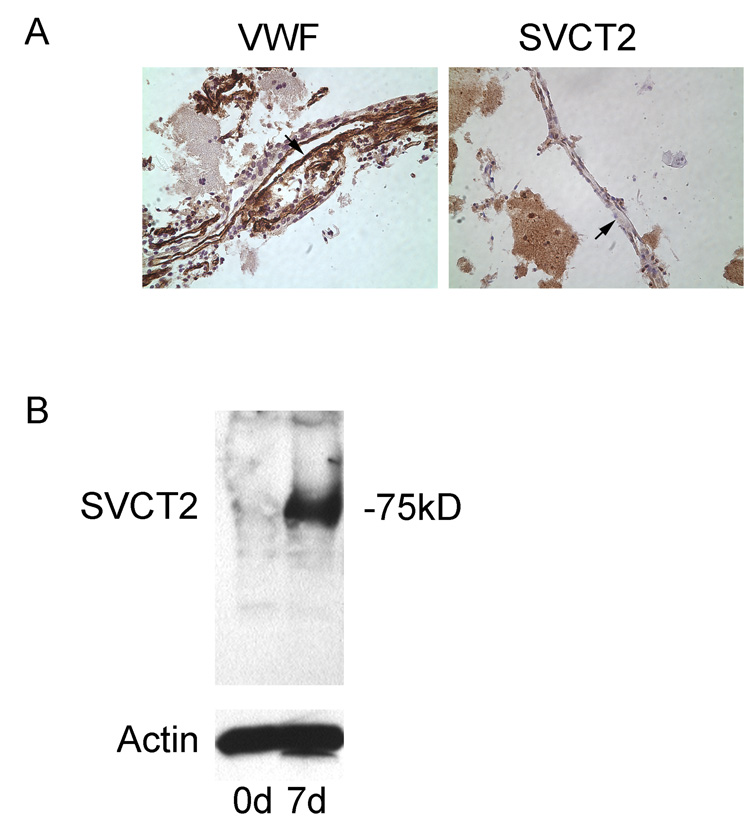
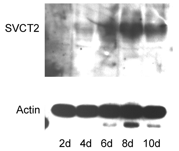
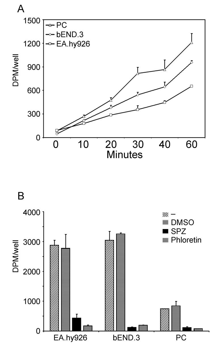
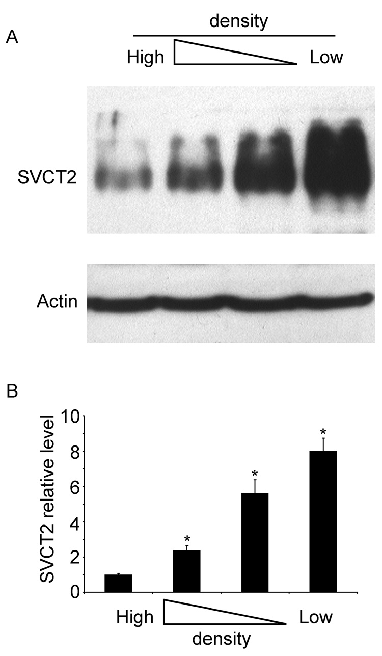
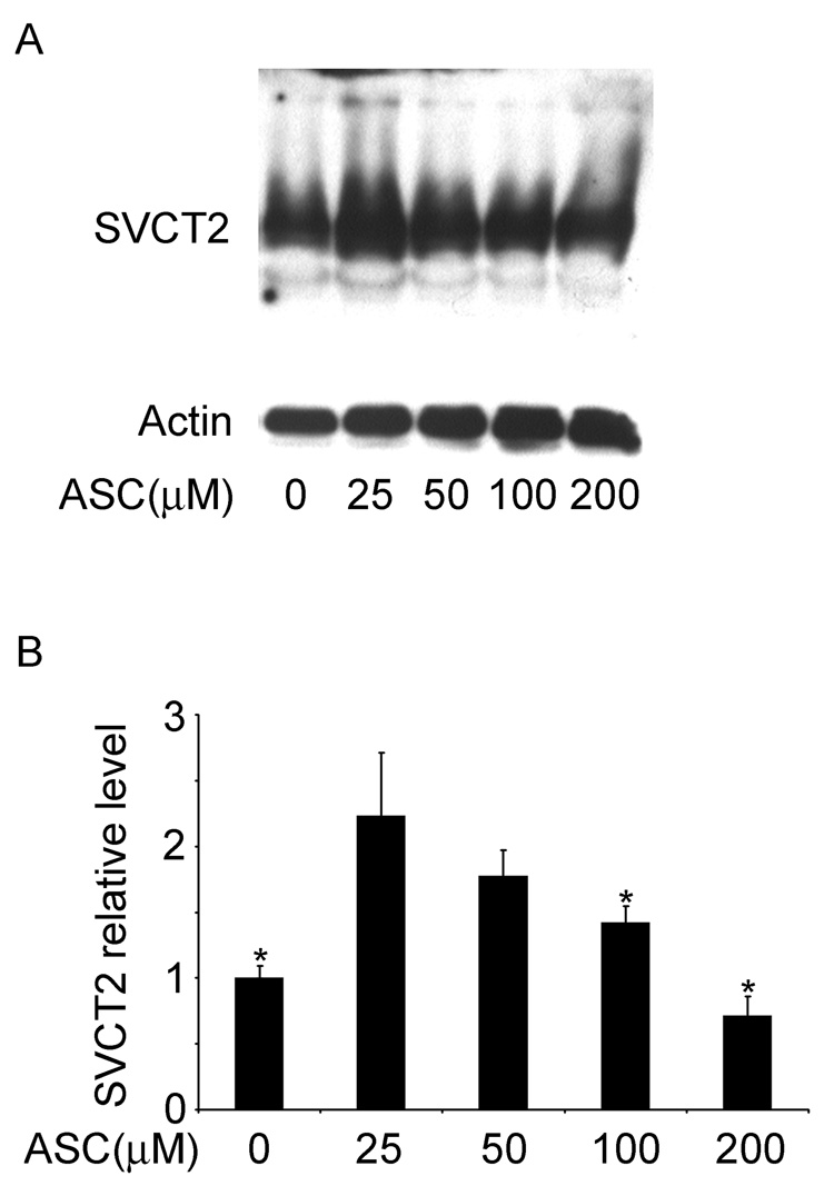
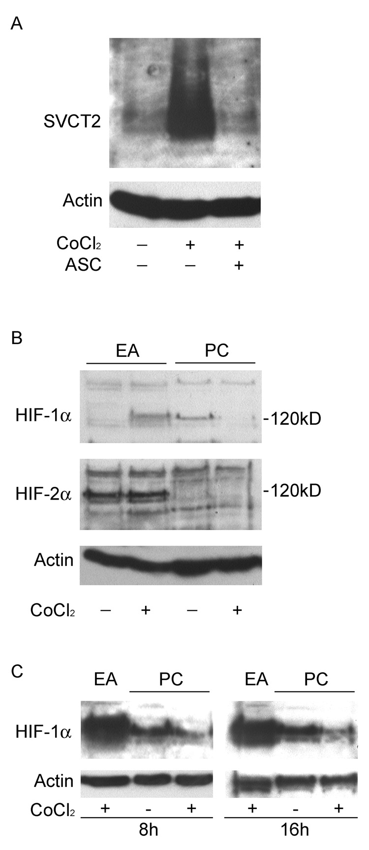
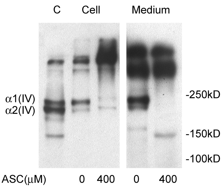
Ascorbic acid in its reduced form is not transported across the capillary endothelial cell blood-brain barrier. This is thought to be due to absence of the SVCT2, a specific transporter for ascorbate. To assess this directly we prepared primary cultures of mouse cortical microvascular endothelial cells. When still in the capillaries, these cells did not express the SVCT2 protein as assessed by immunocytochemistry and by immunoblotting. However, during several days in culture, they developed SVCT2 expression and showed ascorbate transport rates comparable to those in immortalized endothelial cell lines. SVCT2 expression was inversely proportional to cell density, was enhanced by culture at low physiologic plasma ascorbate concentrations, was inhibited by ascorbate concentrations expected in the brain interstitium, and was stimulated by cobalt ions. Expression of the SVCT2 was associated with ascorbate-dependent maturation and release of type IV collagen by the cells in culture. Although the SVCT2 is induced by culture of cortical capillary endothelial cells, its absence in vivo remains perplexing, given the need for intracellular ascorbate to facilitate type IV collagen maturation and release by endothelial cells.
Figures
Similar articles
-
Ascorbate transport by primary cultured neurons and its role in neuronal function and protection against excitotoxicity.J Neurosci Res. 2007 Apr;85(5):1046-56. doi: 10.1002/jnr.21204. J Neurosci Res. 2007. PMID: 17304569
-
Cobalt-induced oxidant stress in cultured endothelial cells: prevention by ascorbate in relation to HIF-1alpha.Biofactors. 2009 May-Jun;35(3):306-13. doi: 10.1002/biof.43. Biofactors. 2009. PMID: 19396871 Free PMC article.
-
Oxidized LDL up-regulates the ascorbic acid transporter SVCT2 in endothelial cells.Mol Cell Biochem. 2010 Oct;343(1-2):217-22. doi: 10.1007/s11010-010-0516-4. Epub 2010 Jun 13. Mol Cell Biochem. 2010. PMID: 20549544 Free PMC article.
-
SVCT1 and SVCT2: key proteins for vitamin C uptake.Amino Acids. 2008 Apr;34(3):347-55. doi: 10.1007/s00726-007-0555-7. Epub 2007 Jun 1. Amino Acids. 2008. PMID: 17541511 Review.
-
Vitamin C function in the brain: vital role of the ascorbate transporter SVCT2.Free Radic Biol Med. 2009 Mar 15;46(6):719-30. doi: 10.1016/j.freeradbiomed.2008.12.018. Epub 2009 Jan 6. Free Radic Biol Med. 2009. PMID: 19162177 Free PMC article. Review.
Cited by
-
Unwinding the potentials of vitamin C in COVID-19 and other diseases: An updated review.Nutr Health. 2023 Sep;29(3):415-433. doi: 10.1177/02601060221139628. Epub 2022 Nov 29. Nutr Health. 2023. PMID: 36445072 Free PMC article. Review.
-
Vitamin C Transporters, Recycling and the Bystander Effect in the Nervous System: SVCT2 versus Gluts.J Stem Cell Res Ther. 2014 May 19;4(5):209. doi: 10.4172/2157-7633.1000209. J Stem Cell Res Ther. 2014. PMID: 25110615 Free PMC article.
-
Increased expression of SVCT2 in a new mouse model raises ascorbic acid in tissues and protects against paraquat-induced oxidative damage in lung.PLoS One. 2012;7(4):e35623. doi: 10.1371/journal.pone.0035623. Epub 2012 Apr 30. PLoS One. 2012. PMID: 22558179 Free PMC article.
-
Transfer of ascorbic acid across the vascular endothelium: mechanism and self-regulation.Am J Physiol Cell Physiol. 2009 Jul;297(1):C169-78. doi: 10.1152/ajpcell.00674.2008. Epub 2009 May 6. Am J Physiol Cell Physiol. 2009. PMID: 19419995 Free PMC article.
-
Ascorbic acid and the brain: rationale for the use against cognitive decline.Nutrients. 2014 Apr 24;6(4):1752-81. doi: 10.3390/nu6041752. Nutrients. 2014. PMID: 24763117 Free PMC article. Review.
References
-
- Astuya A, Caprile T, Castro M, Salazar K, García MD, Reinicke K, Rodríguez F, Vera JC, Millán C, Ulloa V, Low M, Martínez F, Nualart F. Vitamin C uptake and recycling among normal and tumor cells from the central nervous system. J. Neurosci. Res. 2005;79:146–156. - PubMed
-
- Berger UV, Hediger MA. The vitamin C transporter SVCT2 is expressed by astrocytes in culture but not in situ. Neuroreport. 2000;11:1395–1399. - PubMed
-
- Berger UV, Lu XC, Liu W, Tang Z, Slusher BS, Hediger MA. Effect of middle cerebral artery occlusion on mRNA expression for the sodium-coupled vitamin C transporter SVCT2 in rat brain. J. Neurochem. 2003;86:896–906. - PubMed
Publication types
MeSH terms
Substances
Grants and funding
LinkOut - more resources
Full Text Sources
Medical
Molecular Biology Databases
