Comparative Study
. 2008 May 15;317(2):467-79.
doi: 10.1016/j.ydbio.2008.02.031. Epub 2008 Mar 4.

Multiple roles for Med12 in vertebrate endoderm development

Affiliations
Comparative Study

Multiple roles for Med12 in vertebrate endoderm development

Chong Hyun Shin et al. Dev Biol. .

Abstract

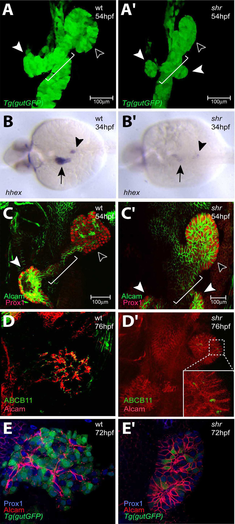
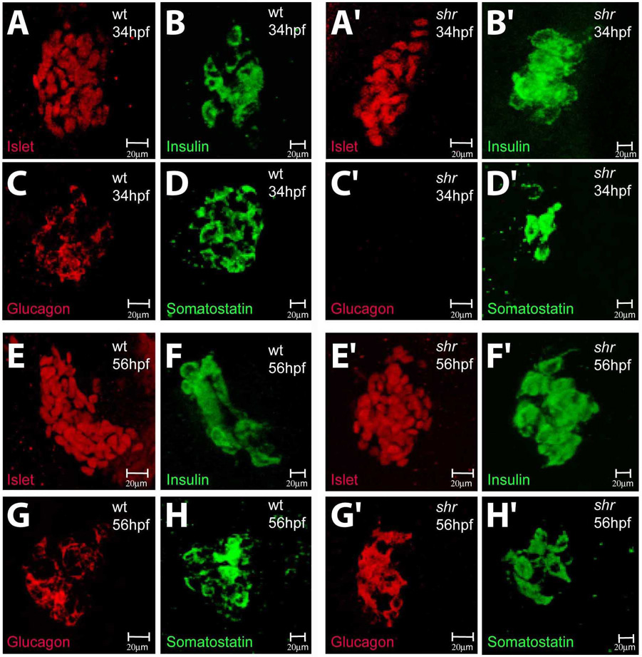
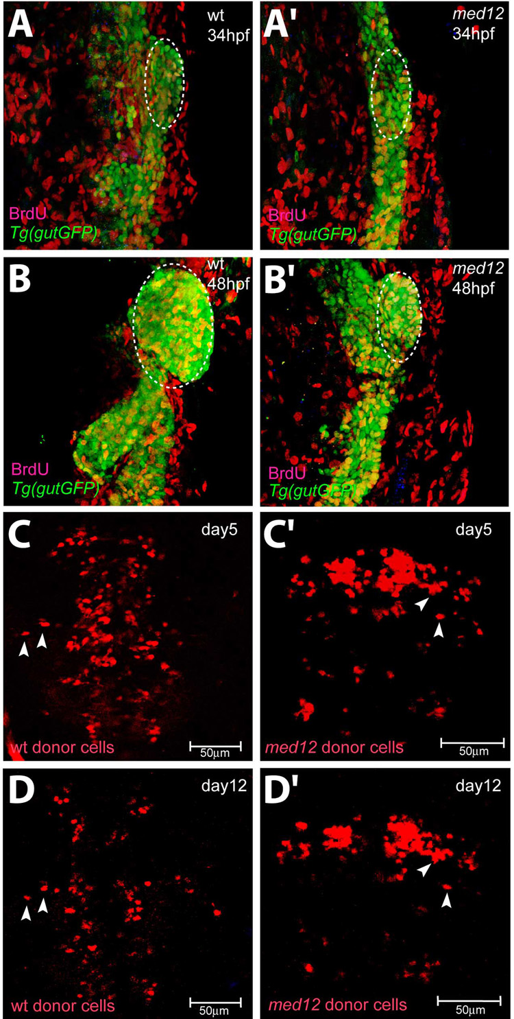
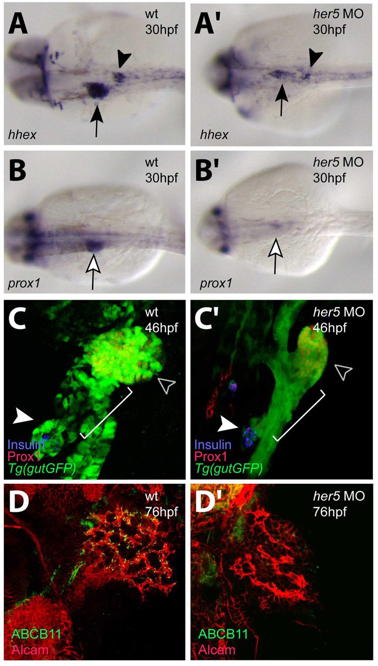
In zebrafish, the endoderm originates at the blastula stage from the most marginal blastomeres. Through a series of complex morphogenetic movements and differentiation events, the endodermal germ layer gives rise to the epithelial lining of the digestive tract as well as its associated organs such as the liver, pancreas, and swim bladder. How endodermal cells differentiate into distinct cell types such as hepatocytes or endocrine and exocrine pancreatic cells remains a major question. In a forward genetic screen for genes regulating endodermal organ development, we identified mutations at the shiri locus that cause defects in the development of a number of endodermal organs including the liver and pancreas. Detailed phenotypic analyses indicate that these defects are partially due to a reduction in endodermal expression of the hairy/enhancer of split-related gene, her5, at mid to late gastrulation stages. Using the Tg(0.7her5:EGFP)(ne2067) line, we show that her5 is expressed in the endodermal precursors that populate the pharyngeal region as well as the organ-forming region. We also find that knocking down her5 recapitulates some of the endodermal phenotypes of shiri mutants, further revealing the role of her5 in endoderm development. Positional cloning reveals that shiri encodes Med12, a regulatory subunit of the transcriptional Mediator complex recently associated with two human syndromes. Additional studies indicate that Med12 modulates the ability of Casanova/Sox32 to induce sox17 expression. Thus, detailed phenotypic analyses of embryos defective in a component of the Mediator complex have revealed new insights into discrete aspects of vertebrate endoderm development, and provide possible explanations for the craniofacial and digestive system defects observed in humans with mutations in MED12.


Figures

Fig. 1
Fig. 1
shr is required for liver and ventral pancreas development. Confocal projections of wild-type and shrs435 mutant embryos in the Tg(gutGFP)s854 line at 54 hpf (A, A’), hhex expression in wild-type and shrs435 mutant embryos at 34 hpf (B, B’), confocal projections of Alcam (green) and Prox1 (red) expression at 54 hpf (C, C’), of Alcam (red) and ABCB11 (green) expression at 76 hpf (D, D’), and of Alcam (red) and Prox1 (blue) expression in the Tg(gutGFP)s854 line at 72 hpf (E, E’). (A, A’) The size of the liver (black arrowhead) and pancreas (white arrowhead) is often reduced in shrs435 mutants compared to wild-type. By 54 hpf, the formation of the hepatopancreatic duct (white bracket) is initiated in wild-type embryos, whereas shrs435 mutants exhibit an ambiguous duct morphology (white bracket) and two ventral pancreatic buds instead of one (white arrowheads). (B, B’) hhex is clearly expressed in the wild-type liver (black arrow) and pancreatic islet (black arrowhead) while its expression can be detected in only a few cells in the liver and pancreas regions of shrs435 mutants. (C, C’) Prox1 is expressed in the liver (black arrowhead) and pancreas (white arrowhead) in wild-type and shrs435 mutant embryos. At 54hpf, Alcam has started to show restricted localization in wild-type liver, while it remains distributed along the entire surface of liver cells in shrs435 mutants. To better visualize hepatic Alcam expression in the mutants, a 1.2X magnified image is shown in C’. (D, D’) ABCB11 expression is almost absent in shrs435 mutants. To better visualize hepatic ABCB11 expression in the mutants, a magnified image is shown in an inset. (E, E’) By 72 hpf, Alcam localizes on or near the apical side of hepatocytes in wild-type, while it remains distributed along the entire surface of liver cells in shrs435 mutants. All images except B, B’ are ventral views, anterior to the top; B, B’ are dorsal views, anterior to the left.
Fig. 2
Fig. 2
shr is required for dorsal pancreas development. Confocal projections of wild-type and shrs435 mutant embryos at 34 (A–D’) and 56 (E–H’) hpf stained for Islet (red, A, A’, E, E’), Insulin (green, B, B’, F, F’), Glucagon (red, C, C’, G, G’) and Somatostatin (green, D, D’, H, H’) expression. The pancreatic expression of Islet and Insulin appears unaffected in shrs435 mutants at 34 and 56 hpf (A’, B’, E’, F’). The expression of Somatostatin appears reduced (D’) and that of Glucagon almost completely absent (C’) in shrs435 mutants at 34 hpf, but restored to wild-type levels by 56 hpf (G’, H’). All images are ventral views, anterior to the top.
Fig. 3
Fig. 3
shr encodes Med12. (A) Genetic map of the shr region on chromosome 14. Numbers below the SSLP markers indicate the number of recombination events in 1400 diploid embryos tested. (B) Sequencing of two shr mutant alleles reveals the molecular lesions (leading to the premature truncation of Med12). L, leucine-rich domain; LS, leucine-serine-rich domain; PQL, proline-glutamine-leucine-rich domain; OPA, opposite-paired domain. Numbering corresponds to the amino acid number. (C) Knock-down of med12 by the injection of 2 ng of a splice-site MO into the Tg(gutGFP)s854 line recapitulates the shr mutant phenotypes at 36 (bright field; lateral view) and 48 hpf (confocal projections of Tg(gutGFP)s854 line (green), black arrowhead points to the liver and white bracket indicates the hepatopancreatic duct; ventral views, anterior to the top).
Fig. 4
Fig. 4
Med12 does not appear to be essential for embryonic cell proliferation or viability. (A–B’) Confocal projections of wild-type and med12s435 mutant embryos in the Tg(gutGFP)s854 line (green) at 34 (A, A’) and 48 (B, B’) hpf in conjunction with BrdU (red) antibody staining. Yellow cells are endodermal cells (green) with BrdU (red) incorporation and white dashed lines surround the area used to count BrdU-positive cells. The overall level of endodermal BrdU incorporation in wild-type and med12s435 mutant embryos appears similar (Table 1). (C–D’) Confocal projections of wild-type host embryos transplanted with wild-type (C, D) or med12s435 mutant (C’, D’) donor cells, visualized at days 5 (C, C’) and 12 (D, D’). Most wild-type and med12s435 mutant donor cells survive until at least day 12 (D, D’). White arrowheads point to individual rhodamine dextran-labeled donor cells. The same embryo is shown in C and D, and another in C’ and D’. Images A–B’ are ventral views, anterior to the top; C–D’ are dorsal views, anterior to the top.
Fig. 5
Fig. 5
Med12 regulates her5 expression in endodermal cells during gastrulation and somitogenesis. (A, A’) in situ hybridization at 90% epiboly shows specific reduction of her5 expression in endodermal cells in med12s432 mutants compared to wild-type siblings. The V shaped stripe represents the domain of her5 expression in the midbrain-hindbrain boundary. (B–D’) Injection of med12 MO into the Tg(−0.7her5:EGFP)ne2067 line caused decreased GFP expression. Note that the Tg(−0.7her5:EGFP)ne2067 line shows GFP expression in the pharyngeal endoderm as well as the organ-forming region during late somitogenesis stages (C, D). White bracket in C–D’ indicates pharyngeal region. Exposure time for imaging B, B’ was 300ms; C, C’ 250ms; D, D’ 250ms. Dorsal views, anterior to the top.
Fig. 6
Fig. 6
Her5 is required for the development of the endodermal organs. Wild-type and her5 MO-injected animals at 30 (A–B’), 46 (C, C’), and 76 (D, D’) hpf were analyzed for hhex (A, A’) and prox1 (B, B’) expression, confocal projections in the Tg(gutGFP)s854 line in conjunction with Prox1 (red) and Insulin (blue) expression (C, C’), and Alcam (red) and ABCB11 (green) expression (D,D’). (A, A’) hhex expression in the liver (black arrow) and pancreatic islet (black arrowhead) regions could be detected in only a few cells in her5 MO-injected embryos. (B, B’) prox1 expression in the liver region (white arrow) could only be detected at low levels in her5 MO-injected embryos. (C, C’) At 46 hpf, the formation of the hepatopancreatic duct (white bracket) is initiated in wild-type embryos, whereas in her5 MO-injected embryos, this process was perturbed, resulting in an ambiguous ductal morphology. Note the lack of engulfment of the Insulin-positive endocrine cells (white arrowhead) by the ventral pancreatic bud derived tissue in MO-injected embryos. Black arrowhead points to the liver. (D, D’) ABCB11 expression was almost absent in her5 MO-injected larvae. N=20 in each set of MO-injections; the percentage of MO-injected embryos/larvae exhibiting the phenotype shown in C’ was 60% (n=12/20), and the one in D’ was 60% (n=12/20). Images A–B’ are dorsal views, anterior to the left; C–D’ are ventral views, anterior to the top.
Fig. 7
Fig. 7
Med12 functions with Cas/Sox32 and Foxa2 to regulate endodermal organ development. (A–C) Embryos from med12s432 heterozygote incrosses injected with cas mRNA were harvested at 90% epiboly and analyzed for sox17 expression. med12s432 homozygous mutant embryos overexpressing cas mRNA showed decreased induction of sox17 expression (C) compared to med12 heterozygous (B) and wild-type (A) embryos. (D–H’) Embryos from wild-type crosses and med12s432 heterozygote incrosses injected with foxa2 MO were harvested at 48 hpf and analyzed for foxa3 expression. Wild-type foxa3 expression reveals structures such as the liver (black arrow), pancreas (black arrowhead), and swim bladder (white arrowhead) (D). med12s432 mutants show an ambiguous ductal morphology (black bracket) and defective development of endodermal organs (E). foxa2 MO injected embryos showed slight perturbation of organ development (F), while med12s432 heterozygous embryos injected with foxa2 MO showed a more severe malformation of organs (G), and med12s432 homozygous mutant embryos injected with foxa2 MO showed an almost complete absence of accessory organs, exhibiting a solid endodermal rod (H, H’). (I–K) Confocal projections of larvae from med12s432 heterozygote outcrosses in the Tg (fabp1a:dsRed, elastase:GFP; insulin:dsRed) heterozygote line injected with foxa2 MO (J and K) and analyzed at 96 hpf. While foxa2 MO injected larvae showed slight perturbation of liver and exocrine pancreas (J), med12s432 heterozygous larvae with foxa2 MO injection showed a more severe malformation of these organs (K). Images A–C are dorsal views, anterior to the top; D–H’ are dorsal views, anterior to the left; I–K are ventral views, anterior to the top.

References

    1. Alexander J, Stainier DY, Yelon D. Screening mosaic F1 females for mutations affecting zebrafish heart induction and patterning. Dev Genet. 1998;22:288–299. - PubMed
    1. Alexander J, Rothenberg M, Henry GL, Stainier DY. Casanova plays an early and essential role in endoderm formation in zebrafish. Dev Biol. 1999;215:343–357. - PubMed
    1. Aoki TO, Mathieu J, Saint-Etienne L, Rebagliati MR, Peyrieras N, Rosa FM. Regulation of nodal signaling and mesendoderm formation by TARAM-A, a TGFbeta-related type I receptor. Dev Biol. 2002;241:273–288. - PubMed
    1. Bally-Cuif L, Goutel C, Wassef M, Wurst W, Rosa F. Coregulatin of anterior and posterior mesendodermal development by a hairy-related transcriptional repressor. Genes Dev. 2000;14:1664–1677. - PMC - PubMed
    1. Biemar F, Argenton F, Schmidtke R, Epperlein S, Peers B, Driever W. Pancreas development in zebrafish: early dispersed appearance of endocrine hormone expressing cells and their convergence to form the definitive islet. Dev Biol. 2001;230:189–203. - PubMed

Publication types

MeSH terms

LinkOut - more resources