Comparative Study
. 2008 Jun;211(2):362-9.
doi: 10.1016/j.expneurol.2008.01.025. Epub 2008 Feb 14.

Human neural stem cells migrate along the nigrostriatal pathway in a primate model of Parkinson's disease

Affiliations
Comparative Study

Human neural stem cells migrate along the nigrostriatal pathway in a primate model of Parkinson's disease

Kimberly B Bjugstad et al. Exp Neurol. 2008 Jun.

Abstract

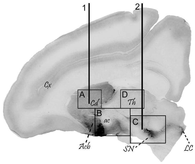
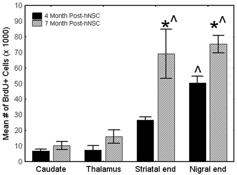
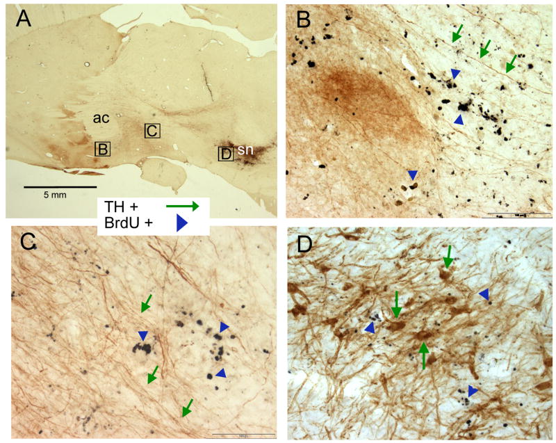
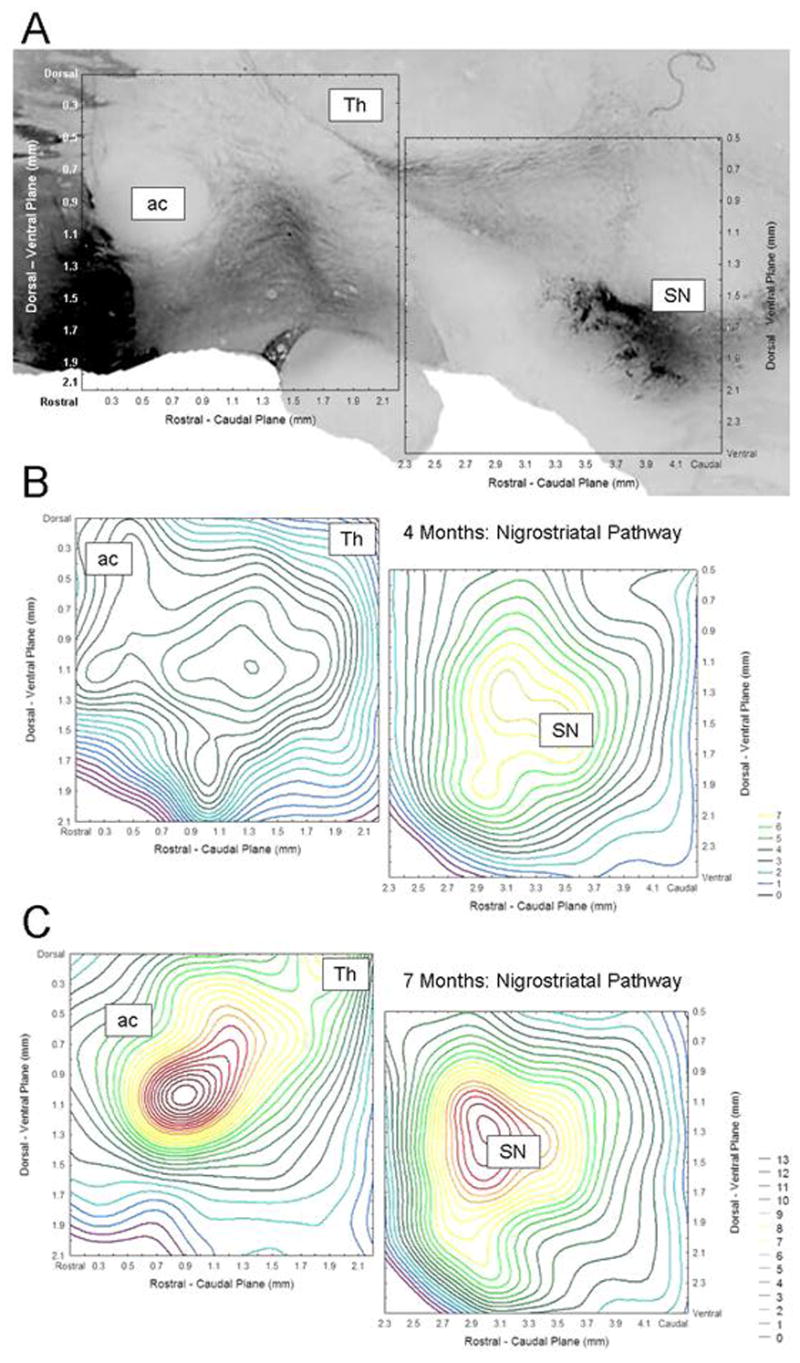
Although evidence of damage-directed neural stem cell (NSC) migration has been well-documented in the rodent, to our knowledge it has never been confirmed or quantified using human NSC (hNSC) in an adult non-human primate modeling a human neurodegenerative disease state. In this report, we attempt to provide that confirmation, potentially advancing basic stem cell concepts toward clinical relevance. hNSCs were implanted into the caudate nucleus (bilaterally) and substantia nigra (unilaterally) of 7, adult St. Kitts African green monkeys (Chlorocebus sabaeus) with previous exposure to systemic 1-methyl-4-phenyl-1,2,3,6-tetrahydropyridine (MPTP), a neurotoxin that disrupts the dopaminergic nigrostriatal pathway. A detailed quantitative analysis of hNSC migration patterns at two time points (4 and 7 months) following transplantation was performed. Density contour mapping of hNSCs along the dorsal-ventral and medial-lateral axes of the brain suggested that >80% of hNSCs migrated from the point of implantation to and along the impaired nigrostriatal pathway. Although 2/3 of hNSCs were transplanted within the caudate, <1% of 3x10(6) total injected donor cells were identified at this site. The migrating hNSC did not appear to be pursuing a neuronal lineage. In the striatum and nigrostriatal pathway, but not in the substantia nigra, some hNSCs were found to have taken a glial lineage. The property of neural stem cells to align themselves along a neural pathway rendered dysfunctional by a given disease is potentially a valuable clinical tool.


Figures

FIGURE 1
FIGURE 1
Parasagittal section of a monkey brain, with boxes depicting the four areas in which counts of BrdU+ cells were made. The caudate nucleus (A) was implanted bilaterally (line 1) with BrdU pre-labeled hNSCs, while the SN (C) was implanted unilaterally only (line 2). Most BrdU-positive cells appeared in the areas between the caudate and the SN, along the nigrostriatal pathway (boxes B and C). Cell counts were done for the anterior portion of the pathway (B: Striatal Half) and for the posterior portion of the pathway, which included the SN (C: Nigral Half). The thalamus (D) also was included as a control area which is not a component of the nigrostriatal dopamine system, although the SN implant track (line 2) penetrated the right thalamus. This parasagittal section was immunostained for tyrosine hydroxylase (TH, black immunoreactive regions) to better depict the SN, the nigrostriatal pathway, and innervation of the CD. Cx- cerebral cortex, Cd- caudate, ac- anterior commissure, Th- thalamus, SN- substantia nigra, LC- locus coeruleus.
FIGURE 2
FIGURE 2
The total number of BrdU-positive cells found in each area for MPTP-lesioned monkeys 4 months and 7 months post-transplantation. There were significantly more BrdU positive cells found along the nigrostriatal pathway (striatal end and nigral end) than in the caudate nucleus, an area specifically implanted with hNSC [F(3, 30)= 27.00, p< 0.0005]. As many cells were found in the thalamus, an unimplanted site, as were found in the caudate nucleus (p> 0.05). The 7 month animals had significantly more BrdU+ cells along the nigrostriatal pathway than the 4 month animals [F(3,30)= 2.90, p< 0.05]. There was no difference between the groups in the number of cells found in the caudate and thalamus. * Significantly greater than the same region in the 4 month animals. ^ Significantly different than the other brain areas. Significance level p< 0.05.
FIGURE 3
FIGURE 3
Overview the nigrostriatal pathway and the BrdU-positive hNSC-derived cells found there. hNSCs were labeled with BrdU ex vivo prior to transplantation. Immunohistochemistry against BrdU and TH was used to identify the location of the hNSCs (black nuclei) and the dopamine neurons and their fibers (brown), respectively. [A] A low power view of the nigrostriatal pathway illustrating where the photomicrographs of [B-D] were taken. The anterior commissure (ac) and substantia nigra (sn) are labeled. [B] Anterior portion of the nigrostriatal pathway. hNSCs (black nuclei, blue arrowheads) were found ventral to the anterior commissure along with TH positive fibers originating from the midbrain (brown fibers, green arrows). [C] Posterior portion of the nigrostriatal pathway. BrdU-positive cells were found (black nuclei, blue arrowheads) along with many TH-positive fibers (brown, green arrows). [D] The endogenous TH+ neurons of the SN (brown, green arrows) were surrounded by BrdU-positive cells (black nuclei, blue arrowheads). A density contour map of these cells is shown in Fig. 4 and a higher-power view using dual immunofluorescence of the relative positions of donor hNSC-derived cells and host TH+ cells is presented in Figure 5.
FIGURE 4
FIGURE 4
hNSC density contour mapping. The mean number of BrdU-positive cells at each dorsal-ventral / rostral-caudal location (counting frames of 400 × 400 μm) was plotted to create a hNSC density contour map to show the changes in density of hNSC across the two planes. [A] An overlay of the X and Y axis onto a photomicrograph of the areas being described. [B] Contour mapping of the striatal and nigral half of the nigrostriatal pathway for the 4 month-post transplant data. The greatest density of hNSCs was found in the SN with >7 BrdU+ cells per 400 μm2 (yellow contour lines). [C] By comparison, the 7 month post-transplant data had a high density of BrdU+ cells at the posterior/ventral side of the ac and in the SN, with almost twice the density (red contour lines). ac= anterior commissure, Th= thalamus, SN= substantia nigra.
FIGURE 5
FIGURE 5
Dual immunohistochemistry for BrdU+ and TH+ cells demonstrating the intimate association between donor hNSC-derived cells (red nuclei, arrowheads) and host SN dopaminergic neurons (green somata and processes, arrows), respectively. Note that no TH+ neurons in the SN co-labeled with BrdU, suggesting that dopaminergic lineage was not the preferred fate of these undifferentiated hNSCs in the SN.
FIGURE 6
FIGURE 6
Some striatal astrocytes double-label for BrdU suggesting that they are of donor origin. GFAP was used to identify astrocytes (brown) while BrdU was used to identify the implanted hNSC (black nuclei). Most astrocytes did not double label for BrdU (brown cells, white nucleus, closed-headed black arrows). The nuclei of these cells are void of BrdU labeling. An occasional astrocyte appeared with a BrdU labeled nucleus (brown cell with black nucleus, open-headed arrow), however most BrdU labeled cells did not co-localize with GFAP (black nuclei, black arrow heads).

References

    1. Aboody KS, Brown A, Rainov NG, Bower KA, Liu S, Yang W, Small JE, Herrlinger U, Ourednik V, Black PM, Breakefield XO, Snyder EY. Neural stem cells display extensive tropism for pathology in adult brain: evidence from intracranial gliomas. Proc Natl Acad Sci USA. 2000;97:12846–51. - PMC - PubMed
    1. An YH, Wang HY, Gao ZX, Wang ZC. Differentiation of rat neural stem cells and its relationship with environment. Biomed Environ Sci. 2004;17:1–7. - PubMed
    1. Behrstock S, Ebert A, McHugh J, Vosberg S, Moore J, Schneider B, Capowski E, Hei D, Kordower J, Aebischer P, Svendsen CN. Human neural progenitors deliver glial cell line-derived neurotrophic factor to parkinsonian rodents and aged primates. Gene Ther. 2006;13:379–88. - PubMed
    1. Betarbet R, Turner R, Chockkan V, DeLong MR, Allers KA, Walters J, Levey AI, Greenamyre JT. Dopaminergic neurons intrinsic to the primate striatum. J Neurosci. 1997;17:6761–6768. - PMC - PubMed
    1. Bezard E, Gross CE. Compensatory mechanisms in experimental and human parkinsonism: Towards a dynamic approach. Prog Neurobiol. 1998;55:93–116. - PubMed

Publication types

MeSH terms