. 2008 Apr 8;18(7):507-13.
doi: 10.1016/j.cub.2008.02.076.

Cell-polarity dynamics controls the mechanism of lumen formation in epithelial morphogenesis

Affiliations

Cell-polarity dynamics controls the mechanism of lumen formation in epithelial morphogenesis

Fernando Martín-Belmonte et al. Curr Biol. .

Erratum in

  • Curr Biol. 2008 Apr 22;18(8):630. Ewald, Andrew [corrected to Ewald, Andrew J]
  • Curr Biol. 2008 Jul 8;18(13):1016

Abstract

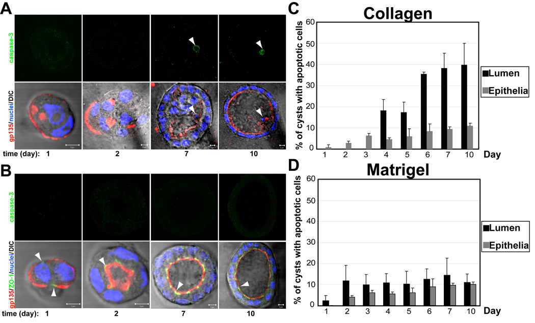
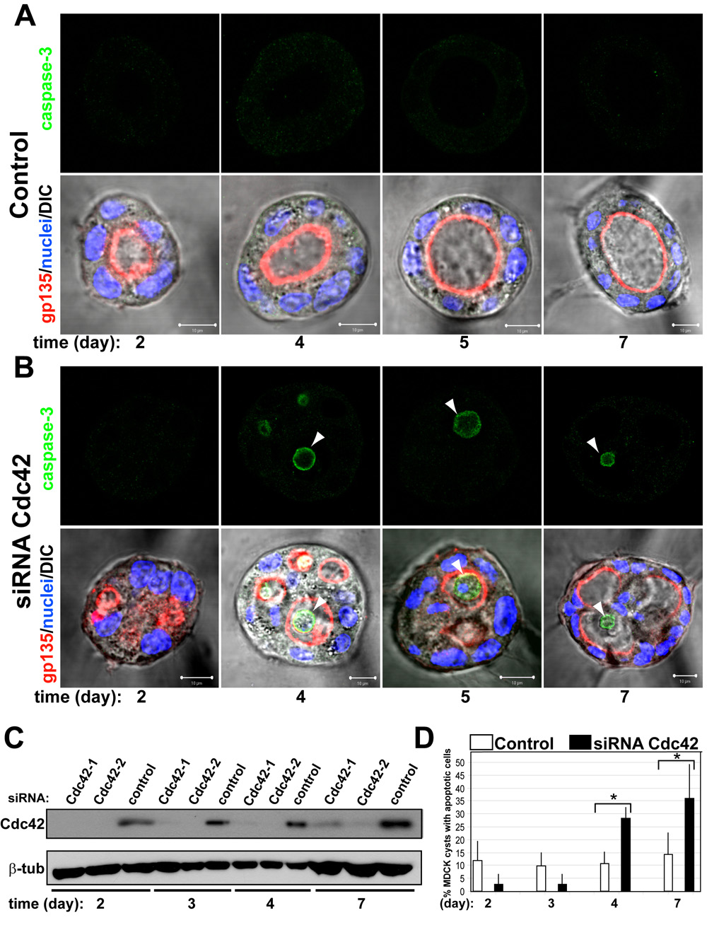
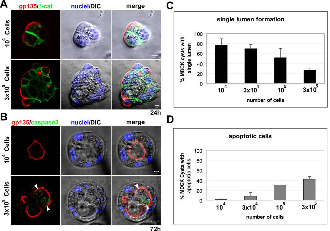
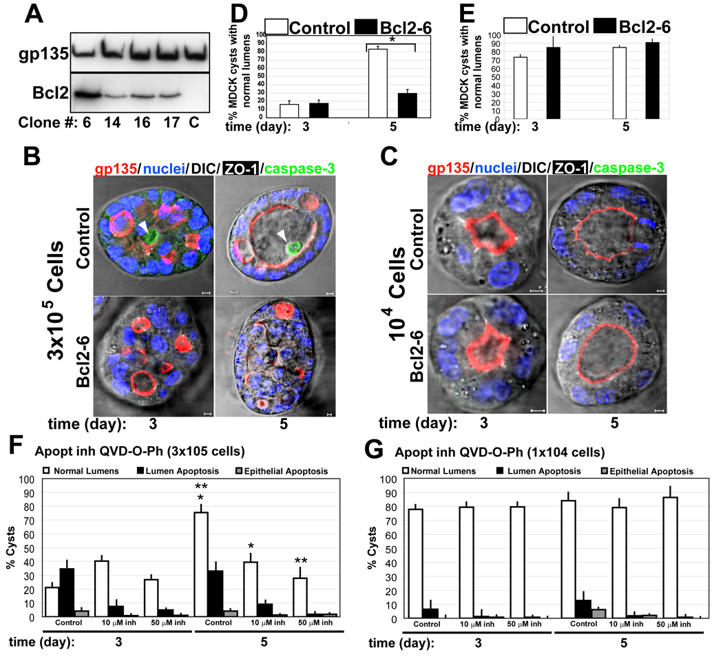
Many organs consist of tubes of epithelial cells enclosing a central lumen. How the space of this lumen is generated is a key question in morphogenesis. Two predominant mechanisms of de novo lumen formation have been observed: hollowing and cavitation. In hollowing, the lumen is formed by exocytosis and membrane separation, whereas, in cavitation, the lumen is generated by apoptosis of cells in the middle of the structure [1, 2]. Using MDCK cells in three-dimensional cultures, we found an inverse correlation between polarization efficiency and apoptosis. When cells were grown in collagen, where cells polarized slowly, apoptosis was needed for lumen formation. However, in the presence of Matrigel, which allowed rapid polarization, lumens formed without apoptosis. If polarization in Matrigel was perturbed by blocking formation of the apical surface by RNAi of Cdc42, lumens formed by apoptosis. In a complementary approach, we plated cells at high density so that aggregates formed with little polarity. These aggregates required apoptosis to form lumens, whereas cells plated at low density formed cysts with rapidly polarizing cells and did not need apoptosis to form lumens. The mechanism of lumen formation in the 3D-MDCK model can shift between hollowing and cavitation, depending on cell polarization.


Figures

Fig 1
Fig 1. The presence of Laminin in the ECM reduces apoptosis in lumen clearance
(A) and (B) Effect of laminin on polarity, lumen formation and apoptosis. MDCK cells were plated to form cysts in Collagen (A) or LGF Matrigel (B). Cells were fixed at 1, 2, 3, 4, 5, 6, 7, and 10 days and stained to detect gp135 (red), ZO-1 (green) and nuclei (lower panels merged with DIC); and caspase3 (green, upper panels). Arrowheads indicate apoptotic cells in the lumen of the cysts. Scale bars 5 µm. (C) and (D) Quantitation of cysts with apoptotic cells in the lumen (black bars) or in the epithelial wall (grey bars) in cells plated from 1 to 10 days in collagen (C) or LGF Matrigel (D). Values shown are mean ±SD from 4 different experiments. Statistical analysis of the data demonstrates that apoptosis occurs specifically in the lumen (P<0.005)
Fig 2
Fig 2. Cdc42 siRNA-depletion delays lumen formation in MDCK cyst
(A) and (B) Effect of Cdc42-1 siRNA on lumen formation and apoptosis. MDCK cells were transfected with Cdc42 siRNA (B) or siRNA control (A) and plated to form cysts. Cells were fixed at 2, 3, 4, 5 and 7 days and stained to detect gp135 (red) and nuclei (lower panels merged with DIC); and caspase3 (green, upper panels). Arrowheads indicate apoptotic cells in the lumen of the cysts. Scale bars 5 µm. (C) Down-regulation of Cdc42 by siRNA. Cells were transfected with siRNAs Cdc42-1 and Cdc42-2 against canine Cdc42 or with control siRNA, allowed to form cysts for 2, 3, 4 or 7 days and then total cell lysates western blotted for Cdc42 and β-tubulin (control). (D) Quantitation of cysts with apoptotic cells in the lumen in cells transfected with control siRNA (white bars), specific siRNA Cdc42-1 (black bars). Values shown are mean ±SD from 4 different experiments. * P≤0.01; P<0.01
Fig 3
Fig 3. Cell density determines apoptosis requirement and single lumen formation
(A) and (B) Effect of cell density on lumen formation and apoptosis. MDCK cells were plated at a cell density of 104 (upper panels) or 3 × 10⁵ cells/ml (lower panels) and allowed to form cysts for 24 (A) or 72h (B). Cells were fixed and stained to detect gp135 (red), β-catenin (green, upper panels) or caspase3 (green, lower panels), and nuclei (middle panels merged with DIC). Arrowheads indicate apoptotic cells in the lumen of the cysts. Scale bars 5 µm. (C) and (D) Quantitation of cysts with normal lumens (C) and apoptotic cells in the lumen (D) in cells plated at different cell density. Values shown are mean ±SD from 4 different experiments. Statistical analysis of the data demonstrates a direct relationship between cell density and apoptosis (P<0.005); and an inverse relationship between single lumen formation and cell density (P<0.005)
Fig 4
Fig 4. Blc2 expression, prevents apoptosis in MDCK cysts at high density, but has no effect at low density
(A) Western blot of stable expression of Bcl2. Extracts from different clones of MDCK Bcl2 were inmunoblotted with gp135 (upper panel) as a control and anti-Bcl2 (lower panel) to detect endogenous and transfected proteins. (B) Effect of Bcl2 expression on lumen formation and apoptosis in high density MDCK cells. MDCK cells expressing Bcl2 (lower panels) or not (upper panels) were plated at high density (3×10⁵ cell/ml) to form cysts for 3 and 5 days. Cells were fixed and stained to detect gp135 (red), caspase3 (green), ZO-1 (white) and nuclei (merged with DIC). Arrowheads indicate apoptotic cells in the lumen of the cysts. Scale bars 5 µm. (C) Effect of Bcl2 expression on lumen formation and apoptosis in low density. MDCK cells expressing Bcl2 (lower panels) or not (upper panels) were plated at low density (104 cell/ml) to form cysts for 3 or 5 days. Cells were visualized as in panel B. Scale bars 5 µm. (D) Quantitation of cysts with normal lumens in high density MDCK cells transfected with Bcl2 (black bars) or control (white bars). Values shown are mean ±SD from 4 different experiments. * P <0.01 (E) Quantitation of cysts with normal lumens in low density MDCK cells transfected with Bcl2 (black bars) or control (white bars). Values shown are mean ±SD from 4 different experiments. (F) Quantitation of cysts with normal lumens (white bars) in high density MDCK cells treated with Q-VD-OPh at different concentrations: control (0) 10µM and 50µM. Apoptosis in luminal (black bars) and epithelial cells (grey bars) were also quantified. Values shown are mean ±SD from 2 different experiments. *P<0.005; **P<0.005 (G) Quantitation of cysts with normal lumens (white bars) in low density MDCK cells treated with Q-VD-OPh at different concentrations control (0), 10µM and 50µM. Apoptosis in luminal (black bars) and epithelial cells (grey bars) were also quantified. Values shown are mean ±SD from 2 different experiments

References

    1. Lubarsky B, Krasnow MA. Tube morphogenesis: making and shaping biological tubes. Cell. 2003;112:19–28. - PubMed
    1. O'Brien LE, Zegers MM, Mostov KE. Opinion: Building epithelial architecture: insights from three-dimensional culture models. Nat Rev Mol Cell Biol. 2002;3:531–537. - PubMed
    1. Lin HH, Yang TP, Jiang ST, Yang HY, Tang MJ. Bcl-2 overexpression prevents apoptosis-induced Madin-Darby canine kidney simple epithelial cyst formation. Kidney Int. 1999;55:168–178. - PubMed
    1. O'Brien LE, Jou TS, Pollack AL, Zhang Q, Hansen SH, Yurchenco P, Mostov KE. Rac1 orientates epithelial apical polarity through effects on basolateral laminin assembly. Nat Cell Biol. 2001;3:831–838. - PubMed
    1. Yu W, Datta A, Leroy P, O'Brien LE, Mak G, Jou TS, Matlin KS, Mostov KE, Zegers MM. Beta1-integrin orients epithelial polarity via Rac1 and laminin. Mol Biol Cell. 2005;16:433–445. - PMC - PubMed

Publication types