. 2008 Apr;28(4):571-80.
doi: 10.1016/j.immuni.2008.02.016.

NKG2D-deficient mice are defective in tumor surveillance in models of spontaneous malignancy

Affiliations

NKG2D-deficient mice are defective in tumor surveillance in models of spontaneous malignancy

Nadia Guerra et al. Immunity. 2008 Apr.

Erratum in

  • Immunity. 2008 May;28(5):723. Greenberg, Norman R [corrected to Greenberg, Norman M]

Abstract

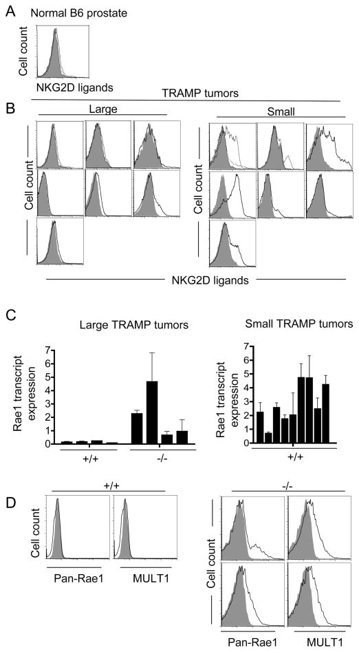
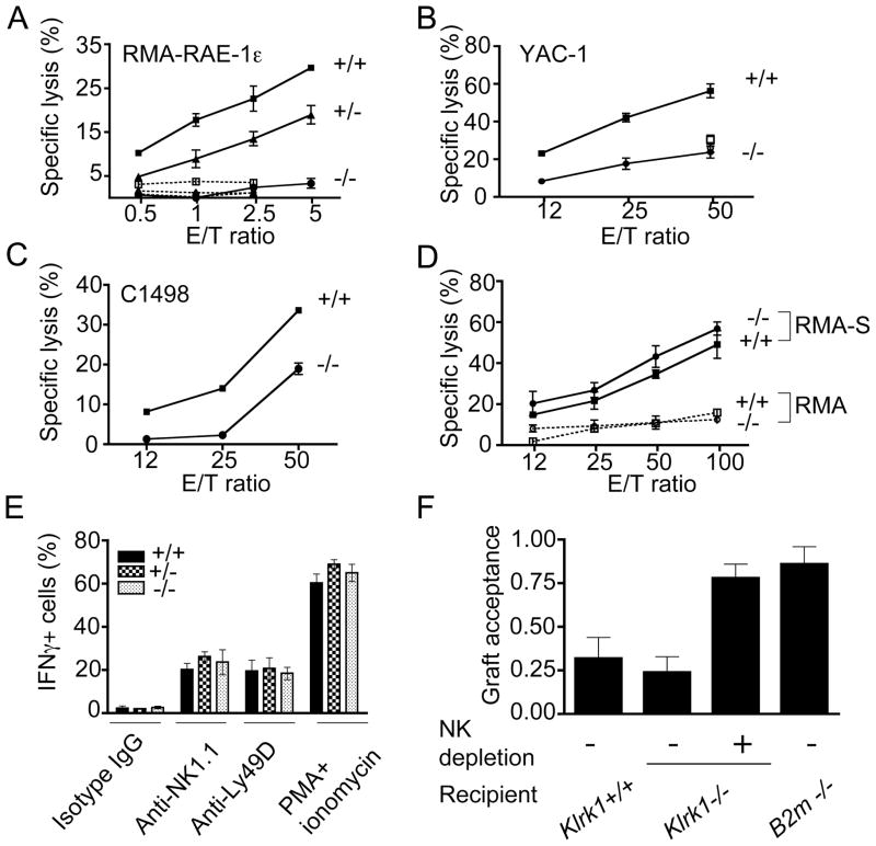
Ligands for the NKG2D stimulatory receptor are frequently upregulated on tumor lines, rendering them sensitive to natural killer (NK) cells, but the role of NKG2D in tumor surveillance has not been addressed in spontaneous cancer models. Here, we provided the first characterization of NKG2D-deficient mice, including evidence that NKG2D was not necessary for NK cell development but was critical for immunosurveillance of epithelial and lymphoid malignancies in two transgenic models of de novo tumorigenesis. In both models, we detected NKG2D ligands on the tumor cell surface ex vivo, providing needed evidence for ligand expression by primary tumors. In a prostate cancer model, aggressive tumors arising in NKG2D-deficient mice expressed higher amounts of NKG2D ligands than did similar tumors in wild-type mice, suggesting an NKG2D-dependent immunoediting of tumors in this model. These findings provide important genetic evidence for surveillance of primary tumors by an NK receptor.


Conflict of interest statement

COMPETING INTERESTS STATEMENT

The authors declare that they have no competing financial interests.

Figures

Figure 1.
Figure 1.. NK cell development is preserved in NKG2D-deficient (Klrk1−/−) mice.
Analysis of NK subsets in the bone marrow (BM, n=3, panel A) and spleen (n=3–5, panel B) from Klrk1−/− (neo cassette deleted) mice and Klrk1+/+ littermate controls. The top groups in panels A and B represent gated NK1.1+CD3 cells, whereas the remaining panels represent gated CD3 cells. The numbers represent mean percentages (±SD) of cells expressing the indicated markers. (C) Representative NKG2D staining on freshly isolated NK1.1+CD3 splenic NK cells is shown for Klrk1−/− mice with the neo cassette deleted and littermate controls. Isotype control stain is shown as shaded histogram. The bone marrow analysis was repeated in one additional independent experiment and the spleen cell analysis was repeated in two additional independent experiments, with similar results.
Figure 2.
Figure 2.. Increased incidence of large, early prostate carcinomas in Klrk1−/− TRAMP mice.
(A) Each square represents the weight of the prostate tissue and age at necropsy of individual Klrk1+/+ TRAMP (n=33) (upper panel) or Klrk1−/− TRAMP littermates (n=43) (lower panel) mice. Large-early tumors (> 2 SEM greater than the mean weight, circled) were more frequent in Klrk1−/− mice (p=0.029 by Fishers exact test). (B) The average weights of prostate tumors at necropsy is depicted for both cohorts (p=0.0184 by Mann Whitney test).
Figure 3.
Figure 3.. Expression of NKG2D ligands by prostate carcinomas ex vivo.
(A, B) Reduced cell surface expression of NKG2D ligands on large, early-arising B6-TRAMP prostate tumors as compared to smaller, late-arising ones. Freshly dissociated prostate tissue from a representative non-transgenic Klrk1+/+ B6 mouse (A) and sets of large and smaller tumors isolated from Klrk1+/+ B6-TRAMP mice (B) were compared. Labeled NKG2D tetramers (plain line) were used to detect all NKG2D ligands and streptavidin-PE served as a negative control (shaded histogram). In a few cases, the specificity of staining was proved by inhibition with unlabeled NKG2D tetramers (dotted line). (C) Reduced amounts of Rae1 transcripts in large (>2.7 g) Klrk1+/+ TRAMP tumors as compared to large Klrk1−/− TRAMP tumors (p=0.0286 with the Mann-Whitney test) (left panel) or smaller Klrk1+/+ TRAMP tumors (right panel). Values determined by quantitative RT-PCR with primers that detect all Rae1 isoforms were normalized to HPRT transcript amounts, and these data were normalized to the amount in nontransgenic B6 prostates (average of 5 independent B6 mice). Graphs show the mean ± SD of 2–6 separate qPCR assays for each sample. (D) Cell surface staining of Rae1 and MULT1 (solid line) on two dissociated large Klrk1−/− TRAMP tumors and a large tumor from an Klrk1+/+ TRAMP littermate. Isotype control stains are shown as shaded histograms. Gated CD45-negative (non-hematopoietic), PI-negative (live) cells were examined.
Figure 4.
Figure 4.. Accelerated onset of myc-driven lymphomas in Klrk1−/− mice.
(A) Kaplan-Meier representation of tumor progression among Klrk1−/− Eμ-myc transgenic (n=24) and Klrk1+/+ -myc transgenic mice (n=34). p=0.005 by the log-rank test. (B) Ex vivo analysis of lymph node cell suspensions isolated from Klrk1+/+ Eμ-myc, Klrk1−/− Eμ-myc, and non-transgenic Klrk1+/+ mice, as indicated. Dot plots show B220 and IgM expression on the blast (high forward scatter) population from independent mice which were examined after the onset of illness. The age (weeks) at necropsy is specified for each mouse as well as the percentage of cells included in the gate. The histograms show pan-Rae1 and MULT1 expression (solid line) on the gated populations indicated in each dot plot. The shaded histograms represent the staining with an isotype control antibody.
Figure 5.
Figure 5.. Comparable incidence of MCA-induced fibrosarcomas in NKG2D-deficient mice and wild-type littermates.
Klrk1−/− (dashed line) and wild-type littermates (solid line) (neo cassette-deleted) were injected s.c. with 25μg (A) or 5μg (B) of 3-methylcholanthrene (MCA) and monitored for tumor development. Tumor bearing mice were defined by the presence of a mass of at least 7mm diameter growing upon 2 consecutive measurements. These data were compiled from 2 independent experiments. p values are based on the log-rank test.
Figure 6.
Figure 6.. NKG2D-deficiency does not impair NK cell functions in vitro or in vivo.
(A-C) IL-2 activated splenic NK cells (sorted NK1.1+CD3 cells in panel A, unsorted cells in panels B-C from Klrk1−/− mice fail to lyse RMA-Rae1ε target cells, and show reduced lysis of YAC-1 and C1498 target cells. Lysis in the presence of NKG2D mAb is shown for Klrk1+/+ (open squares) and Klrk1−/− (open circles) effector cells in panels A and B. (D) Normal lysis of class I-low RMAS cells by Klrk1−/− IL-2 activated NK cells. Results represent means ±SD obtained with Klrk1−/− mice (neo cassette-retained) and littermate controls. (E) Splenocytes from Klrk1−/− mice (neo cassette deleted) and littermate controls (n=5 for each genotype) were stimulated in vitro for 5 hours on plates coated with NK1.1 mAb (PK136, 40 μg/ml), Ly49D mAb (SED85, 10 μg/ml) or control mouse IgG (40 μg/ml) in the presence of Golgi-plug before staining and analysis. In parallel, NK cells were stimulated with or without a mixture of PMA and ionomycin. Intracellular IFN-γ was detected by flow cytometry on gated NK1.1+CD3 cells, except for the anti-NK1.1-stimulated cells, which were gated on DX5+CD3 cells. Results represent means +/− SD (n=5). (F) In vivo rejection of B2m−/− bone marrow cells by Klrk1−/− mice. A mixture of CFSE (5 μM) labeled BM cells from C57Bl/6-Ly5.2 and B2m−/− C57Bl/6-Ly5.1 mice was injected i.v. in irradiated Klrk1+/+ (n=4), Klrk1−/− (n=6), negative control C57Bl/6 B2m−/− (n=2), or NK-depleted Klrk1−/− (n=3) recipients. Similar results were obtained in one additional independent experiment.

Comment in

References

    1. Adams JM, Harris AW, Pinkert CA, Corcoran LM, Alexander WS, Cory S, Palmiter RD, Brinster RL. The c-myc oncogene driven by immunoglobulin enhancers induces lymphoid malignancy in transgenic mice. Nature. 1985;318:533–538. - PubMed
    1. Bartkova J, Horejsi Z, Koed K, Kramer A, Tort F, Zieger K, Guldberg P, Sehested M, Nesland JM, Lukas C, et al. DNA damage response as a candidate anti-cancer barrier in early human tumorigenesis. Nature. 2005;434:864–870. - PubMed
    1. Bauer S, Groh V, Wu J, Steinle A, Phillips JH, Lanier LL, Spies T. Activation of NK cells and T cells by NKG2D, a receptor for stress-inducible MICA. Science. 1999;285:727–729. - PubMed
    1. Bix M, Liao NS, Zijlstra M, Loring J, Jaenisch R, Raulet D. Rejection of class I MHC-deficient hemopoietic cells by irradiated MHC-matched mice. Nature. 1991;349:329–331. - PubMed
    1. Cerwenka A, Bakker ABH, McClanahan T, Wagner J, Wu J, Phillips JH, Lanier LL. Retinoic acid early inducible genes define a ligand family for the activating NKG2D receptor in mice. Immunity. 2000;12:721–727. - PubMed

Publication types

MeSH terms

Substances