Comparative Study
. 2008 Apr;134(4):1083-93.
doi: 10.1053/j.gastro.2008.01.036. Epub 2008 Jan 18.

Progenitors of interstitial cells of cajal in the postnatal murine stomach

Affiliations
Comparative Study

Progenitors of interstitial cells of cajal in the postnatal murine stomach

Andrea Lorincz et al. Gastroenterology. 2008 Apr.

Abstract

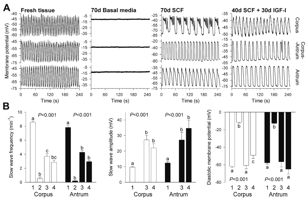
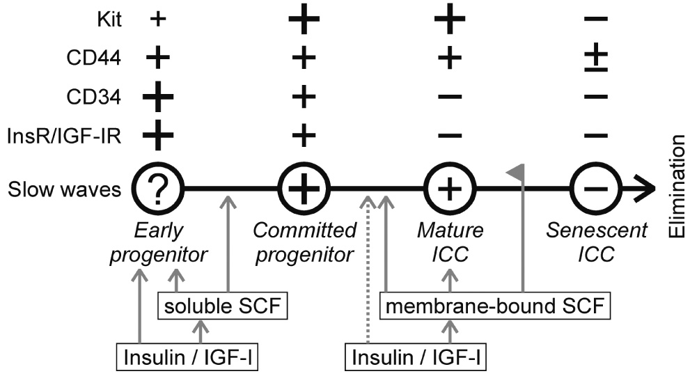
Background & aims: Maintaining the integrity of networks of interstitial cells of Cajal (ICC) is essential to preserve orderly contractile activity and neuroregulation in the gastrointestinal tract and to restore these functions after tissue damage or surgeries. Maintenance of ICC requires insulin-dependent or insulin-like growth factor I (IGF-I)-dependent production of membrane-bound stem cell factor (SCF) and may involve regeneration from local progenitors. Our goal was to identify ICC precursors in postnatal murine gastric muscles.

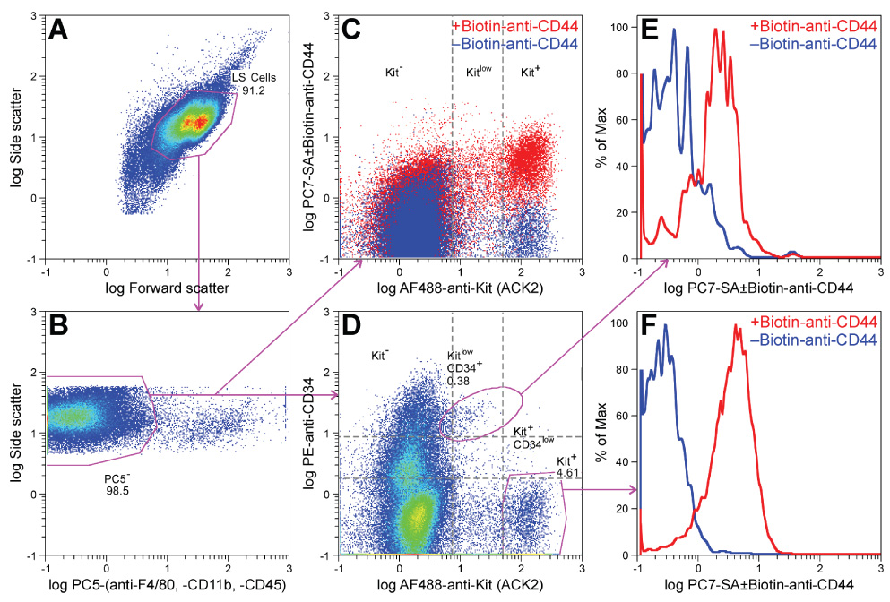
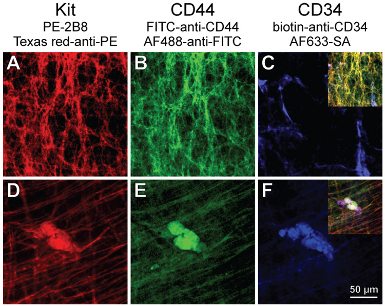
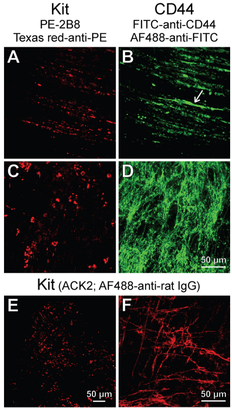
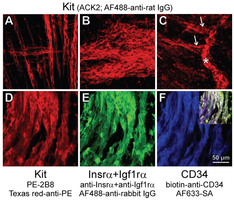
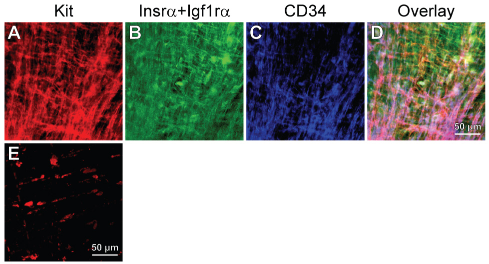
Methods: We used flow cytometry and immunohistochemistry to examine freshly dissected and cultured muscles for cells expressing CD34, an adhesion molecule expressed by stromal tumors; CD44, which occurs on mesenchymal stem cells; and receptors for SCF (Kit), insulin (Insr), and IGF-I (Igf1r). Slow waves were studied by intracellular recording.

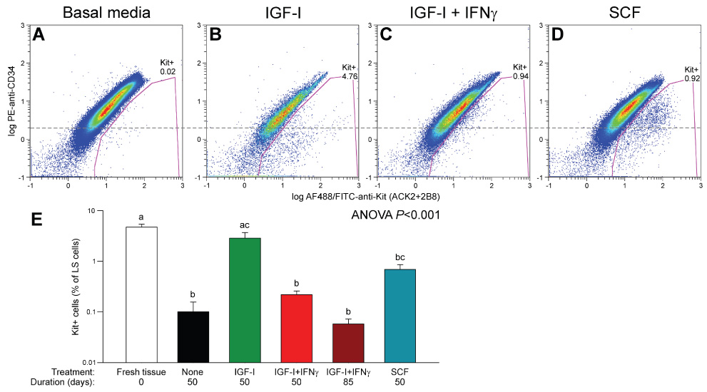
Results: In gastric muscles, we identified rare, Kit(low)CD44(+)CD34(+)Insr(+)Igf1r(+) cells resembling common embryonic precursors of ICC and smooth muscle. These putative progenitors were absent from organotypic cultures lacking mature ICC (Kit(+)CD44(+)CD34(-)Insr(-)Igf1r(-)) due to prolonged insulin/IGF-I deprivation but were rescued by IGF-I that also prevented ICC loss. Soluble SCF failed to prevent the loss of mature ICC but dramatically expanded the putative progenitors, which supported robust slow wave activity despite retaining an immature, Kit(+)CD44(+)CD34(+)Insr(+)Igf1r(+) phenotype. Differentiation of these cells into mature, network-forming ICC required IGF-I. Conversely, restoration of ICC networks by IGF-I after prolonged insulin and IGF-I deprivation required the survival of the presumed progenitors.

Conclusions: Kit(low)CD44(+)CD34(+)Insr(+)Igf1r(+) cells may be local progenitors for gastric ICC and stromal tumors. Loss of these cells may contribute to gastrointestinal dysmotilities.


Figures

Figure 1
Figure 1
Identification of KitlowCD44+CD34+ presumed ICC progenitors in dispersed postnatal murine gastric corpus+antrum muscles by flow cytometry. A representative of 10 experiments is shown. Numbers indicate cell frequencies (%). (A) Gates used for selecting cells with light scatter properties characteristic of live cells (LS cells). (B) Gates used for detecting cells that do not express macrophage markers (F4/80, CD11b) and the general hematopoietic marker CD45 (phycoerythrine-cyanine 5 (PC5) cells). (C) Expression of CD44 in the PC5 population shown by an overlay of two aliquots of the same sample labeled either with biotin-anti-CD44 plus phycoerythrine-cyanine 7 (PC7)-streptavidin (SA) (red) or with PC7-SA only (control; blue). Kit was detected with Alexa Fluor (AF) 488-anti-Kit antibodies. (D) Relationship between Kit and CD34 expression (detected with phycoerythrine (PE)-anti-CD34 antibodies) in the PC5 population. (E, F) Histogram representation of CD44 expression in the KitlowCD34+ subset and in the Kit+ ICC, respectively.
Figure 2
Figure 2
Identification of cells co-expressing Kit (A,D), CD44 (B,E) and CD34 (C,F) in the myenteric region of the gastric antrum of juvenile mice by immunohistochemistry. FITC, fluorescein isothiocyanate. Insets in C and F show digital overlays. (A–C) Mature ICC-MY expressing Kit and CD44 but not CD34. (D–F) Co-localization of CD34 with variable levels of Kit and CD44 in a rare cluster of small, round cells.
Figure 3
Figure 3
Loss of Kit+ cells in long-term growth factor-deprived cultures. (A–D) Juvenile gastric corpus+antrum tissues maintained with unsupplemented basal media for 85 days. Kit (A,C) and CD44 (B,D) immunofluorescence in the same fields-of-view. Note KitCD44+ cells in the circular muscle (arrow in B) and networks of KitCD44+ cells in the myenteric region (D). (E,F) Kit immunofluorescence from adult gastric corpus+antrum tissues cultured with unsupplemented, normoglycemic basal media for 32 days. Residual Kit+ ICC-MY were only found in the mid-corpus (F).
Figure 4
Figure 4
Flow cytometry analysis of Kit and CD34 in organotypic cultures of juvenile gastric corpus+antrum muscles. Tissues were maintained with unsupplemented basal media (A; n=5), 100 ng/ml IGF-I (B; n=5), 100 ng/ml IGF-I+100 U/ml IFNγ (C; n=3) or 50 ng/ml SCF (D; n=3) for 50 days. Gating was performed as in Figure 1A,B; only LS+PC5 cells are shown. Numbers indicate cell frequencies (%). Dashed line represents the approximate boundary between CD34+ and CD34 cells. Most Kit+ cells were CD34 in the IGF-I-treated tissues (B) and CD34+ in the SCF-treated muscles (D). (E) Statistical comparison of Kit+ cells in the above groups, freshly dissected juvenile muscles (n=5) and tissues treated with IGF-I+IFNγ for 85 days (n=3) by ANOVA following arcsin square root transformation. Groups not sharing the same superscript were different by all-pairwise Tukey test.
Figure 5
Figure 5
Effects of in vitro treatments on gastric electrical slow wave activity in organotypic cultures of adult gastric corpus+antrum muscles. (A) Representative recordings from freshly dissected (n=5) and cultured tissues (n=3 each) maintained for 70 days with basal media, 100 ng/mL SCF or sequential application of 100 ng/mL SCF (40 days) and 100 ng/mL IGF-I (30 days). (B) Quantitative analysis of slow wave parameters. Bars are labeled as follows: (1) fresh tissue (corpus: n=31 recordings; antrum: n=46); (2) basal media (corpus: n=13; antrum: n=9); (3) SCF (corpus: n=16; antrum: n=12); (4) SCF followed by IGF-I (corpus: n=14; antrum: n=9). Growth factor-deprived cultures, which only displayed sporadic, irregular slow waves in 2 out of 22 recordings, were excluded from the analysis of slow wave amplitudes. P values are from one-way ANOVA or Kruskal–Wallis one-way ANOVA on ranks. Groups not sharing the same superscripts were different by post-hoc multiple comparisons.
Figure 6
Figure 6
Loss of mature ICC and expansion of clusters of presumed ICC progenitors in organotypic cultures maintained with soluble SCF. (A,B) Presumed ICC precursors in adult gastric muscles cultured for 36 days with 100 ng/ml SCF. (A) Precursors forming chords within the circular and longitudinal muscle layers in the orad corpus. (B) Cluster of presumed precursors in the myenteric region of the antrum. (C) Presumed precursors in a day 14 distal antrum cultured with 50 ng/ml SCF for 50 days. Note cluster on the submucosal surface (asterisk) giving rise to chords extending along muscle fibers (arrows) and the lack of mature ICC. (D–F) Immature phenotype of presumed subserosal ICC progenitors in an adult tissue (corpus) maintained with 100 ng/ml SCF for 61 days. Note dominance of the the presumed precursors, the lack of processes and the continuing co-expression of Kit (D), Insr/Igf1r (E), and CD34 (F). Inset in F shows digital overlay.
Figure 7
Figure 7
IGF-I promotes the differentiation of ICC from its precursors. (A–D) IGF-I-induced conversion into mature ICC of presumed ICC progenitors following stimulation with soluble SCF. Kit (A), Insr/Igf1rα (B), and CD34 (C) immunofluorescence and their digital overlay (D) in an adult gastric corpus maintained with 100 ng/mL IGF-I for 23 days following a 41-day pretreatment with 100 ng/mL SCF and stained as in Figure 6D–F. The mature, interconnected Kit+ ICC networks lacked Insrα and Igf1rα although some cells continued to express CD34. (E) Failure of IGF-I treatment to maintain or rescue Kit+ ICC when applied after a period of growth factor deprivation. Adult tissue cultured with unsupplemented basal media for 42 days before replacing IGF-I for 28 days.
Figure 8
Figure 8
Proposed model for ICC turnover in the postnatal murine gastric corpus and antrum. +, present; −, absent. The size of the + signs is proportional to the approximate degree of expression. Gray arrows indicate maintenance or stimulation; dashed arrow, possible stimulation. Line with flag indicates inhibition.

Comment in

Similar articles

Cited by

References

    1. Sanders KM, Koh SD, Ward SM. Interstitial cells of cajal as pacemakers in the gastrointestinal tract. Annu Rev Physiol. 2006;68:307–343. - PubMed
    1. Huizinga JD. Gastrointestinal peristalsis: joint action of enteric nerves, smooth muscle, and interstitial cells of Cajal. Microsc Res Tech. 1999;47:239–247. - PubMed
    1. Ward SM, Sanders KM. Interstitial cells of Cajal: primary targets of enteric motor innervation. Anat Rec. 2001;262:125–135. - PubMed
    1. Fox EA, Phillips RJ, Martinson FA, et al. C-Kit mutant mice have a selective loss of vagal intramuscular mechanoreceptors in the forestomach. Anat Embryol (Berl) 2001;204:11–26. - PubMed
    1. Vanderwinden JM, Rumessen JJ. Interstitial cells of Cajal in human gut and gastrointestinal disease. Microsc Res Tech. 1999;47:344–360. - PubMed

Publication types

MeSH terms