. 2008 Apr;38(4):1138-47.
doi: 10.1002/eji.200737898.

TLR4 and S1P receptors cooperate to enhance inflammatory cytokine production in human gingival epithelial cells

Affiliations

TLR4 and S1P receptors cooperate to enhance inflammatory cytokine production in human gingival epithelial cells

Mehmet A Eskan et al. Eur J Immunol. 2008 Apr.

Abstract

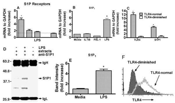
Toll-like receptors (TLR) are pattern recognition receptors for highly conserved microbial molecular patterns. Activation of TLR is a pivotal step in the initiation of innate, inflammatory, and immune defense mechanisms. Recent findings indicate that G protein-coupled receptors (GPCR) may modulate TLR signaling, but it is unclear which GPCR are involved in this process. One such cooperation between GPCR and TLR can be attributed to the sphingosine 1-phosphate (S1P) receptor family. The S1P receptors (S1P1-5) are a family of GPCR with a high affinity for S1P, a serum-borne bioactive lipid associated with diverse biological activities such as inflammation and healing. In this study, we show that pro-inflammatory cytokine production, including IL-6 and IL-8, was increased with LPS and concomitant S1P stimulation. Furthermore, elevated cytokine production following LPS and S1P challenge in human gingival epithelial cells (HGEC) was significantly reduced when TLR4, S1P1 or S1P3 signaling was blocked. Our study also shows that S1P1 and S1P3 expression was induced by LPS in HGEC, and this elevated expression enhanced the influence of S1P in its cooperation with TLR4 to increase cytokine production. This cooperation between TLR4 and S1P1 or S1P3 demonstrates that TLR4 and GPCR can interact to enhance cytokine production in epithelial cells.


Conflict of interest statement

Conflict of interest: The authors declare no financial or commercial conflict of interest.

Figures

Figure 1
Figure 1
Cytokine production following S1P/LPS stimuli in epithelial cells. Primary HGEC were challenged with LPS (1 μg/mL), S1P (100 nM), or a combination of LPS + S1P for 24 h at 37°C. Induction of IL-6 (A) and IL-8 (B) was determined in TLR4-normal and TLR4-diminished epithelial cell culture supernatants by Luminex following S1P, LPS or LPS + S1P challenge. TLR4-normal cells were challenged with 1, 2, or 4 μM SK inhibitor (inh) or medium only for 24 h. Then IL-6 production was determined in the challenged groups (C). The cells were challenged with LPS + S1P in the presence or absence of SK (inh) for 24 h. Subsequently, IL-6 production was measured by ELISA (D). Data are presented as the mean ± standard deviation (SD) of triplicate determinations, from one of three independent sets of cell cultures that yielded similar findings. Statistically significant (p <0.05) induction of cytokine release is indicated by an asterisk in TLR4-normal cells (NS; not statistically significant from basal level, LPS or S1P challenge).
Figure 2
Figure 2
S1P does not cooperate with TLR2 for the production of cytokines. HGEC were challenged with S1P (100 nM), FSL-1 (1 μg/mL) or a combination of FSL-1 + S1P for 24 h at 37°C. The production of IL-6 (A) and IL-8 (B) was assayed by Luminex following FSL-1, S1P or FSL-1 + S1P challenge for 24 h. Data are presented as the mean ± SD of triplicate determinations from one of three independent sets of cell cultures that yielded similar findings (NS; not statistically significant from FSL-1 challenge).
Figure 3
Figure 3
S1P1 is induced by LPS in a time- and dose-dependent fashion in epithelial cells. TLR4-normal cells were challenged with 100, 200, 500, 1000 and 2000 ng/mL of protein-free E. coli LPS or medium only for 24 h at 37°C (A). TLR4-normal cells were challenged with 1 μg/mL of protein-free E. coli LPS or medium only for 0.5, 1, 2, 4, 6, 8 or 24 h at 37°C (B). Following the challenge assay, relative expression of S1P1 was determined by real-time PCR. The ratio of S1P1 was normalized to GAPDH mRNA in cells following LPS challenge. Statistically significant (p <0.05) expression of S1P1 is indicated by an asterisk.
Figure 4
Figure 4
S1P1 is induced by TLR4 ligation in human epithelial cells. TLR4-normal HGEC were challenged with LPS (1 μg/mL) for 24 h. Relative expression of S1P1–5 was determined by real-time PCR after normalizing their expression to GAPDH (A). TLR4-normal HGEC were challenged with protein-free E. coli LPS (1 μg/mL), TLR2 agonist (FSL-1, 1 μg/mL) or IL-1β (5 ng/mL) for 24 h at 37°C. Real-time PCR was performed and the ratio of S1P1 was normalized to GAPDH mRNA in TLR4-normal gingival epithelial cells following LPS, FSL-1 or IL-1β challenge (B). The ratios of TLR4 and S1P1 were normalized to GAPDH in TLR4-normal and TLR4-diminished gingival epithelial cells after LPS challenge (C). Protein expression of S1P1 in TLR4-normal epithelial cells following LPS challenge was detected by Western blotting after S1P1 immunoprecipitation (IgH and IgL were included for specificity, the control β-actin is not shown) (D) and band intensity was determined using the ratio of the increase of S1P1 over endogenous control (β-Actin) (E). Both cells types (TLR-4-normal and TLR-4-diminished) were stained with PE-conjugated monoclonal anti-human TLR4 antibody or its isotype control (IgG2a, data not shown) for 20 min at 4°C. The stained cells were analyzed by flow cytometry using a BD FACSCalibur and CellQuest software (F). Data presented are the means ± SD of triplicate determinations from one of three independent sets of cell cultures that yielded similar findings. Statistically significant (p <0.05) expression of TLR4 or S1P1 is indicated by an asterisk.
Figure 5
Figure 5
TLR4 cooperation is required for cytokine production in HGEC. The TLR4-normal epithelial cells were transfected with 100 pmol of anti-sense RNA to TLR4 or laminin, an irrelevant gene silencer; 3 μL of the transfection reagent FuGene 6 was diluted using 95 μL of serum-free media, and 100 pmol of siTLR4 or siLaminin was added and incubated at room temperature for 15 min. The reaction was carried out overnight and the medium was replaced with fresh medium. After 48 h of silencing, the cells were challenged with LPS (1 μg/mL) in the presence of S1P (100 nM) for 24 h. The expression of TLR4, and S1P1 following the silencing, was determined by real-time PCR (A). The protein induction of IL-8 (B) and IL-6 (C) was determined in culture supernatants by Luminex. Data are presented as the mean ± SD of triplicate determinations. Statistically significantly (p <0.05) induced or reduced cytokine production is indicated by two asterisks or an asterisk, respectively.
Figure 6
Figure 6
S1P1 is specifically involved in the production of cytokines in gingival epithelial cells. TLR4-normal epithelial cells were silenced for S1P1 or a scramble (scr) gene. The reaction was carried out as described in the Materials and methods section. After 48 h of silencing, the cells were challenged with LPS (1 μg/mL) in the presence of S1P (100 nM) for 24 h. The expression of S1P1 was determined by real-time PCR performed with an ABI 7500 system following the silencing (A). The cytokines IL-8 (B) and IL-6 (C) were determined in culture supernatants by Luminex technology. TLR4-normal epithelial cells were challenged with LPS (1 μg/mL), SEW (3 μM), or a combination of LPS + SEW for 24 h at 37°C. Production of IL-6 was determined in the cell culture supernatants by ELISA following SEW, LPS or LPS + SEW stimuli (D). The cells were incubated with pertussis toxin (PTx) prior to LPS and S1P challenge. The induction of IL-6 (E) and IL-8 (F) was determined in culture supernatants by Luminex. Data are shown as means ± SD of triplicate determinations. Statistical significance, declared at p <0.05 for molecule induction or reduction, is shown by asterisks or an asterisk, respectively.
Figure 7
Figure 7
S1P3 is also involved in the cytokine production. TLR4-normal cells were treated with 0.2, 0.4, 0.5, 1, or 2 mg/mL suramin or medium only for 8 h. Subsequently, the production of IL-6 was determined by ELISA (A). The cells were challenged with LPS + S1P for 8 h after 1 h of suramin (0.4 mg/mL) treatment. The IL-6 (B) and IL-8 (C) induction was measured by ELISA. Statistical significance was declared at p <0.05, and cytokine induction or reduction is shown by an asterisk or asterisks, respectively.
Figure 8
Figure 8
ERK1/2 activation was enhanced by S1P in LPS-challenged cells. The cells were incubated with inhibitory reagent against ERK1/2 (U0126) prior to LPS and S1P challenge. The induction of IL-6 (A) and IL-8 (B) was determined in culture supernatants by Luminex technology. The level of phosphorylated ERK1/2 was determined by FACE (Active Motif), as described in the Material and methods section, in cells following LPS or S1P/LPS challenge (C). Data are presented as means ± SD of triplicate determinations. Statistically significant (p <0.05) cytokine induction is indicated by two asterisks, and significantly reduced phosho-ERK/12 or induction of cytokines is indicated by an asterisk.

References

    1. Rimoldi M, Chieppa M, Salucci V, Avogadri F, Sonzogni A, Sampietro GM, Nespoli A, et al. Intestinal immune homeostasis is regulated by the crosstalk between epithelial cells and dendritic cells. Nat Immunol. 2005;6:507–514. - PubMed
    1. Kawai T, Akira S. Pathogen recognition with Toll-like receptors. Curr Opin Immunol. 2005;17:338–344. - PubMed
    1. Kaisho T, Akira S. Critical roles of Toll-like receptors in host defense. Crit Rev Immunol. 2000;20:393–405. - PubMed
    1. Medzhitov R, Preston-Hurlburt P, Janeway CA., Jr A human homologue of the DrosophilaToll protein signals activation of adaptive immunity. Nature. 1997;388:394–397. - PubMed
    1. Takeuchi O, Hoshino K, Kawai T, Sanjo H, Takada H, Ogawa T, Takeda K, Akira S. Differential roles of TLR2 and TLR4 in recognition of gram-negative and gram-positive bacterial cell wall components. Immunity. 1999;11:443–451. - PubMed

Publication types

MeSH terms

LinkOut - more resources