. 2008 Oct;125(2):161-9.
doi: 10.1111/j.1365-2567.2008.02837.x. Epub 2008 Apr 4.

T helper cell type 1 (Th1), Th2 and Th17 responses to myelin basic protein and disease activity in multiple sclerosis

Affiliations

T helper cell type 1 (Th1), Th2 and Th17 responses to myelin basic protein and disease activity in multiple sclerosis

Chris J Hedegaard et al. Immunology. 2008 Oct.

Erratum in

  • Immunology. 2008 Nov;125(3):438

Abstract

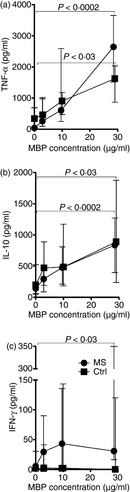
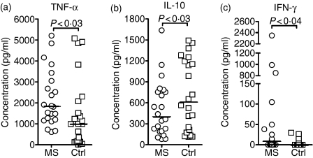
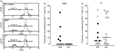
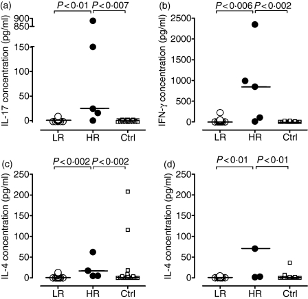
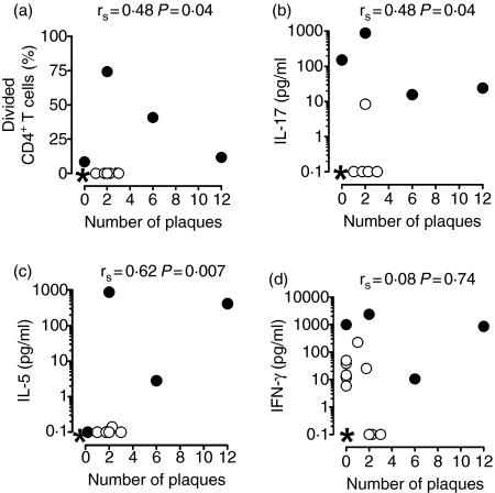
Autoreactive T cells are thought to play an essential role in the pathogenesis of multiple sclerosis (MS). We examined the stimulatory effect of human myelin basic protein (MBP) on mononuclear cell (MNC) cultures from 22 patients with MS and 22 sex-matched and age-matched healthy individuals, and related the patient responses to disease activity, as indicated by magnetic resonance imaging. The MBP induced a dose-dependent release of interferon-gamma (IFN-gamma), tumour necrosis factor-alpha (TNF-alpha) and interleukin-10 (IL-10) by patient-derived MNCs. The patients' cells produced higher amounts of IFN-gamma and TNF-alpha, and lower amounts of IL-10, than cells from healthy controls (P<0.03 to P<0.04). Five patients with MS and no controls, displayed MBP-induced CD4+ T-cell proliferation. These high-responders exhibited enhanced production of IL-17, IFN-gamma, IL-5 and IL-4 upon challenge with MBP, as compared with the remaining patients and the healthy controls (P<0.002 to P<0.01). A strong correlation was found between the MBP-induced CD4+ T-cell proliferation and production of IL-17, IFN-gamma, IL-5 and IL-4 (P<0.0001 to P<0.01) within the patient group, and the production of IL-17 and IL-5 correlated with the number of active plaques on magnetic resonance images (P=0.04 and P=0.007). These data suggest that autoantigen-driven CD4+ T-cell proliferation and release of IL-17 and IL-5 may be associated with disease activity. Larger studies are needed to confirm this.


Figures

Figure 1
Figure 1
Dose-dependent cytokine responses to myelin basic protein (MBP). Mononuclear cells from 13 patients with multiple sclerosis (MS; circles) and six healthy controls (Ctrl; squares) were grown in medium containing autologous serum (30% v/v) and increasing concentrations of purified human MBP. The levels of tumour necrosis factor-α (a) and interleukin-10 (b) in the culture supernatants after 1 day of incubation, and the levels of interferon-γ after 7 days of incubation (c) are shown as median and interquartile ranges.
Figure 2
Figure 2
Myelin basic protein (MBP)-elicited cytokine production by patient and control cells. Mononuclear cells from 22 untreated patients with multiple sclerosis [MS; 20 with relapsing–remitting MS (RRMS); two with clinically isolated syndrome (CIS)] and 22 healthy controls (Ctrl) were grown in the presence of human MBP (30 μg/ml) or no antigen. The resulting net production of tumour necrosis factor-α (TNF-α; a) and interleukin-10 (IL-10; b) at day 1, and interferon-γ at day 7 (IFN-γ; c), after subtraction of the background production in the absence of stimulating antigen is shown. The backround production was similar in patients and controls, < 34 pg/ml for TNF-α, < 60 pg/ml for IL-10 and < 3 pg/ml for IFN-γ. Horizontal bars represent median values.
Figure 3
Figure 3
Myelin basic protein (MBP)-elicited CD4+ T-cell proliferation. Carboxyfluorescein-diacetate-succinimidyl-ester (CFSE)-labelled mononuclear cells were incubated for 10 days with human MBP (30 μg/ml), or no antigen, and divisions of CD4+ T cells were tracked by flow cytometry. (a) Histograms showing undivided CD4+ T cells (G0) and cells having undergone more than one division, as identified by a reduction in fluorescence intensity. The upper panel shows unstimulated cells, the middle panel shows cells stimulated with MBP, and the lower panel shows tetanus toxoid (TT)-stimulated cells. (b) The proportion of divided CD4+ T cells from 22 patients with multiple sclerosis (MS) and 22 sex-matched and age-matched healthy controls (Ctrl) after 10 days of stimulation with MBP is shown. (c) The corresponding proportion of divided CD4+ T cells from 13 randomly selected patients with multiple sclerosis (MS) and 15 healthy controls (Ctrl) after 10 days of stimulation with a control antigen, TT. Closed circles in (b) and (c) represent the five high-responders to MBP. In all cases, the proportion of cells in division was less than 0.5% in the absence of antigenic stimulation.
Figure 4
Figure 4
Myelin basic protein (MBP)-induced production of interleukin-17 (IL-17), interferon-γ (IFN-γ), IL-4 and IL-5. Mononuclear cells (MNCs) from patients with multiple sclerosis (MS), 17 low-responders (LR) and five high-responders (HR), and from 22 healthy, sex-matched and age-matched controls (Ctrl) were incubated with MBP for 7 days. The resulting net production of IL-17 (a), IFN-γ (b), IL-4 (c) and IL-5 (d), after subtraction of the background production in the absence of stimulating antigen, is shown. The background production was similar in the patient and control groups, being 0 pg/ml for IL-17, < 3 pg/ml for IFN-γ, < 3 pg/ml for IL-4 and < 2 pg/ml for IL-5.
Figure 5
Figure 5
Correlation between myelin basic protein (MBP)-induced T-cell proliferation, interleukin-17 (IL-17) production and disease activity. Mononuclear cell (MNC) cultures from 18 patients with multiple sclerosis (MS) were incubated with human MBP for 10 days. The proportion of divided CD4+ T cells at day 10 (a), and the content of IL-17 (b), IL-5 (c), and interferon-γ (IFN-γ) (d) in the culture supernatants at day 7 correlated with the number of brain plaques, as assessed by magnetic resonance imaging. Closed circles represent the high-responders who were scanned. *Represents multiple values of 0, 0 [nine in (a–c), six in (d)].

References

    1. Sospedra M, Martin R. Immunology of multiple sclerosis. Annu Rev Immunol. 2005;23:683–747. - PubMed
    1. Gausas J, Paterson PY, Day ED, Dal Canto MC. Intact B-cell activity is essential for complete expression of experimental allergic encephalomyelitis in Lewis rats. Cell Immunol. 1982;72:360–6. - PubMed
    1. Willenborg DO, Prowse SJ. Immunoglobulin-deficient rats fail to develop experimental allergic encephalomyelitis. J Neuroimmunol. 1983;5:99–109. - PubMed
    1. Myers KJ, Sprent J, Dougherty JP, Ron Y. Synergy between encephalitogenic T cells and myelin basic protein-specific antibodies in the induction of experimental autoimmune encephalomyelitis. J Neuroimmunol. 1992;41:1–8. - PubMed
    1. Mosmann TR, Cherwinski H, Bond MW, Giedlin MA, Coffman RL. Two types of murine helper T cell clone. I. Definition according to profiles of lymphokine activities and secreted proteins. J Immunol. 1986;136:2348–57. - PubMed

Publication types