. 2008 Jun;41(3):506-20.
doi: 10.1111/j.1365-2184.2008.00530.x. Epub 2008 Apr 7.

Cell proliferation, apoptosis and mitochondrial damage in rat B50 neuronal cells after cisplatin treatment

Affiliations

Cell proliferation, apoptosis and mitochondrial damage in rat B50 neuronal cells after cisplatin treatment

M G Bottone et al. Cell Prolif. 2008 Jun.

Abstract

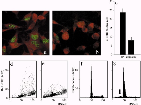
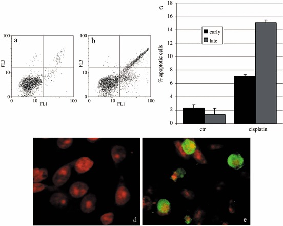
Objectives: Cisplatin (cisPt) is used as a chemotherapeutic agent for the treatment of a variety of human tumours; more recently, it has been demonstrated that tumour cell exposure to cisPt ultimately results in apoptosis, but the mechanism by which nuclear cisPt/DNA generates the cytoplasmic cascade of events involved has not been clarified. We have investigated the effects of cisPt on proliferation in the neuronal cell line B50, with particular attention being given to understand whether mitochondria are a target of cisPt and their involvement in the apoptotic process.

Materials and methods: Rat neuronal B50 cells were used to investigate the mechanisms of cisPt-induced cytotoxicity; this line has been used as a model system for neurotoxicity in vivo.

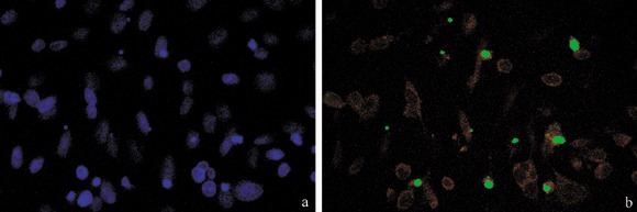
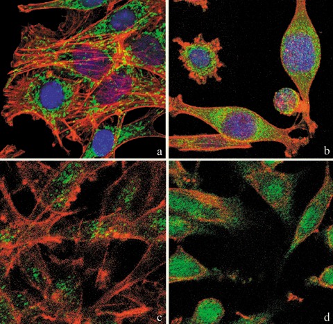
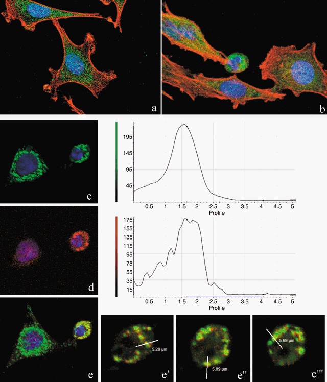
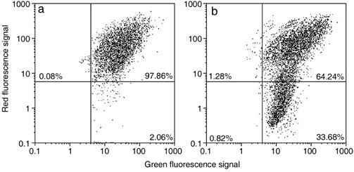
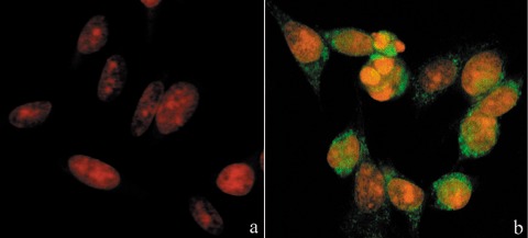
Results: Changes in proliferation, induction of apoptosis, activation of caspase-3 and DNA fragmentation were observed in the cells, as well as morphological and biochemical alterations of mithocondria. Activation of caspase-9 confirmed that mitochondria are a target of cisPt.

Conclusion: CisPt exerts cytotoxic effects in the neuronal B50 cell line via a caspase-dependent pathway with mitochondria being central relay stations.


Figures

Figure 1
Figure 1
Immunocytochemical reaction for BrdU in B50 cells: positive (green fluorescence) control sample (a) and after treatment with cisPt (b). Cytofluorimetric analysis of BrdU in B50 control cells (d) and treated with cisPt (e). The histogram represents the average of five independent experiments (c). Cytofluorimetric histograms of DNA content after propidium iodide (PI) staining in control B50 cells (f). The sub‐G1 peak (arrow) demonstrates that apoptotic cells are present after cisPt treatment (g).
Figure 2
Figure 2
Dual parameter cytogram of FITC‐labelled annexin V (in abscissa) versus propidium iodide (PI) staining (ordinate). Double‐negative, non‐apoptotic cells fall in the lower left quadrant, while early and late apoptotic cells fall in the lower and upper right quadrant, respectively. There was an increase of early and late apoptotic cells in cisPt‐treated cells (b). The histogram represents the average of five independent experiments (c). Immunocytochemical reaction for activated caspase‐3 in control (d) and cisPt‐treated cells (e, green fluorescence). DNA was counterstained with PI.
Figure 3
Figure 3
Treatment with cisPt induced caspase activation, and DNA fragmentation demonstrated by TUNEL assay. TUNEL‐positive cells (b, green fluorescence) were counterstained with Evan's blue (red fluorescence); in (a) DNA was counterstained with Hoechst 33258.
Figure 4
Figure 4
Confocal microscopy of dual immunolabelling of mitochondria (green fluorescence) and actinic cytoskeleton (red fluorescence) in B50 cells. Compared to untreated controls (a), in cisPt‐treated cells (b), mitochondria clustered around the nucleus and formed dense masses in the cytoplasm. Control cells (a) revealed a filamentous actin skeleton; in (b) cisPt induced disruption of filamentous actin structures and assembly of depolymerized actin in peripheral regions of cytoplasm close to the cell membrane. DNA was counterstained with Hoechst 33258. Confocal microscopy: immunocytochemical detection of Bcl‐2 (green fluorescence) in control cells (c) and after cisPt treatment (d). After treatment with cisPt, Bcl‐2 protein levels increased significantly in the nucleus. The cytoskeleton was labelled with Alexa 594‐conjugated phalloidin (red fluorescence).
Figure 5
Figure 5
Confocal microscopy. Immunocytochemical detection of Bax (green fluorescence) in control cells (a) and after cisPt treatment (b). Cytoskeleton was labelled with Alexa 594‐conjugated phalloidin (red fluorescence) and DNA was counterstained with Hoechst 33258. Double immunoreaction for mitochondria (green fluorescence, c) and Bax (red fluorescence, d) in cisPt‐treated B50 cells, DNA counterstained with Hoechst 33258. The three different sections (e′, e″, e′″) represent the fluorescence distribution determined for sections of the cell, as indicated in the red/green fusion image (e). After treatment, Bax translocates from its predominantly cytoplasmic location to mitochondria and induces activation of apoptosis.
Figure 6
Figure 6
Cytometric analysis of green‐versus‐red fluorescence of JC‐1 showing effects of cisPt treatment (b) on mitochondrial potential: compared to control (a), there is an increase of cell fraction with high green fluorescence and a relatively low red fluorescence.
Figure 7
Figure 7
Immunocytochemical detection of activated caspase‐9 (green fluorescence) in cisPt‐treated cells (b). In (a) untreated cells, DNA counterstained with propidium iodide (PI).

References

    1. Antonsson B, Conti F, Ciavatta A, Montessuit S, Lewis S, Martinou I, Bernasconi L, Bernard A, Mermod JJ, Mazzei G, Maundrell K, Gambale F, Sadoul R, Martinou JC (1997) Inhibition of Bax channel‐forming activity by Bcl‐2. Science 277, 370–372. - PubMed
    1. Antonsson B, Montessuit S, Sanchez B, Martinou JC (2001) Bax is present as a high molecular weight oligomer/complex in the mitochondrial membrane of apoptotic cells. J. Biol. Chem. 276, 11615–11623. - PubMed
    1. Avella D, Pisu MB, Roda E, Gravati M, Bernocchi G (2006) Reorganization of the rat cerebellar cortex during postnatal development following cisplatin treatment. Exp. Neurol. 201, 131–143. - PubMed
    1. Bedner E, Li X, Kunicki J, Darzynkiewicz Z (2000) Translocation of Bax to mitochondria during apoptosis measured by laser scanning cytometry. Cytometry 41, 83–88. - PubMed
    1. Bernocchi G, Scherini E, Nano R (1990) Developmental patterns in the rat cerebellum after cis‐dichlorodiammineplatinum treatment. Neuroscience 39, 179–188. - PubMed

Publication types

MeSH terms