. 2008 Jul 15;17(14):2118-31.
doi: 10.1093/hmg/ddn110. Epub 2008 Apr 7.

A sensitized mutagenesis screen identifies Gli3 as a modifier of Sox10 neurocristopathy

Affiliations

A sensitized mutagenesis screen identifies Gli3 as a modifier of Sox10 neurocristopathy

Ivana Matera et al. Hum Mol Genet. .

Abstract

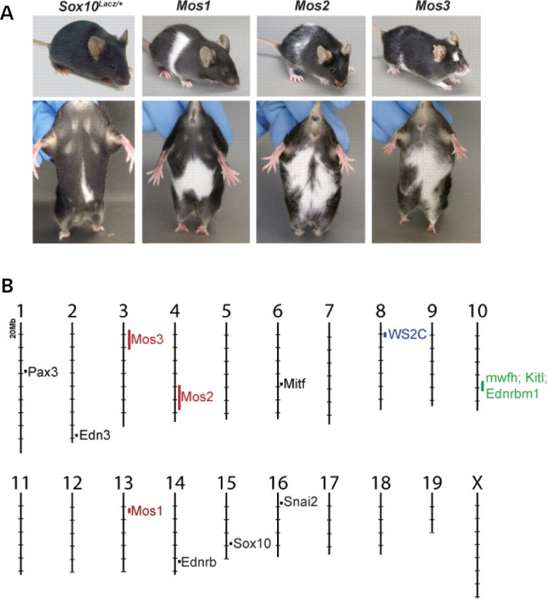
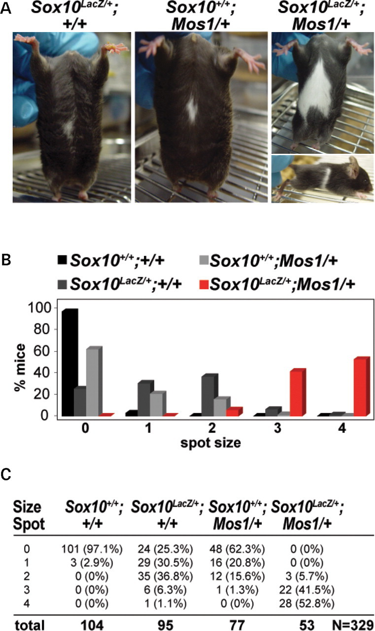
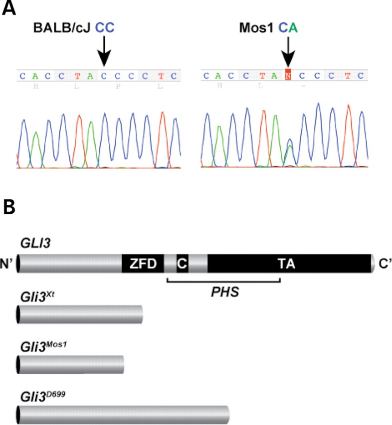
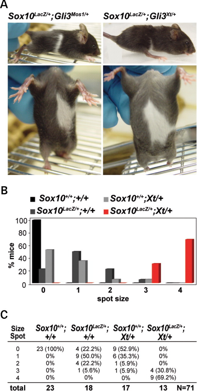
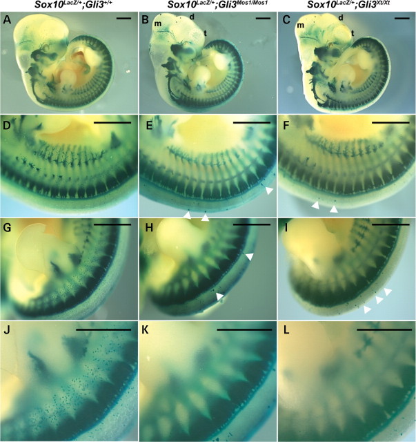
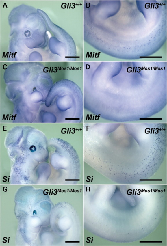
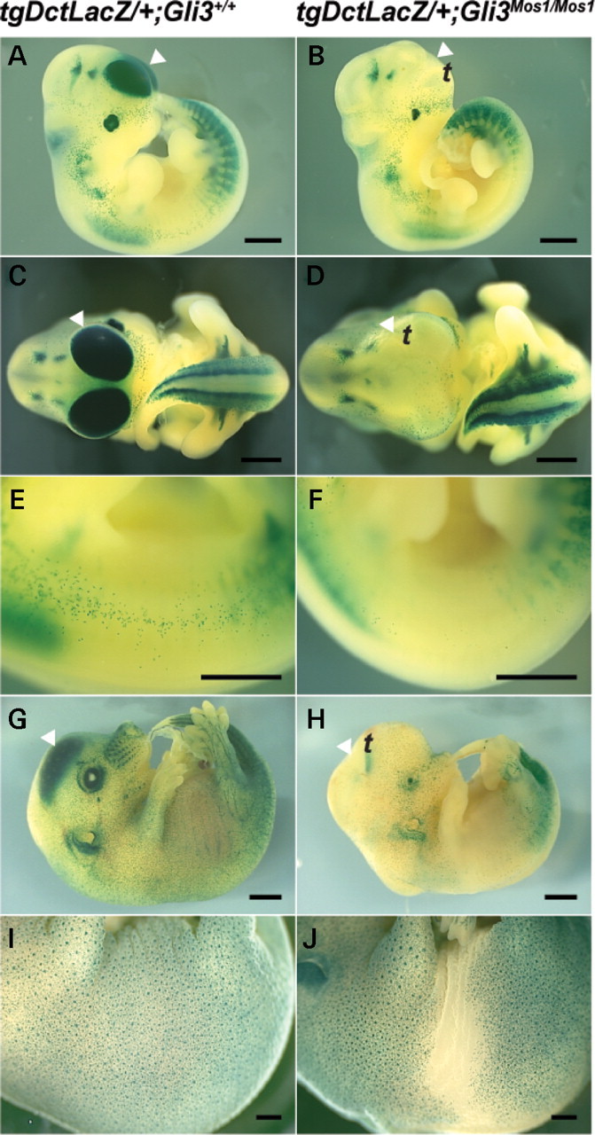
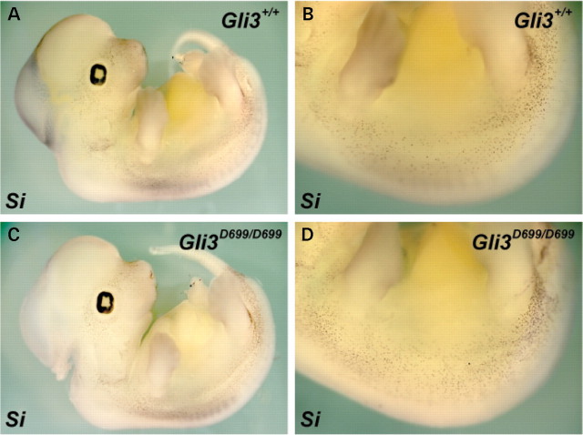
Haploinsufficiency for the transcription factor SOX10 is associated with the pigmentary deficiencies of Waardenburg syndrome (WS) and is modeled in Sox10 haploinsufficient mice (Sox10(LacZ/+)). As genetic background affects WS severity in both humans and mice, we established an N-ethyl-N-nitrosourea (ENU) mutagenesis screen to identify modifiers that increase the phenotypic severity of Sox10(LacZ/+) mice. Analysis of 230 pedigrees identified three modifiers, named modifier of Sox10 neurocristopathies (Mos1, Mos2 and Mos3). Linkage analysis confirmed their locations on mouse chromosomes 13, 4 and 3, respectively, within regions distinct from previously identified WS loci. Positional candidate analysis of Mos1 identified a truncation mutation in a hedgehog(HH)-signaling mediator, GLI-Kruppel family member 3 (Gli3). Complementation tests using a second allele of Gli3 (Gli3(Xt-J)) confirmed that a null mutation of Gli3 causes the increased hypopigmentation in Sox10(LacZ/+);Gli3(Mos1/)(+) double heterozygotes. Early melanoblast markers (Mitf, Sox10, Dct, and Si) are reduced in Gli3(Mos1/)(Mos1) embryos, indicating that loss of GLI3 signaling disrupts melanoblast specification. In contrast, mice expressing only the GLI3 repressor have normal melanoblast specification, indicating that the full-length GLI3 activator is not required for specification of neural crest to the melanocyte lineage. This study demonstrates the feasibility of sensitized screens to identify disease modifier loci and implicates GLI3 and other HH signaling components as modifiers of human neurocristopathies.


Figures

Figure 1.
Figure 1.
(A) Three hypopigmentation phenotypes, Mos1, Mos2, and Mos3, were identified in an ENU screen that increase the severity of hypopigmentation in Sox10LacZ/+ mice. Sox10LacZ/+;Mos1/+ double heterozygote, Mos2/+ heterozygotes, and Sox10LacZ/+;Mos3/+ double heterozygote individuals are shown after several generations of outcrossing to C57BL/6J. (B) The murine genomic locations of Mos1, 2, 3 map independently from previously identified WS loci. The locations of Mos1, Mos2, and Mos3 (red) are indicated on the mouse physical map relative to the locations of cloned WS genes (black), a regions of conserved synteny for a human WS modifier (WS2C in blue) (61), and a modifier of mouse Sox10 white forelock hypopigmentation (mwfh in green) (62) which overlaps modifiers of mouse EdnrbS hypopigmentation (Kitl and Ednrbm1 in green) (63).
Figure 2.
Figure 2.
Mos1 increases hypopigmentation in Sox10LacZ heterozygous mice. (A) Heterozygous Sox10LacZ/+ mice display a small region of ventral hypopigmentation (left) similar to that observed in heterozygous Mos1/+ mice (middle). Double heterozygous Sox10LacZ/+; Mos1/+ mice exhibit extensive hypopigmentation that frequently extends dorsally as a partial belt (right). (B, C) Quantitative data for the extent of ventral hypopigmentation in each of four genotype classes including wild-type (Sox10+/+;Mos1/+), Sox10LacZ heterozygotes (Sox10LacZ/+;Mos1+/+), Mos1 heterozygotes (Sox10+/+;Mos1/+), and Sox10;Mos1 double heterozygotes (Sox10LacZ/+;Mos1/+). (B) shows a graphical representation of the data, and (C) shows the total numbers and percentages within each genotype/phenotype class (total N = 329).
Figure 3.
Figure 3.
A nucleotide substitution in Mos1 lies in the N-terminal region of the Gli3 coding sequence. (A) Sequencing of BALB/cJ control and Mos1 heterozygote genomic DNA reveals a C to A substitution at nucleotide position 1148 (arrows) resulting in a nonsense mutation at codon 350 (Tyr350Stop) in the Mos1 allele. The heterozygous Mos1 trace shown contains both C (wild-type sequence of BALB/cJ) and A (mutant sequence of Mos1) at position 1148. (B) Graphic representation of full-length GLI3 protein and 3 truncated proteins resulting from mutant mouse alleles of Gli3. The zinc finger domain (ZFD) (34) and proteolytic cleavage site (C) (26) are indicated along with a region of the protein important for transactivation (TA) that spans several fragments independently shown to function in transactivation (–66). While the Gli3Xt-J allele results in a truncated protein that lacks the full ZFD and is a loss of function allele, Gli3D699 retains the ZFD, and thus functions as a transcriptional repressor. Mutations in the middle third of the human GLI3 gene (bracketed) are predicted to produce truncated functional repressor proteins causing Pallister-Hall syndrome (PHS). The location of the Mos1 mutation would result in a truncated protein lacking the ZFD, and is predicted to be a loss of function allele similar to Gli3Xt-J, and also to human Greig cephalopolysyndactyly, which is caused by mutations in the human gene that fall outside the bracketed PHS region (19).
Figure 4.
Figure 4.
The Gli3Xt-J allele increases hypopigmentation in Sox10LacZ heterozygous mice similar to the ENU mutation Mos1, showing that Mos1 is a Gli3 allelic variant. (A) Double heterozygous Sox10LacZ/+;Gli3Xt-J/+ mice show enhanced ventral and dorsal hypopigmentation (right) very similar to Sox10LacZ/+;Mos1/+ mice (left). (B, C) Representative data for ventral hypopigmentation phenotypes for each four genotype classes including wild-type (Sox10+/+;Gli3+/+), Sox10LacZ heterozygous mutants (Sox10LacZ/+;Gli3+/+), Gli3Xt-J heterozygous mutants (Sox10+/+;Gli3Xt-J/+), and double heterozygotes (Sox10LacZ/+;Gli3Xt-J/+). (B) Shows a graphical representation of the data, and (C) shows the total numbers and percentages within each genotype/phenotype class (total N = 71).
Figure 5.
Figure 5.
Sox10LacZ expression in Gli3Mos1/Mos1 embryos reveals reduced melanoblasts and ectopic Sox10LacZ-expressing cells. Sox10LacZ/+;Gli3+/+ (A, D, G, J), Sox10LacZ/+;Gli3Mos1/Mos1 (B, E, H, K), and Sox10LacZ/+;Gli3Xt-J/Xt-J (C, F, I, L) embryos at E11.5 after X-gal staining. Shown are lateral (A –C) views of E11.5 embryos and the corresponding trunk (D–F) and hind limb regions (G–L). Microphthalmia, as well as malformation of the mesencephalon (m), diencephalon (d) and telencephalon (t) are visible in Sox10LacZ/+;Gli3Mos1/Mos1 (B) and Sox10LacZ/+;Gli3Xt-J/Xt-J embryos (C). The Sox10LacZ-expressing sympathetic, cranial and DRG appeared unaffected in Sox10LacZ/+;Gli3Mos1/Mos1 (B) and Sox10LacZ/+;Gli3Xt-J/Xt-J (C) embryos as compared with Sox10Lacz/+ control embryos (A). Ectopic Sox10LacZ-expressing cells adjacent to the neural tube in Sox10LacZ/+;Gli3Mos1/Mos1 (E, Hbold>) and Sox10LacZ/+;Gli3Xt-J/Xt-J (F, I) embryos are indicated by arrowheads. The number of Sox10LacZ-expressing melanoblasts was reduced in Sox10LacZ/+;Gli3 Mos1/Mos1 (E, H, K) and Sox10LacZ/+;Gli3Xt-J/Xt-J (F, I, L) embryos within the medial lateral trunk region as compared with Sox10Lacz/+ control embryos (D, G, J).
Figure 6.
Figure 6.
GLI3 deficiency disrupts melanocyte specification in the trunk. Shown are lateral views of E11.5 embryos after whole-mount in situ hybridizations of wild-type (A, B, E, F) and Gli3Mos1/Mos1 (C, D, G, H) embryos with probes specific for the melanoblast markers Mitf (A–D), and Si (E–H). Gli3Mos1/Mos1 embryos show Mitf and Si expression patterns similar to wild-type embryos in the head and in cervical and tail regions (A, C, E, G). However, in the trunk region between forelimb and hind limb, Gli3Mos1/Mos1 embryos show very few melanoblasts (D, H) as compared with wild-type embryos (B, F).
Figure 7.
Figure 7.
Tg (Dct-LacZ) expression reveals reduction of melanoblasts in Gli3Mos1/Mos1 embryos. Lateral (A, B, E–J) or frontal (C, D) views of Gli3+/+;Tg(Dct-LacZ) (A, C, E, G, I) and Gli3Mos1/Mos1;Tg(Dct-LacZ) (B, D, F, H, J), embryos after X-gal staining. The trunk region extending from the ventral to the dorsal surface of Gli3Mos1/Mos1 embryos shows a severe reduction of melanoblast numbers at E11.5 (B, F) and E14.0 (H) compared with Gli3+/+;Tg (Dct-LacZ) embryos at E11.5 (A, E), and E14.0 (G). Lateral view of E16.0 Gli3Mos1/Mos1; Tg(Dct-LacZ) embryo (J) shows a reduction of melanoblast numbers of the ventral and lumbar trunk regions compared with Gli3+/+;Tg(Dct-LacZ) E16.0 embryos (I). Arrowheads (A–D, G and H) point toward Tg(Dct-LacZ) expressing cells of the telencephalon (t). A complete absence of Tg(Dct-LacZ) expression was noted in the telencephalon (t) of Gli3Mos1/Mos1;Tg(Dct-LacZ) embryos at E11.5 (B, D) and E14.0 (H) compared with Gli3+/+;Tg(Dct-LacZ) embryos (A, C at E11.5; G at E14.0).
Figure 8.
Figure 8.
GLI3 repressor is sufficient for melanoblast specification in the trunk. Shown are lateral views of E11.5 embryos after whole-mount in situ hybridizations of wild-type (A, B) and Gli3D699/D699 (C, D) embryos with probes specific for the melanoblast marker Si. Gli3D699/D699 embryos show Si expression patterns similar to wild-type embryos. We did not observe a drastic reduction in trunk melanoblasts as was observed in Gli3Mos1/Mos1 embryos (Fig. 6) that lack both GLI3 activator and repressor function.

Similar articles

Cited by

References

    1. Baldwin C.T., Hoth C.F., Amos J.A., da-Silva E.O., Milunsky A. An exonic mutation in the HuP2 paired domain gene causes Waardenburg’s syndrome. Nature. 1992;355:637–638. - PubMed
    1. Bondurand N., Dastot-Le Moal F., Stanchina L., Collot N., Baral V., Marlin S., Attie-Bitach T., Giurgea I., Skopinski L., Reardon W., et al. Deletions at the SOX10 gene locus cause Waardenburg syndrome types 2 and 4. Am. J. Hum. Genet. 2007;81:1169–1185. - PMC - PubMed
    1. Tassabehji M., Read A.P., Newton V.E., Harris R., Balling R., Gruss P., Strachan T. Waardenburg’s syndrome patients have mutations in the human homologue of the Pax-3 paired box gene. Nature. 1992;355:635–636. - PubMed
    1. Tassabehji M., Newton V.E., Read A.P. Waardenburg syndrome type 2 caused by mutations in the human microphthalmia (MITF) gene. Nat. Genet. 1994;8:251–255. - PubMed
    1. Sanchez-Martin M., Rodriguez-Garcia A., Perez-Losada J., Sagrera A., Read A.P., Sanchez-Garcia I. SLUG (SNAI2) deletions in patients with Waardenburg disease. Hum. Mol. Genet. 2002;11:3231–3236. - PubMed

Publication types

MeSH terms