. 2008 Apr;29(4):681-7.
doi: 10.3174/ajnr.A0901.

Arteriovenous shunt visualization in arteriovenous malformations with arterial spin-labeling MR imaging

Affiliations

Arteriovenous shunt visualization in arteriovenous malformations with arterial spin-labeling MR imaging

R L Wolf et al. AJNR Am J Neuroradiol. 2008 Apr.

Abstract

Background and purpose: A reliable quantitative technique for measuring arteriovenous (AV) shunt in vascular malformations is not currently available. Here, we evaluated the hypothesis that continuous arterial spin-labeled (CASL) perfusion MR imaging can be used to detect and measure AV shunt in patients with arteriovenous malformations (AVMs).

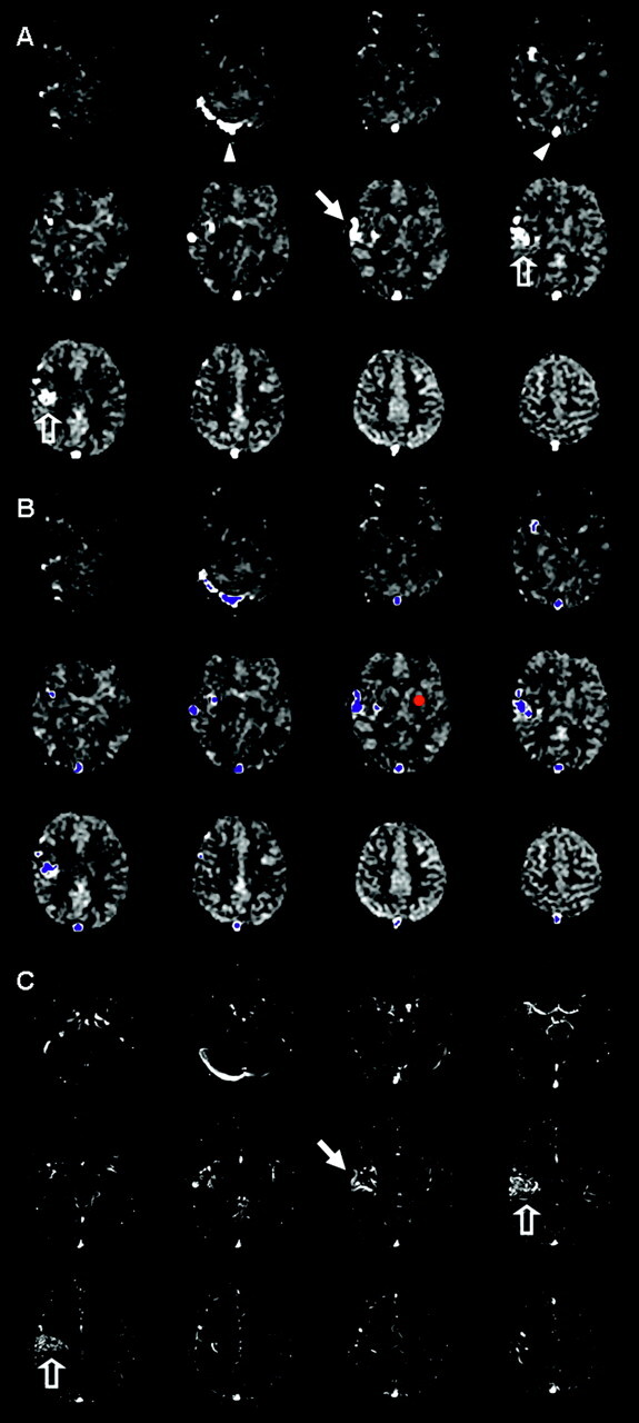
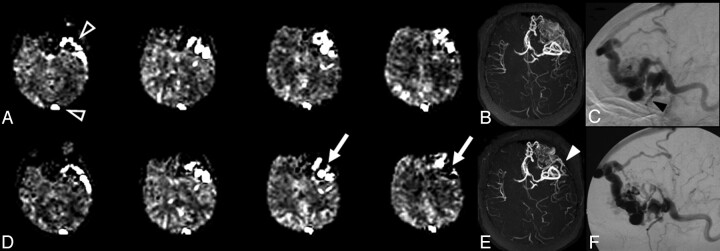
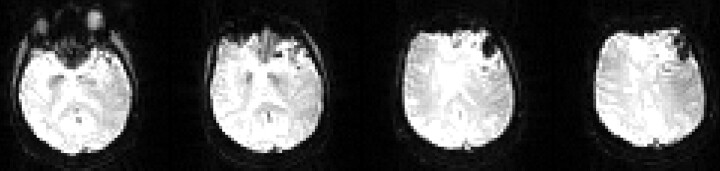
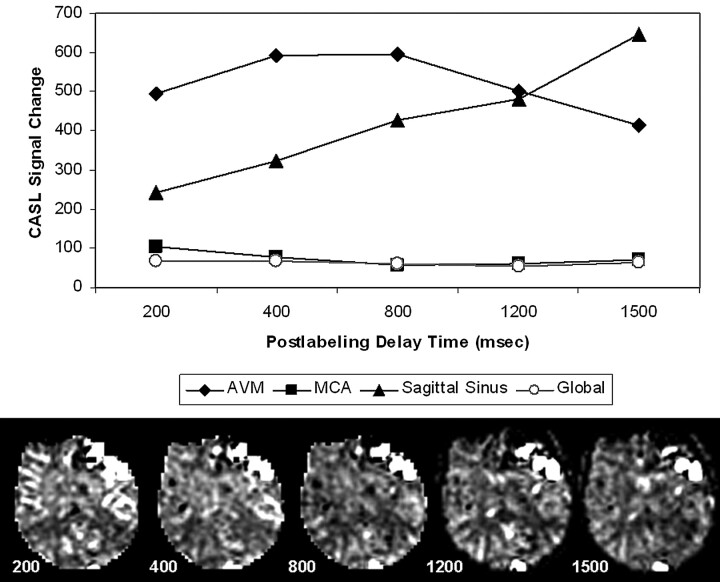
Materials and methods: CASL perfusion MR imaging was performed in 7 patients with AVMs. Semiquantitative AV shunt estimates were generated based on a thresholding strategy by using signal-intensity difference (DeltaM) images to avoid potential errors in cerebral blood flow (CBF) calculation related to abnormal transit times and nonphysiologic blood-tissue water exchange in and around the AVMs. The potential for measuring CBF in regions distant from and near the AVM was explored, as was the relationship of CBF changes related to the size of the shunt.

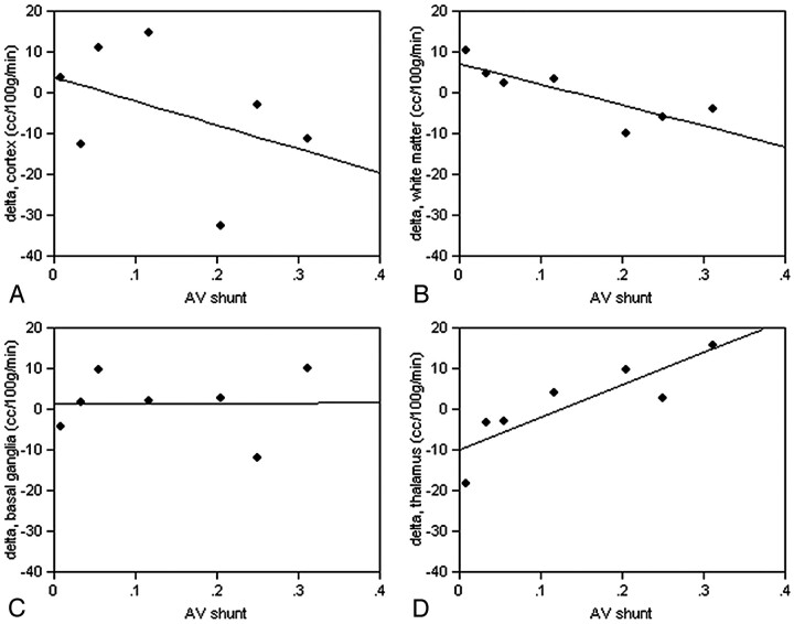
Results: In all 7 cases, striking increased intensity was seen on CASL perfusion DeltaM maps in the nidus and venous structures draining the AVM. Shunt estimates ranged from 30% to 0.6%. Mean CBF measurements in structures near the AVMs were not significantly different from the contralateral measurements. However, CBF in adjacent ipsilateral white matter increased relative to the contralateral side as the percent shunt increased (P = .02). Cortical gray matter CBF Delta (contralateral-ipsilateral) values demonstrated the same effect, but the correlation was weak and not significant. Thalamic CBF decreased ipsilaterally with increasing percent AV shunt (P = .01), indicating a possible steal effect. Basal ganglia Delta values showed little change in CBF with the size of the AV shunt.

Conclusion: CASL perfusion MR imaging can demonstrate AV shunting, providing high lesion conspicuity and a novel means for evaluating AVM physiology.


Figures

Fig 1.
Fig 1.
Image evaluation and shunt approximation for a right hemisphere AVM. A, CASL perfusion ΔM maps show bright signal intensity in the sagittal sinus and right transverse sinus (arrowheads) and in draining veins (arrows) adjacent to the nidus (open arrows). B, A small region of interest (red circle) placed in the left basal ganglia is used for each patient to measure mean ΔM (BGmean) and SD (BGSD). A threshold is generated (empirically chosen as threshold = BGmean + 8 *BGSD) above which voxels are labeled as shunt voxels to create a mask (blue voxels) for the AVM and draining vessels. The AV shunt fraction is estimated by multiplying the mean ΔM in the mask by the number of voxels in the mask, then dividing by the mean ΔM in the entire brain multiplied by all labeled voxels. C, Registered 2D TOF MRA source images (no superior saturation band) are reviewed to verify reasonable selection of AVM and draining vessels (open and closed arrows).
Fig 2.
Fig 2.
AVM before (top) and 2 days after (bottom) partial embolization (patient 1). CASL ΔM images (A, D) show decrease in AV shunt (arrows, D), estimated decrease from 23% to 20%. Superficial venous drainage is identified, including the superior sagittal sinus and sphenobasal sinus on both studies (open arrowheads, A). Of interest, despite intense signal intensity in the sphenobasal sinus, this corresponds to a relatively small venous structure on DSA (black arrowhead, C). DSA lateral projections (C, F) and 3D TOF MRA collapsed maximum intensity projection images (B, E) confirm decreased flow in a portion of AVM, especially the posterior-superior portion (white arrowhead, E).
Fig 3.
Fig 3.
Susceptibility artifact resulting from the presence of embolic material, thrombus, and/or hemorrhage (patient 1). CASL source images from 4 imaging locations are shown.
Fig 4.
Fig 4.
Multiple postlabeling delay times (200–1500 ms): effect on ASL signal intensity in the AVM (patient 1). ΔM (T1-corrected) measured in regions of interest for the AVM (diamond), sagittal sinus (triangle), right MCA territory (box), and entire brain (circle) at each delay is plotted (above), with ΔM maps from 1 imaging location at multiple delays for comparison (below). ASL signal intensity is relatively stable in global measures over the entire brain and in the MCA at longer delays as expected. Signal intensity decreases in AVM and increases in sagittal sinus as delay increases.
Fig 5.
Fig 5.
Regional changes in CBF compared with extent of AV shunt. Δ = contralateral − ipsilateral CBF in measured regions of interest in white matter near the AVM, the cortex near the AVM, the thalamus, and the basal ganglia. A, Cortical gray matter CBF Δ values are negatively but weakly correlated with percent AV shunt and are not significant. (B) Δ values in white matter (R2 = 0.70, F = 11.58, P = .02) are negatively correlated (blood flow in adjacent ipsilateral white matter increases as percent shunt increased). Basal ganglia Δ values (C) show little change in CBF with the size of the AV shunt, but thalamic CBF (D) appears to decrease ipsilaterally as percent AV shunt increases (R2 = 0.74, F = 14.19, P = .01).

Similar articles

Cited by

References

    1. Greenberg MS. Handbook of Neurosurgery, 4th ed. Lakeland, FL: Greenberg Graphics;1997. :871
    1. Crawford PM, West CR, Chadwick DW, et al. Arteriovenous malformations of the brain: natural history in unoperated patients. J Neurol Neurosurg Psychiatry 1986;49:1–10 - PMC - PubMed
    1. Ondra SL, Troupp H, George ED, et al. The natural history of symptomatic arteriovenous malformations of the brain: a 24-year follow-up assessment. J Neurosurg 1990;73:387–91 - PubMed
    1. Carroll TJ, Grist TM. Technical developments in MR angiography. Radiol Clin North Am 2002;40:921–51 - PubMed
    1. Langer DJ, Song JK, Niimi Y, et al. Transarterial embolization of vein of Galen malformations: the use of magnetic resonance imaging noninvasive optimal vessel analysis to quantify shunt reduction—report of two cases. J Neurosurg 2006;104:41–45 - PubMed

Publication types

LinkOut - more resources