

Low-dose nonenhanced head CT protocol for follow-up evaluation of children with ventriculoperitoneal shunt: reduction of radiation and effect on image quality
- PMID: 18397968
- PMCID: PMC7978202
- DOI: 10.3174/ajnr.A0923
Low-dose nonenhanced head CT protocol for follow-up evaluation of children with ventriculoperitoneal shunt: reduction of radiation and effect on image quality
Abstract
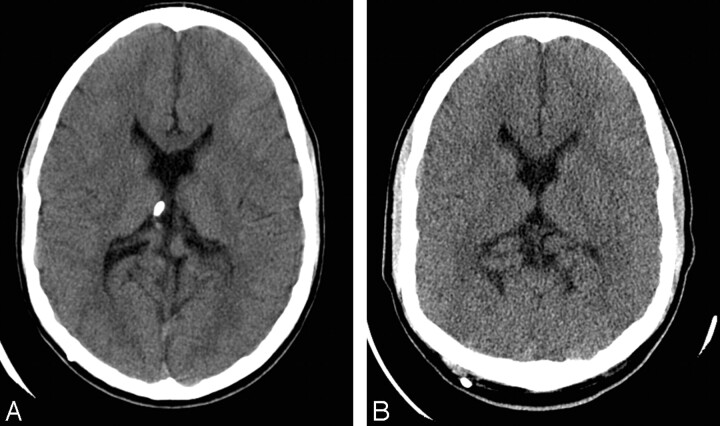
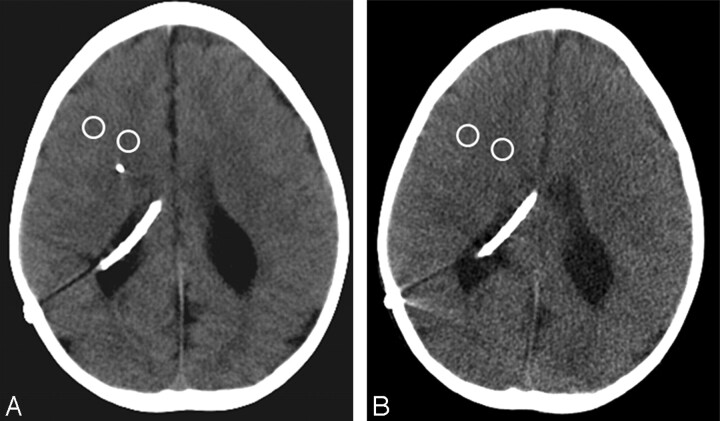
Background and purpose: Children with a shunt for hydrocephalus often undergo multiple follow-up head CT scans, increasing the risk for long-term effects of ionizing radiation. The purpose of our study was to evaluate if an unenhanced low-dose head CT could consistently provide acceptable image quality and diagnostic information.
Materials and methods: Ninety-two children (mean age, 9 years; range, 8 months to 21 years; 45 boys and 47 girls) with a shunt for hydrocephalus and no clinical evidence of shunt malfunction who were referred for a follow-up nonenhanced head CT were included in the study. All studies were performed on a 4-section multidetector CT. Two CT studies were selected retrospectively for each patient, 1 performed at standard dose (220 mA) and 1 at low dose (80 mAs). Two radiologists independently evaluated and graded both standard-dose and low-dose studies for various image quality parameters. Attenuation and noise levels were measured, and gray-white differentiation and contrast-to-noise ratio (CNR) were calculated.
Results: Low-dose CT resulted in 63% mean dose reduction. All low-dose CT scans were diagnostically acceptable. Image quality parameters were significantly lower at low dose (P = .0001) except for the parameters for streak artifacts (P = .46) and need for further imaging (P = .47), which were higher. Mean noise levels were significantly higher (P = .001) in low-dose studies, whereas CNR was significantly higher in standard dose CT (P = .001). A moderate to perfect agreement was noted between the 2 readers with regard to image quality assessment (65%-99%).
Conclusion: Low-dose nonenhanced head CT consistently provides diagnostically acceptable images with relevant diagnostic information in children with VP shunts resulting in substantial dose savings.
Figures
References
-
- Bondurant CP, Jimenez DF. Epidemiology of cerebrospinal fluid shunting. Pediatr Neurosurg 1995;23:254–58; discussion 259 - PubMed
-
- Browd SR, Ragel BT, Gottfried ON, et al. Failure of cerebrospinal fluid shunts: part I: Obstruction and mechanical failure. Pediatr Neurol 2006;34:83–92 - PubMed
-
- Mills DM, Tsai S, Meyer DR, et al. Pediatric ophthalmic computed tomographic scanning and associated cancer risk. Am J Ophthalmol 2006;142:1046–53 - PubMed
-
- Rice HE, Frush DP, Farmer D, et al. Review of radiation risks from computed tomography: essentials for the pediatric surgeon. J Pediatr Surg 2007;42:603–07 - PubMed
-
- Hall EJ. Lessons we have learned from our children: cancer risks from diagnostic radiology. Pediatr Radiol 2002;32:700–06 - PubMed
MeSH terms
LinkOut - more resources
Full Text Sources
Medical