. 2008 Apr 15;105(15):5868-73.
doi: 10.1073/pnas.0801775105. Epub 2008 Apr 8.

Artificial miRNAs mitigate shRNA-mediated toxicity in the brain: implications for the therapeutic development of RNAi

Affiliations

Artificial miRNAs mitigate shRNA-mediated toxicity in the brain: implications for the therapeutic development of RNAi

Jodi L McBride et al. Proc Natl Acad Sci U S A. .

Abstract

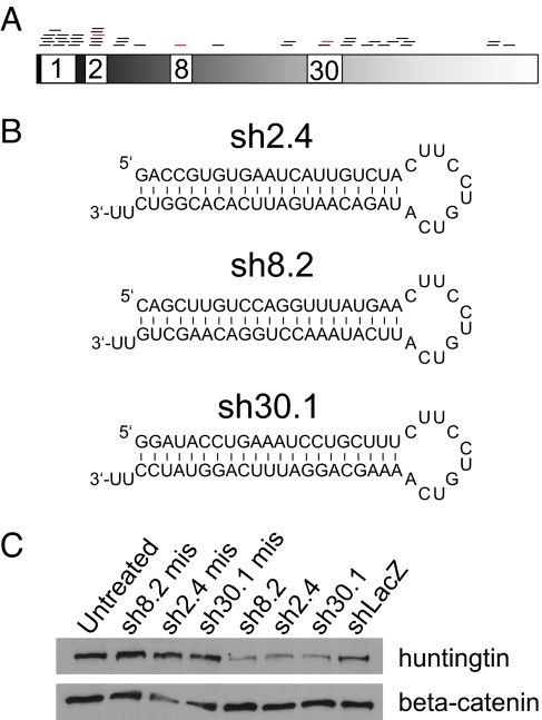
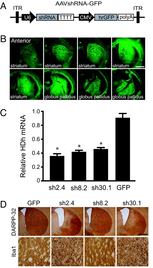
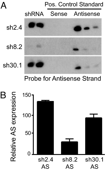
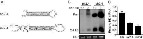
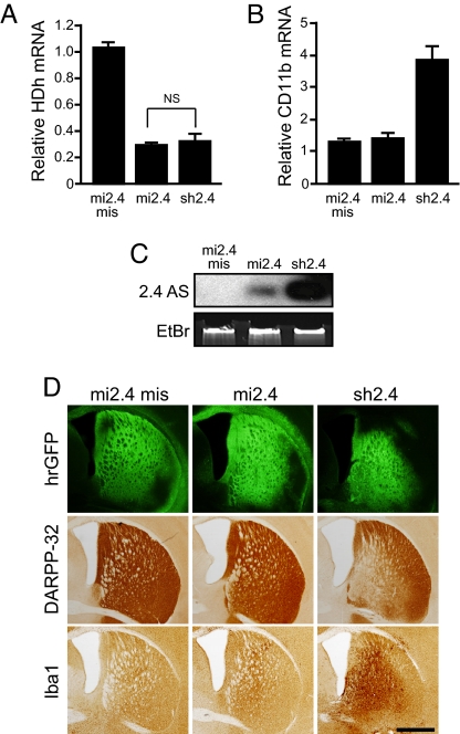
Huntington's disease (HD) is a fatal, dominant neurodegenerative disease caused by a polyglutamine repeat expansion in exon 1 of the HD gene, which encodes the huntingtin protein. We and others have shown that RNAi is a candidate therapy for HD because expression of inhibitory RNAs targeting mutant human HD transgenes improved neuropathology and behavioral deficits in HD mouse models. Here, we developed shRNAs targeting conserved sequences in human HD and mouse HD homolog (HDh) mRNAs to initiate preclinical testing in a knockin mouse model of HD. We screened 35 shRNAs in vitro and subsequently narrowed our focus to three candidates for in vivo testing. Unexpectedly, two active shRNAs induced significant neurotoxicity in mouse striatum, although HDh mRNA expression was reduced to similar levels by all three. Additionally, a control shRNA containing mismatches also induced toxicity, although it did not reduce HDh mRNA expression. Interestingly, the toxic shRNAs generated higher antisense RNA levels, compared with the nontoxic shRNA. These results demonstrate that the robust levels of antisense RNAs emerging from shRNA expression systems can be problematic in the mouse brain. Importantly, when sequences that were toxic in the context of shRNAs were placed into artificial microRNA (miRNA) expression systems, molecular and neuropathological readouts of neurotoxicity were significantly attenuated without compromising mouse HDh silencing efficacy. Thus, miRNA-based approaches may provide more appropriate biological tools for expressing inhibitory RNAs in the brain, the implications of which are crucial to the development of RNAi for both basic biological and therapeutic applications.


Conflict of interest statement

Conflict of interest statement: B.L.D. was a consultant for Sirna Therapeutics, Inc.

Figures

Fig. 1.
Fig. 1.
In vitro screening of shRNAs targeting human HD and mouse HDh transcripts. (A) Thirty-five shRNAs (bars above cartoon) targeting conserved sequences (Table S1) spanning human HD and mouse HDh mRNAs were generated with consideration for sequences that promote proper loading of the antisense strands into the RISC. Plasmids expressing U6-driven shRNAs were transfected into HEK 293 cells, and HD gene silencing was evaluated by QPCR and protein dot blot analyses 48 h after transfection. (B) Three candidate shRNAs targeting sequences in exons 2 (sh2.4), 8 (sh8.2), and 30 (sh30.1) were chosen for further study (red bars above cartoon in A). (C) shRNA expression plasmids were transfected into mouse C2C12 cells, and endogenous huntingtin protein levels were evaluated by Western blot analyses 48 h after transfection. Mismatch (mis) controls contain 4-bp changes that render the shRNAs ineffective. β-Catenin serves as the loading control.
Fig. 2.
Fig. 2.
HD shRNAs cause sequence-specific striatal toxicity in mice. (A) Diagram of the recombinant AAV2/1 viral vectors containing shRNA and hrGFP expression cassettes. (B) Photomicrographs represent the rostral-to-caudal distribution of hrGFP-positive cells in mouse brain after direct injection of virus into the striatum. (Scale bar: 500 μm.) (C) QPCR analysis measuring HDh mRNA levels in shRNA-treated mouse striata demonstrates similar silencing efficacies among sh2.4, sh8.2, and sh30.1. Mice were injected into the striatum with AAVsh2.4-GFP, AAVsh8.2-GFP, AAVsh30.1-GFP, or AAV-GFP, and RNA was harvested 4 months later from GFP-positive striata. All values were normalized to β-actin and are shown relative to AAV-GFP-treated brains. (D) Immunohistochemistry reveals that sh2.4 and sh30.1 induce striatal toxicity in mice. Mice were injected with the indicated AAVshRNA-GFP or AAV-GFP into the striatum, and histological analyses were performed on brains harvested at 4 months after treatment. Representative photomicrographs for immunohistochemical staining of DARPP-32-positive neurons (Upper) and IbaI-positive microglia (Lower) are shown for each treatment group. (Scale bar: 500 μm for Upper; 100 μm for Lower.)
Fig. 3.
Fig. 3.
The nontoxic sh8.2 generates lower levels of processed antisense RNA. (A) Small transcript Northern blot was performed to assess AS RNA levels present in mouse striata treated with the indicated AAVshRNA-GFP. (Left) Two separately treated striatal tissue samples. (Center and Right) Positive controls loaded as standards [10-fold dilutions for both S (Center) or AS (Right) strands]. (B) Densitometry analysis was used to quantify the relative levels of HD AS RNAs. Signals were quantified by using Image J software, and expression is shown as femtomoles per microgram of total RNA.
Fig. 4.
Fig. 4.
An artificial miRNA approach naturally reduces precursor and mature inhibitory RNAs. (A) Sequences and comparison of sh2.4 and mi2.4 containing the core HD2.4 sequence (shaded boxes). Each transcript starts with the +1-G nucleotide natural to the U6 promoter. The major Drosha and Dicer cleavage sites are shown by hash marks. (B) HEK 293 cells were transfected with HD2.4 RNAi expression plasmids at the indicated amounts, and small-transcript Northern blot was performed 48 h later. Results demonstrate that sh2.4 generates abundant levels of unprocessed precursor (Pre-) and processed antisense RNAs (2.4AS) even at a 10-fold-lower dose relative to mi2.4. Ethidium bromide (EtdBr) staining is shown as the loading control. (C) HD2.4 RNAi expression plasmids were transfected into HEK 293 cells, and QPCR analysis was performed 48 h later to measure endogenous HD mRNA levels. Results demonstrate that mi2.4 silences HD transcripts efficiently, relative to sh2.4, despite being expressed at considerably lower levels.
Fig. 5.
Fig. 5.
Artificial miRNAs mitigate striatal toxicity in mice. (A and B) QPCR analyses were performed to measure mouse HDh (A) and CD11b (B) mRNA levels in AAV-RNAi-injected striata harvested 4 months after treatment (NS, not significant). Samples were normalized to β-actin. Results, shown relative to uninjected striata, demonstrate that mi2.4 silences HD transcripts as effectively as sh2.4, but avoids induction of CD11b, a marker for microglial activation. (C) Small-transcript Northern blot analysis for mature HD2.4 AS RNAs present in AAV-RNAi-treated striatal lysates reveals a robust disparity between the levels generated from sh2.4 and mi2.4 vectors. EtdBr staining is shown as the loading control. (D) Histological analyses demonstrate the improved safety profile of mi2.4. Mice were injected with the indicated AAV-RNAi-GFP viruses into the striatum, and histological analyses were performed on brains harvested at 4 months after treatment. Photomicrographs representing hrGFP (Top), immunohistochemical staining of DARPP-32-positive neurons (Middle), and IbaI-positive microglia (Bottom) are shown for each treatment group. (Scale bar: 500 μm.)

Similar articles

Cited by

References

    1. Fire A, et al. Potent and specific genetic interference by double-stranded RNA in caenorhabditis elegans. Nature. 1998;391:806–811. - PubMed
    1. Reinhart BJ, et al. The 21-nucleotide let-7 RNA regulates developmental timing in Caenorhabditis elegans. Nature. 2000;403:901–906. - PubMed
    1. Lagos-Quintana M, Rauhut R, Lendeckel W, Tuschl T. Identification of novel genes coding for small expressed RNAs. Science. 2001;294:853–858. - PubMed
    1. Lau NC, Lim LP, Weinstein EG, Bartel DP. An abundant class of tiny RNAs with probable regulatory roles in Caenorhabditis elegans. Science. 2001;294:858–862. - PubMed
    1. Lee RC, Ambros V. An extensive class of small RNAs in Caenorhabditis elegans. Science. 2001;294:862–864. - PubMed

Publication types

LinkOut - more resources