

Preconditioning mediated by sublethal oxygen-glucose deprivation-induced cyclooxygenase-2 expression via the signal transducers and activators of transcription 3 phosphorylation
- PMID: 18398416
- PMCID: PMC2645802
- DOI: 10.1038/jcbfm.2008.26
Preconditioning mediated by sublethal oxygen-glucose deprivation-induced cyclooxygenase-2 expression via the signal transducers and activators of transcription 3 phosphorylation
Abstract
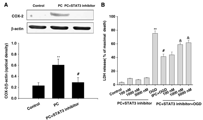
The signal transducers and activators of transcription (STATs) were found to be essential for cardioprotection. However, their role in preconditioning (PC) neuroprotection remains undefined. Previously, our studies showed that PC mediated a signaling cascade that involves activation of epsilon protein kinase C (varepsilonPKC), extracellular signal-regulated kinase (ERK1/2), and cyclooxygenase-2 (COX-2) pathways. However, the intermediate pathway by which ERK1/2 activates COX-2 was not defined. In this study, we investigated whether the PC-induced signaling pathway requires phosphorylation of STAT isoforms for COX-2 expression. To mimic PC or lethal ischemia, mixed cortical neuron/astrocyte cell cultures were subjected to 1 and/or 4 h of oxygen-glucose deprivation (OGD), respectively. The results indicated serine phosphorylation of STAT3 after PC or varepsilonPKC activation. Inhibition of either varepsilonPKC or ERK1/2 activation abolished PC-induced serine phosphorylation of STAT3. Additionally, inhibition of STAT3 prevented PC-induced COX-2 expression and neuroprotection against OGD. Therefore, our findings suggest that PC signaling cascade involves STAT3 activation after varepsilonPKC and ERK1/2 activation. Finally, we show that STAT3 activation mediates COX-2 expression and ischemic tolerance.
Figures


Similar articles
-
Role of the protein kinase C-epsilon-Raf-1-MEK-1/2-p44/42 MAPK signaling cascade in the activation of signal transducers and activators of transcription 1 and 3 and induction of cyclooxygenase-2 after ischemic preconditioning.Circulation. 2005 Sep 27;112(13):1971-8. doi: 10.1161/CIRCULATIONAHA.105.561522. Epub 2005 Sep 19. Circulation. 2005. PMID: 16172266 Free PMC article.
-
Ischemic preconditioning via epsilon protein kinase C activation requires cyclooxygenase-2 activation in vitro.Neuroscience. 2007 Mar 30;145(3):931-41. doi: 10.1016/j.neuroscience.2006.12.063. Epub 2007 Feb 20. Neuroscience. 2007. PMID: 17307294 Free PMC article.
-
Endothelial nitric oxide synthase plays an obligatory role in the late phase of ischemic preconditioning by activating the protein kinase C epsilon p44/42 mitogen-activated protein kinase pSer-signal transducers and activators of transcription1/3 pathway.Circulation. 2007 Jul 31;116(5):535-44. doi: 10.1161/CIRCULATIONAHA.107.689471. Epub 2007 Jul 2. Circulation. 2007. PMID: 17606840 Free PMC article.
-
Repeated Non-Invasive Limb Ischemic Preconditioning Confers Cardioprotection Through PKC-Ԑ/STAT3 Signaling in Diabetic Rats.Cell Physiol Biochem. 2018;45(5):2107-2121. doi: 10.1159/000488047. Epub 2018 Mar 7. Cell Physiol Biochem. 2018. PMID: 29533954
-
Signal transducers and activators of transcription: STATs-mediated mitochondrial neuroprotection.Antioxid Redox Signal. 2011 May 15;14(10):1853-61. doi: 10.1089/ars.2010.3467. Epub 2011 Jan 5. Antioxid Redox Signal. 2011. PMID: 20712401 Free PMC article. Review.
Cited by
-
The role of protein kinase C epsilon in neural signal transduction and neurogenic diseases.Front Med. 2011 Mar;5(1):70-6. doi: 10.1007/s11684-011-0119-9. Epub 2011 Mar 17. Front Med. 2011. PMID: 21681677 Review.
-
Ischemic preconditioning and clinical scenarios.Curr Opin Neurol. 2013 Feb;26(1):1-7. doi: 10.1097/WCO.0b013e32835bf200. Curr Opin Neurol. 2013. PMID: 23197083 Free PMC article. Review.
-
Ischemic conditioning-induced endogenous brain protection: Applications pre-, per- or post-stroke.Exp Neurol. 2015 Oct;272:26-40. doi: 10.1016/j.expneurol.2015.04.009. Epub 2015 Apr 18. Exp Neurol. 2015. PMID: 25900056 Free PMC article. Review.
-
Activation of STAT1 in neurons following spinal cord injury in mice.Neurochem Res. 2011 Dec;36(12):2236-43. doi: 10.1007/s11064-011-0547-6. Epub 2011 Jul 22. Neurochem Res. 2011. PMID: 21833847
-
GABA synapses mediate neuroprotection after ischemic and epsilonPKC preconditioning in rat hippocampal slice cultures.J Cereb Blood Flow Metab. 2009 Feb;29(2):375-84. doi: 10.1038/jcbfm.2008.126. Epub 2008 Oct 29. J Cereb Blood Flow Metab. 2009. PMID: 18957990 Free PMC article.
References
-
- Allport VC, Slater DM, Newton R, Bennett PR. NF-kappaB and AP-1 are required for cyclo-oxygenase 2 gene expression in amnion epithelial cell line (WISH) Mol Hum Reprod. 2000;6:561–565. - PubMed
-
- Aziz MH, Manoharan HT, Verma AK. Protein kinase C epsilon, which sensitizes skin to sun’s UV radiation-induced cutaneous damage and development of squamous cell carcinomas, associates with Stat3. Cancer Res. 2007;67:1385–1394. - PubMed
-
- Bolli R, Dawn B, Xuan YT. Role of the JAK–STAT pathway in protection against myocardial ischemia/reperfusion injury. Trends Cardiovasc Med. 2003;13:72–79. - PubMed
Publication types
MeSH terms
Substances
Grants and funding
LinkOut - more resources
Full Text Sources
Research Materials
Miscellaneous
