. 2008 Apr 9;3(4):e1938.
doi: 10.1371/journal.pone.0001938.

The homeodomain derived peptide Penetratin induces curvature of fluid membrane domains

Affiliations

The homeodomain derived peptide Penetratin induces curvature of fluid membrane domains

Antonin Lamazière et al. PLoS One. .

Abstract

Background: Protein membrane transduction domains that are able to cross the plasma membrane are present in several transcription factors, such as the homeodomain proteins and the viral proteins such as Tat of HIV-1. Their discovery resulted in both new concepts on the cell communication during development, and the conception of cell penetrating peptide vectors for internalisation of active molecules into cells. A promising cell penetrating peptide is Penetratin, which crosses the cell membranes by a receptor and metabolic energy-independent mechanism. Recent works have claimed that Penetratin and similar peptides are internalized by endocytosis, but other endocytosis-independent mechanisms have been proposed. Endosomes or plasma membranes crossing mechanisms are not well understood. Previously, we have shown that basic peptides induce membrane invaginations suggesting a new mechanism for uptake, "physical endocytosis".

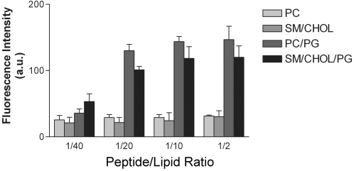
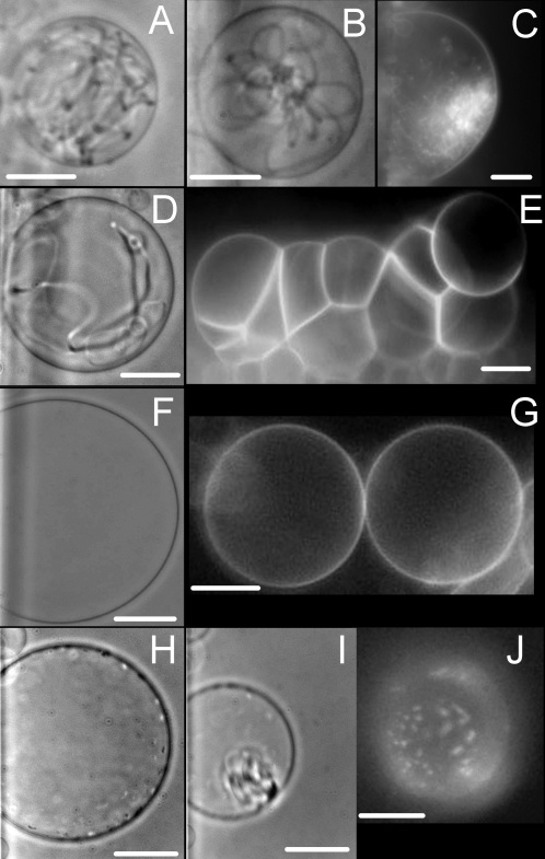
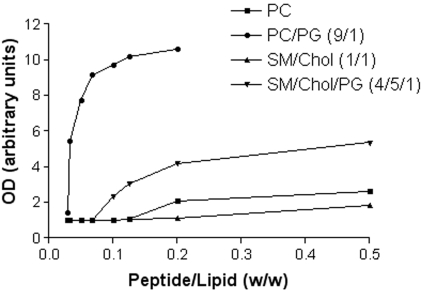
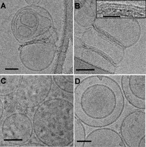
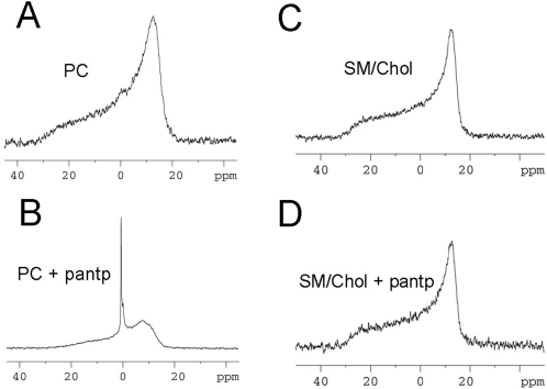
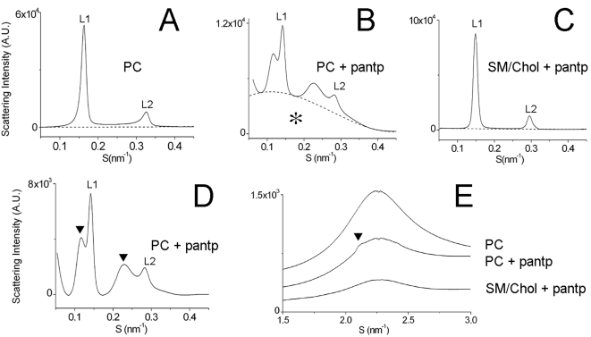
Methodology/principal findings: Herein, we investigate the role of membrane lipid phases on Penetratin induced membrane deformations (liquid ordered such as in "raft" microdomains versus disordered fluid "non-raft" domains) in membrane models. Experimental data show that zwitterionic lipid headgroups take part in the interaction with Penetratin suggesting that the external leaflet lipids of cells plasma membrane are competent for peptide interaction in the absence of net negative charges. NMR and X-ray diffraction data show that the membrane perturbations (tubulation and vesiculation) are associated with an increase in membrane negative curvature. These effects on curvature were observed in the liquid disordered but not in the liquid ordered (raft-like) membrane domains.

Conclusions/significance: The better understanding of the internalisation mechanisms of protein transduction domains will help both the understanding of the mechanisms of cell communication and the development of potential therapeutic molecular vectors. Here we showed that the membrane targets for these molecules are preferentially the fluid membrane domains and that the mechanism involves the induction of membrane negative curvature. Consequences on cellular uptake are discussed.


Conflict of interest statement

Competing Interests: The authors have declared that no competing interests exist.

Figures

Figure 1
Figure 1. Penetratin binding to LUV.
Penetratin binding was followed by detection of tryptophan fluorescence in LUV recovered after centrifugation. Penetratin concentration varied and LUV concentration was maintained constant. Peptide/Lipid ratio is given by weight. In the absence of membranes, no fluorescence was recovered after centrifugation. SM/Chol (1/1), PC/PG (9/1), SM/Chol/PG (4/5/1). (a.u.); arbitrary units. Mean of three independent experiments.
Figure 2
Figure 2. Penetratin effects on GUV in liquid disordered (non-raft) and liquid ordered (raft-like) domains.
GUV obtained by electroformation were incubated with Penetratin or fluorescent Penetratin as described in the methods section. Tubulation was only observed in membranes in the liquid disordered state. DOPC (A), egg yolk PC (B,C) and PC/PG (9/1) (D) GUV. Peptide induced vesicles adhesion was observed in all GUV (only shown for PC/PG) (E). Peptides showed no tubulation effect in membranes in the liquid ordered state (“raft-like” SM/Chol (1/1)) (F,G). In SM/Chol membranes supplemented with 10% PG, Penetratin clusterizes at the GUV surface (H,J) and then provokes a grape-like vesiculation phenomenon (I) see also videos S1,S2,S3 in supplement. Dark field images were obtained with CF-Penetratin. Phase contrast images were obtained with cargo-free Penetratin. Bars 20 µm.
Figure 3
Figure 3. Membrane aggregation by Penetratin.
Curves of LUV aggregation as a function of peptide/lipid ratio. LUV aggregation was measured by turbidimetry at plateau (20 minutes after peptide addition). Representative curves of three experiments.
Figure 4
Figure 4. LUV aggregation and deformation by Penetratin as a function of lipid composition.
LUV of different composition in the presence of Penetratin were observed by cryo-electron microscopy. PC (A), PC/PG (9/1) (B), SM/Chol (1/1) (C), and SM/Chol/PG (4/5/1) (D). Bars 50 nm. Inset in B shows an amplification of the Penetratin membrane bridges. Notice the perturbation of the phospholipid bilayer. Bar for inset 20 nm.
Figure 5
Figure 5. Effect of Penetratin in curvature of multilamellar vesicles.
31P-NMR spectra of MLV of different composition in the absence (A,C) and the presence of Penetratin (B,D) at a Peptide/Lipid weight ratio of 1/7. Typical spectra of lamellar phases are observed for both PC and SM/Chol (1/1) preparations in the absence of peptide. In the presence of Penetratin, an isotropic signal was observed for PC membranes consistent with the presence of highly curved membranes (B). Penetratin showed no effect on the liquid ordered (raft-like) membrane (D).
Figure 6
Figure 6. Effects of Penetratin observed by small angle and wide angle X-ray diffraction.
A to D small angle X-ray diffraction. Diffractograms are not corrected for the base line; (A) PC membranes, (B) PC membranes in the presence of Penetratin showing a “distorted baseline” (star) corresponding to the form factor contribution. The broad signal was tentatively related to the formation of small vesicles, (C) SM/Chol (1/1) membranes in the presence of Penetratin. (D) Diffractogram is subtracted for the distorted baseline. PC membranes in the presence of Penetratin show first (L1) and second (L2) order Bragg peaks of a lamellar phase (d-spacing in the ratio 1∶1/2). The peaks as compared to (A) in the absence of peptide are shifted to the left indicating an increase in the repeat distance from 6.01 to 7.05 nm. In addition, Penetratin induces the splitting of peaks and the separation of a lamellar phase with considerably larger repeat distance (arrowheads). (E) Effects of Penetratin as observed by the wide angle X-ray diffraction. Peaks of PC MLV membranes are shown in the absence (top) and in the presence of Penetratin (middle). Peak of SM/Chol membranes is also shown in the presence of Penetratin (bottom). The arrowhead indicates the unresolved contribution assumed to correspond to the increased inter-acyl chains spacing. All experiments presented were done at 25°C.

Similar articles

Cited by

References

    1. Prochiantz A. Messenger proteins: homeoproteins, TAT and others. Curr Opin Cell Biol. 2000;12:400–406. - PubMed
    1. Prochiantz A, Joliot A. Can transcription factors function as cell-cell signalling molecules? Nat Rev Mol Cell Biol. 2003;4:814–819. - PubMed
    1. Mae M, Langel U. Cell-penetrating peptides as vectors for peptide, protein and oligonucleotide delivery. Curr Opin Pharmacol. 2006;6:509–514. - PubMed
    1. Murriel CL, Dowdy SF. Influence of protein transduction domains on intracellular delivery of macromolecules. Expert Opin Drug Deliv. 2006;3:739–746. - PubMed
    1. Joliot A, Pernelle C, Deagostini-Bazin H, Prochiantz A. Antennapedia homeobox peptide regulates neural morphogenesis. Proc Natl Acad Sci U S A. 1991;88:1864–1868. - PMC - PubMed

Publication types