. 2008 May;118(5):1944-54.
doi: 10.1172/JCI33680.

Impaired microRNA processing causes corpus luteum insufficiency and infertility in mice

Affiliations

Impaired microRNA processing causes corpus luteum insufficiency and infertility in mice

Motoyuki Otsuka et al. J Clin Invest. 2008 May.

Erratum in

  • J Clin Invest. 2008 Jun;118(6):2366

Abstract

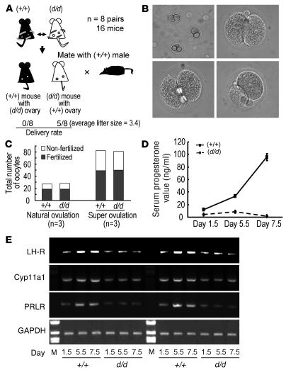
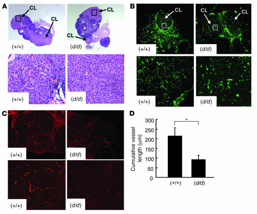
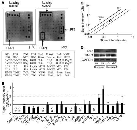
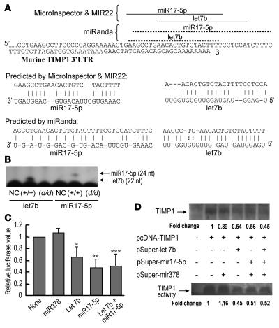
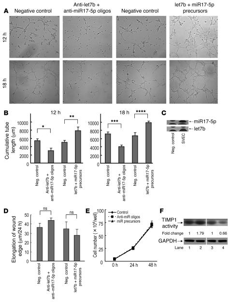
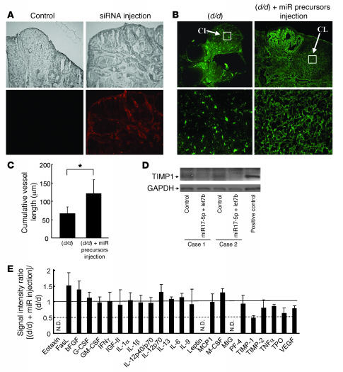
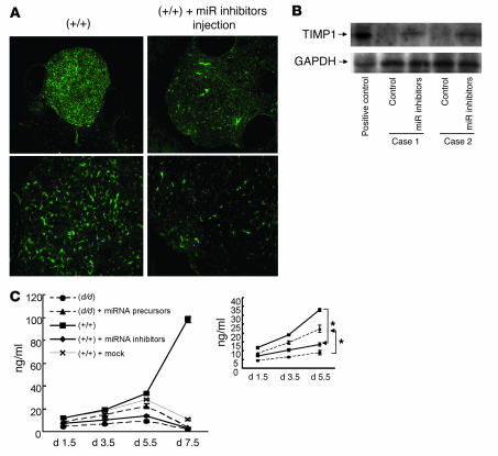
The microRNA (miRNA) processing enzyme Dicer1 is required for zygotic and embryonic development, but the early embryonic lethality of Dicer1 null alleles in mice has limited our ability to address the role of Dicer1 in normal mouse growth and development. To address this question, we used a mouse mutant with a hypomorphic Dicer1 allele (Dicer(d/d)) and found that Dicer1 deficiency resulted in female infertility. This defect in female Dicer(d/d) mice was caused by corpus luteum (CL) insufficiency and resulted, at least in part, from the impaired growth of new capillary vessels in the ovary. We found that the impaired CL angiogenesis in Dicer(d/d) mice was associated with a lack of miR17-5p and let7b, 2 miRNAs that participate in angiogenesis by regulating the expression of the antiangiogenic factor tissue inhibitor of metalloproteinase 1. Furthermore, injection of miR17-5p and let7b into the ovaries of Dicer(d/d) mice partially normalized tissue inhibitor of metalloproteinase 1 expression and CL angiogenesis. Our data indicate that the development and function of the ovarian CL is a physiological process that appears to be regulated by miRNAs and requires Dicer1 function.


Figures

Figure 1
Figure 1. CL insufficiency is the cause of Dicerd/d female mouse infertility.
(A) Fertility of Dicerd/d female mice transplanted with Dicer+/+ ovaries. Ovaries of Dicerd/d and Dicer+/+ littermate mice were exchanged (n = 8 pairs), and the mice were subsequently bred to wild-type fertile males. Delivery rate and average litter size are shown. (B and C) Ovulation and fertilization were normal in Dicerd/d mice. (B) Representative morphologies of the 2 cell embryos collected from the oviducts of Dicerd/d mice on day 1.5 after coitus. Original magnification, ×100 (top), ×400 (bottom). (C) Rates of ovulation and fertilization in Dicerd/d and Dicer+/+ mice were examined on day 1.5 with or without superovulation. (D) Serum progesterone levels were lower in Dicerd/d mice during pregnancy. Serum levels of progesterone in Dicerd/d mice were determined by ELISA on days 1.5, 5.5, and 7.5 of pregnancy and expressed as mean ± SD (n = 3). (E) The expression of genes associated with CL function was lower in Dicerd/d mice. Semiquantitative RT-PCR analyses of luteinizing hormone receptor, prolactin receptor, and cytochrome P450 family 11 subfamily a polypeptide 1 mRNA in ovaries of Dicerd/d and Dicer+/+ mice were conducted on days 1.5, 5.5, and 7.5 after coitus. Results from 2 separate littermate samples are shown.
Figure 2
Figure 2. Impaired angiogenesis in the CLs of Dicerd/d mice.
(A) Ovarian tissue sections obtained from Dicer+/+ and Dicerd/d mice on day 1.5 of pregnancy were stained with H&E (original magnification, ×100). CLs in Dicer+/+ and Dicerd/d mice ovaries is denoted by the boxes and are shown at higher original magnification (×400, bottom). The cells had less cytoplasm and less vascularity in the CLs of Dicerd/d mice. (B) Immunofluorescence analyses of ovaries from Dicerd/d and Dicer+/+ mice on day 1.5 of pregnancy show the localization of type IV collagen — a marker of the basal lamina of endothelial cells. Close examination of the vascular network in the CLs denoted by the boxes (top) is shown at higher magnification (×400; bottom). Dicer+/+ CLs showed more filamentous and punctuated collagen IV staining, which indicated higher vessel densities than those in Dicerd/d mice. (C) PECAM (CD31) immunofluorescence staining of CLs in Dicerd/d and Dicer+/+ mice on day 1.5 of pregnancy. The number and the length of the vessels were greater and longer in Dicer+/+ CLs. Original magnification, ×400. (D) Cumulative vessel length in CLs was determined as the average of the number of vessels per 100 × 100 μm2 multiplied by the vessel length in 3 random fields. The results of type IV collagen immunofluorescence staining from 4 different Dicer+/+ and Dicerd/d pairs were used. The data are expressed as mean ± SD. *P = 0.0012.
Figure 3
Figure 3. TIMP1 expression is elevated in Dicerd/d female mice ovaries.
(A) Mouse angiogenesis-related antibody arrays hybridized with ovarian lysates from Dicer+/+ and Dicerd/d mice on day 1.5 after coitus. Two independent experiments using different Dicer+/+ and Dicerd/d littermate mice pairs were performed. A representative image is shown. The positions of TIMP1 are denoted by boxes and arrows. The positive loading controls are also denoted by boxes and arrows. The map of antibodies spotted on these arrays is shown below. (B) Signal intensity ratios of each gene between Dicer+/+ and Dicerd/d mice ovaries obtained by protein array analyses. The normalized signal intensities of each protein from Dicerd/d mice ovaries were divided by those from Dicer+/+ mice ovaries. Four values for each gene obtained from 2 independent experimental sets were used for the analysis. The data are expressed as mean ± SD. Genes with low signal intensity that could not be analyzed further are expressed as N.D. (not determined). The mean signal intensities of TIMP1 in Dicerd/d mice ovaries are more than 2-fold those in Dicer+/+ mice. (C) The distribution of log10 conversion values of the average of normalized signal intensities (n = 4) from each protein is shown as a scatter plot. The solid lines indicate the position of 2-fold changes in the intensities. The results for TIMP1 and platelet factor 4 are indicated by arrows. (D) Verification of Dicer1 protein deficiency and TIMP1 protein upregulation in Dicerd/d mice ovaries. Dicer1 and TIMP1 protein expression in the ovaries of 2 separate Dicerd/d and Dicer+/+ littermate mice on day 1.5 after coitus are shown. GAPDH was used as a loading control.
Figure 4
Figure 4. miR17-5p and let7b can target 3′-UTR of mTIMP1.
(A) miRNA target positions in the 3′-UTR of mTIMP1 were identified by computational prediction methods, MicroInspector, and miRanda. The same candidate miRNAs, miR17-5p and let7b, were predicted by the different programs; however, the predicted miRNA-targeting sequences in the 3′-UTR of TIMP1 are not exactly the same. (B) Primer extension analyses showed the expression of the indicated miRNAs in Dicer+/+ and Dicerd/d mice ovaries. NC, negative control. (C) miR17-5p and let7b can target 3′-UTR of TIMP1. The 293T cells were transiently transfected with a reporter plasmid (pLuc-TIMP3′-UTR) with or without indicated miRNA expression plasmids, and 36 hours after transfection, a reporter assay was performed. The relative luciferase values were calculated by dividing firefly luciferase values with internal control renilla luciferase values. The value from the negative control was set at 1. Data are shown as mean ± SD from 3 independent experiments. *P = 0.032, **P = 0.00082, ***P = 0.0035. (D) miR17-5p and let7b downregulate TIMP1 expression and activity. The 293T cells were transiently transfected with pcDNA-TIMP1, with or without the indicated miRNA expression plasmids, and pRL-TK. TIMP1 expression was examined by western blotting 36 hours after transfection (top). Cell extracts were normalized based on the luciferase value of cotransfected pRL-TK to avoid the variances of transfection efficiency. Bottom: Result of reverse zymography using the MMP2-containing gels and the concentrated culture supernatant collected from the transfected cells to quantitate the activities of TIMP1 against MMP2 activities. A representative result from 4 independent experiments is shown. Numbers below the images refer to the fold changes of the intensities.
Figure 5
Figure 5. miR17-5p and let7b control endothelial cell angiogenic function.
(A and B) Indicated anti-miRNA oligonucleotides or miRNA precursors were transfected into SVEC. Thirty-six hours after transfection, cells were seeded on Matrigel. Tube length was quantified after 12 and 18 hours. Representative micrographs (A) and statistical summary (B) are shown. Data represent mean ± SD of 3 experiments. *P = 0.0014, **P = 0.0057, ***P = 0.000055, ****P = 0.00067. (C) Primer extension analyses determined the expression of the indicated miRNAs in SVEC. (D) miR17-5p and let7b had no effect on the proliferation of SVEC. Thirty-six hours after transfection of the indicated oligos into SVEC, 5 × 10⁴ cells were seeded, and the number of cells was determined by cell counting at the indicated time points. Data are presented as mean ± SD (n = 4 per group, P = NS). (E) miR17-5p and let7b had no effect on the motility of SVEC. In vitro wound healing was quantified as the average length of the elongation of wound edges at 4 specific points over 24 hours. Data are presented as mean ± SD (n = 3 per group, P = NS). (F) The activities of TIMP1 against MMP2 were modulated by miRNAs. A representative result of reverse zymography is shown using the the SVEC culture supernatant after transfection with the oligos (lane 1, control antisense; lane 2, anti–miRNA17-5p and let7b; lane 3, control miRNA precursors; lane 4, miR17-5p and let7b precursors). The numbers indicate the fold changes of the band intensities. GAPDH expression did not change after transfection of oligos. The experiments were repeated 3 times with similar results.
Figure 6
Figure 6. miR17-5p and let7b recover the vascularity in the CLs in Dicerd/d mouse ovaries in vivo.
(A) Cy-3–labeled siRNA was injected into the ovarian bursa. Injection of transfection reagent alone was used as control. Top: Images in the bright field. Bottom: Same fields in the fluorescent phase. Original magnification, ×100. (B) miR17-5p and let7b recover the vascularity in CLs. miRNA precursors were injected into the ovarian bursa of Dicerd/d mice, and ovaries were stained with anti–type IV collagen, as described in Methods. Top: original magnification, ×100. Close examination of the vascular network denoted by the boxes are shown (original magnification, ×400; bottom). (C) Cumulative vessel length in CLs in the miRNA-injected ovaries [(d/d) + miR precursor injection] and the corresponding control ovaries (d/d) was determined as described in the legend of Figure 2D. Results are from 3 different Dicerd/d mice. Data represent mean ± SD. *P = 0.0031. (D) miR17-5p and let7b suppressed the expression of TIMP1 in the ovaries in vivo. miRNA precursors were injected into the ovaries as described in Methods. TIMP1 protein expression in the ovaries of 2 separate miRNA-injected mice are shown. SVEC lysates were used as a positive control. GAPDH was used as a loading control. (E) The suppression of TIMP1 expression in the ovary was confirmed by an antibody array. Signal intensity ratios of each gene between the miRNA-introduced Dicerd/d ovaries (d/d + miR injection) and the corresponding control Dicerd/d ovaries (d/d) are shown after normalization. Results are the summary of 4 values of each gene from 2 independent experiments using different mice.
Figure 7
Figure 7. Anti–miR17-5p and let7b oligonucleotides impaired the vascularities in the CLs in Dicer+/+ mouse ovaries.
(A) A mixture of anti–miRNA17-5p and let7b oligonucleotides and transfection reagent, or transfection reagent only, were injected into the right or left ovarian bursa of Dicer+/+ mice, respectively, and the patterns of type IV collagen in the CLs were examined as described in the legend of Figure 6B. A representative result from 3 independent mice is shown. (B) Anti–miR17-5p and let7b oligonucleotides enhanced the expression of TIMP1 in the ovaries in vivo. TIMP1 protein expression in the ovaries of mice injected with 2 anti–miR17-5p plus anti-let7b oligonucleotides. SVEC lysates were used as positive control for the detection of TIMP1. GAPDH was used as a loading control. (C) Serum progesterone levels were modified in miRNA precursors or anti-miRNA inhibitor–injected mice. Dicerd/d mice injected with a mixture of miRNA17-5p and let7b precursors into both side ovaries [(d/d) + miRNA precursors] had greater serum progesterone levels than did Dicerd/d mice (d/d) until day 5.5 of pregnancy. In contrast, Dicer+/+ mice injected with a mixture of anti–miRNA17-5p and let7b inhibitors into both ovaries [(+/+) + miRNA inhibitors] had lower serum progesterone levels than did Dicer+/+ mice (+/+). The serum progesterone levels of Dicer+/+ mice injected only with transfection reagents into both side ovaries are also shown [(+/+) + mock]. Serum progesterone levels were determined by ELISA on days 1.5, 3.5, 5.5, and 7.5 of pregnancy and expressed as mean ± SD (n = 3 in each group). A graph of results until day 5.5 is shown in the inset.

Similar articles

Cited by

References

    1. Hannon G.J. RNA interference. Nature. 2002;418:244–251. doi: 10.1038/418244a. - DOI - PubMed
    1. Carmell M.A., Hannon G.J. RNase III enzymes and the initiation of gene silencing. Nat. Struct. Mol. Biol. 2004;11:214–218. doi: 10.1038/nsmb729. - DOI - PubMed
    1. Bartel D.P. MicroRNAs: genomics, biogenesis, mechanism, and function. Cell. 2004;116:281–297. doi: 10.1016/S0092-8674(04)00045-5. - DOI - PubMed
    1. Meister G., Tuschl T. Mechanisms of gene silencing by double-stranded RNA. Nature. 2004;431:343–349. doi: 10.1038/nature02873. - DOI - PubMed
    1. Jing Q., et al. Involvement of microRNA in AU-rich element-mediated mRNA instability. Cell. 2005;120:623–634. doi: 10.1016/j.cell.2004.12.038. - DOI - PubMed

Publication types

MeSH terms