. 2008 Jul;88(3):273-80.
doi: 10.1007/s11060-008-9577-6. Epub 2008 Apr 9.

Selective cancer-germline gene expression in pediatric brain tumors

Affiliations

Selective cancer-germline gene expression in pediatric brain tumors

Joannes F M Jacobs et al. J Neurooncol. 2008 Jul.

Abstract

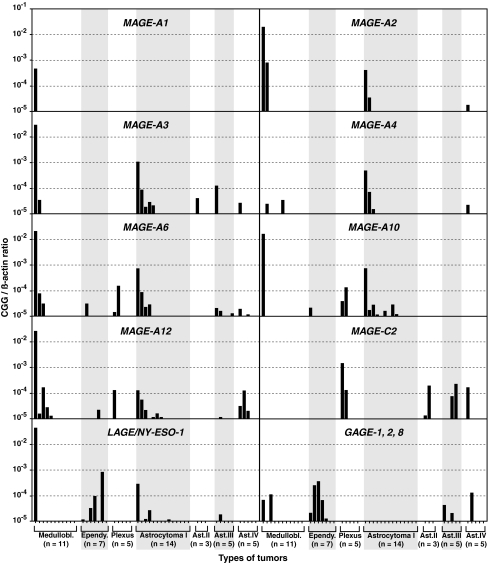
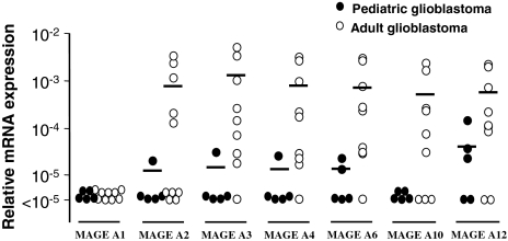
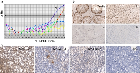
Cancer-germline genes (CGGs) code for immunogenic antigens that are present in various human tumors and can be targeted by immunotherapy. Their expression has been studied in a wide range of human tumors in adults. We measured the expression of 12 CGGs in pediatric brain tumors, to identify targets for therapeutic cancer vaccines. Real Time PCR was used to quantify the expression of genes MAGE-A1, MAGE-A2, MAGE-A3, MAGE-A4, MAGE-A6, MAGE-A10, MAGE-A12, MAGE-C2, NY-ESO-1 and GAGE-1,2,8 in 50 pediatric brain tumors of different histological subtypes. Protein expression was examined with immunohistochemistry. Fifty-five percent of the medulloblastomas (n = 11), 86% of the ependymomas (n = 7), 40% of the choroid plexus tumors (n = 5) and 67% of astrocytic tumors (n = 27) expressed one or more CGGs. Immunohistochemical analysis confirmed qPCR results. With exception of a minority of tumors, the overall level of CGG expression in pediatric brain tumors was low. We observed a high expression of at least one CGG in 32% of the samples. CGG-encoded antigens are therefore suitable targets in a very selected group of pediatric patients with a brain tumor. Interestingly, glioblastomas from adult patients expressed CGGs more often and at significantly higher levels compared to pediatric glioblastomas. This observation is in line with the notion that pediatric and adult glioblastomas develop along different genetic pathways.


Figures

Fig. 1
Fig. 1
Cancer-germline gene (CGG) expression in pediatric brain tumors measured by reverse transcription and quantitative real-time PCR. Each graph shows the results for one CGG in 50 different tumor samples. Samples are arranged in the same order in all graphs. The horizontal axis indicates the tumor type (Medullobl., medulloblastoma; Ependy., ependymoma; Plexus, plexus choroideus; Ast., astrocytic tumors grade I to IV). The bars represent normalized CGG expression values (CGG/ß-actin ratios)
Fig. 2
Fig. 2
MAGE-A expression in pediatric and adult glioblastomas. Normalized MAGE-A expression levels (MAGE-A/ß-actin ratios) of seven MAGE-A genes are shown. Horizontal bars represent mean relative MAGE expression. Pediatric glioblastomas (closed circles) express significantly lower levels of MAGE-A compared to adult glioblastomas (open circles), P = 6.1 × 10−6 calculated with Spearman rank correlation
Fig. 3
Fig. 3
(a) Amplification plots of three pilocytic astrocytomas in duplicate showing high numbers (H), low numbers (L) or no (N) MAGE-A4 cDNA copies. (b) Immunohistochemistry with mAb 57B (anti-MAGE-A4) of the pilocytic astrocytomas shown in a. A section of normal testis is used as a positive control. The intensity of the staining correlates with the amount of MAGE-A4 copies. (c) Immunohistochemistry with mAbs E978 (anti-NY-ESO-1), MA454 (anti-MAGE-A1), 57B (anti-MAGE-A4) and the IgG isotype negative control antibody on sections of medulloblastoma sample 1 (see Fig. 1 for relative mRNA expression). Original magnification 63×. This sample was chosen because of the heterogeneous expression of the MAGE-A4 protein

Similar articles

Cited by

References

    1. Linet MS, Ries LA, Smith MA, et al. Cancer surveillance series: recent trends in childhood cancer incidence and mortality in the United States. J Natl Cancer Inst. 1999;91:1051–1058. doi: 10.1093/jnci/91.12.1051. - DOI - PubMed
    1. Ransohoff RM, Kivisakk P, Kidd G. Three or more routes for leukocyte migration into the central nervous system. Nat Rev Immunol. 2003;3:569–581. doi: 10.1038/nri1130. - DOI - PubMed
    1. Galea I, Bernardes-Silva M, Forse PA, et al. An antigen-specific pathway for CD8 T cells across the blood-brain barrier. J Exp Med. 2007;204:2023–2030. doi: 10.1084/jem.20070064. - DOI - PMC - PubMed
    1. Lampson LA. Brain tumor immunotherapy: an immunologist’s perspective. J Neurooncol. 2003;64:3–11. - PubMed
    1. Khan-Farooqi HR, Prins RM, Liau LM. Tumor immunology, immunomics and targeted immunotherapy for central nervous system malignancies. Neurol Res. 2005;27:692–702. doi: 10.1179/016164105X49490. - DOI - PubMed

Publication types