Review
. 2008;25(6):643-55.
doi: 10.1007/s10585-008-9171-5. Epub 2008 Apr 9.

Phenotypic plasticity of neoplastic ovarian epithelium: unique cadherin profiles in tumor progression

Affiliations
Review

Phenotypic plasticity of neoplastic ovarian epithelium: unique cadherin profiles in tumor progression

Laurie G Hudson et al. Clin Exp Metastasis. 2008.

Abstract

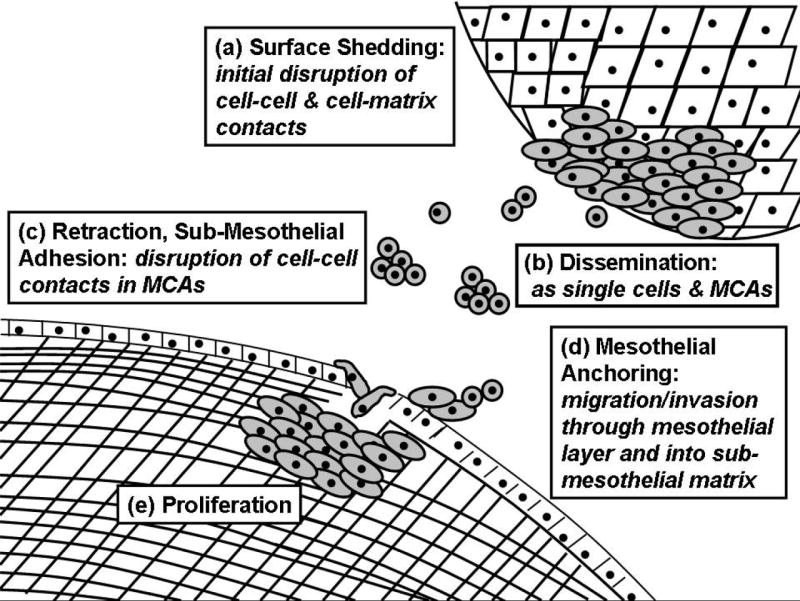
The mesodermally derived normal ovarian surface epithelium (OSE) displays both epithelial and mesenchymal characteristics and exhibits remarkable phenotypic plasticity during post-ovulatory repair. The majority of epithelial ovarian carcinomas (EOC) are derived from the OSE and represent the most lethal of all gynecological malignancies, as most patients (approximately 70%) present at diagnosis with disseminated intra-abdominal metastasis. The predominant pattern of EOC metastasis involves pelvic dissemination rather than lymphatic or hematologic spread, distinguishing EOC from other solid tumors. Acquisition of the metastatic phenotype involves a complex series of interrelated cellular events leading to dissociation (shedding) and dispersal of malignant cells. A key event in this process is disruption of cell-cell contacts via modulation of intercellular junctional components. In contrast to most carcinomas that downregulate E-cadherin expression during tumor progression, a unique feature of primary well-differentiated ovarian cancers is a gain of epithelial features, characterized by an increase in expression of E-cadherin. Subsequent reacquisition of mesenchymal features is observed in more advanced tumors with concomitant loss of E-cadherin expression and/or function during progression to metastasis. The functional consequences of this remarkable phenotypic plasticity are not fully understood, but may play a role in modulation of cell survival in suspension (ascites), chemoresistance, and intraperitoneal anchoring of metastatic lesions.


Figures

Figure 1
Figure 1. Schematic of ovarian cancer metastasis
(a) Early events in ovarian cancer metastasis include alteration of cell adhesive properties leading to shedding of tumor cells from the primary tumor. (b) Exfoliated tumor cells can exist as single cells or multi-cellular aggregates (MCAs) or spheroids in the peritoneal cavity. (c) Tumor cell adhesion to the mesothelium induces rapid retraction, exposing the underlying sub-mesothelial matrix. (d) Tumor cells migrate and invade into the submesothelial matrix to anchor secondary lesions followed by (e) proliferation to establish metastatic lesions within the pelvic/abdominal cavity and organs.
Figure 2
Figure 2. Cadherin profiles in ovarian tumor progression
[1] Genetic changes leading to primary tumor growth are characterized by acquisition of E-cadherin (EC). Early tumors express both epithelial (EC, keratin) and mesenchymal (N-cadherin [NC], vimentin) markers. [2] Tumor cells are shed intra-peritoneally from the primary tumor as single cells and MCAs. It has been speculated that retention of E-cadherin expression enables MCAs to avoid anoikis as cells lose matrix contacts and are suspended in ascites. Unknown mechanisms downregulate EC expression and function in metastatic tumors, although some E-cadherin staining is frequently retained in late stage carcinomas and ascites-derived tumor cells. [3] Intraperitoneal adhesion, invasion and proliferation generate intra-abdominal metastases, but the cadherin composition of the metastatic lesions has not been fully investigated.
Figure 3
Figure 3. Heterogeneity of epithelial and mesenchymal characteristics in ovarian tumors
Ovarian tumors exhibit both epithelial and mesenchymal features, as indicated by initial scoring (grouping 1+ to 3+) of serial sections from an ovarian tissue microarray (n=146). Epithelial markers (E-cadherin and keratin) were evident in 86% and 91% of the tumor samples, respectively. Mesenchymal markers (N-cadherin, vimentin) are expressed in 33% and 37%, respectively. Overall, 28% of ovarian tumors in this analysis are positive for both E-cadherin and N-cadherin, although this was less frequent in clear cell or mucinous histotypes. Representative images are shown to illustrate staining patterns. Serous ovarian carcinoma stage IIIb; endometroid carcinoma stage IIIc; clear cell carcinoma stage Ia; mucinous carcinoma stage Ic. Panels were stained with HECD-1 E-cadherin ectodomain primary antibody; N-cadherin primary antibody (clone 3B9, Zymed), keratin 7 primary antibody (Dako) or vimentin primary antibody (Dako) as indicated on the figure. Tumor tissue microarrays were prepared by the Pathology Core Facility of the Robert H. Lurie Comprehensive Cancer Center at Northwestern University assembled from tumor tissue originally taken for routine diagnostic purposes with Institutional Review Board approval. Immunohistochemical stains were scored by anatomic pathologist Dr. Brian P. Adley.
Figure 4
Figure 4. Cadherin expression in ovarian cancer cell lines
(A) Equal protein from whole cell extracts (WCE) was analyzed by western blot and probed with antibodies to E-cadherin (DakoCytomation clone NCH-38), N-cadherin (Zymed 33-3900), keratin-8 (USBiologicals K0199-10) or vimentin (Chemicon CBL202), followed by incubation with a peroxidase-conjugated secondary antibody and ECL detection. IOSE = IOSE 398, ovarian surface epithelium immortalized with SV40 T antigen [109]. Note that the heterogeneity of epithelial (E-cadherin, keratin) and mesenchymal (N-cadherin, vimentin) markers mirrors that observed in human tumors (Fig. 3). β-actin (Sigma A-5441) was used as a loading control. [Note that the lane containing immortalized normal ovarian surface epithelial cell (IOSE) samples was pasted into lane 1 of the figure from another part of the gel.] (B,C) Phenotypes of cells displaying different cadherin profiles. Phase-contrast microscopy highlights the more epithelial phenotype and tight colony morphology of the E-cadherin expressing cells (OVCA 433) relative to the more fibroblastic phenotype of the N-cadherin expressors (DOV13).
Figure 5
Figure 5. Cadherin expression in paired primary tumors and paired peritoneal metastatic tissue
Tissue was obtained from primary serous tumors localized to the ovary and paired peritoneal metastases from the same patient. Staining for E-cadherin and N-cadherin revealed two predominant patterns illustrated in this figure. One pattern is reduced E-cadherin staining in the metastasis relative to the primary tumor (compare A and B). The other pattern is strong N-cadherin immunoreactivity that is retained in the metastatic lesion (compare C and D). Tumor tissue microarrays were prepared by the Pathology Core Facility of the Robert H. Lurie Comprehensive Cancer Center at Northwestern University assembled from tumor tissue originally taken for routine diagnostic purposes with Institutional Review Board approval. Immunohistochemical stains were scored by anatomic pathologist Dr. Brian P. Adley.
Figure 6
Figure 6. Model for modulation of E-cadherin function in ovarian tumor metastasis
[a] In the well-differentiated primary tumor, cohesion is maintained primarily through E-cadherin. [b] Engagement of the collagen binding integrins (α2β1 or α3β1) leads to upregulation of MMP-9 and E-cadherin cleavage leading to release of the soluble E-cadherin ectodomain into the tumor microenvironment. [c] Destabilization of junctional E-cadherin occurs by multiple mechanisms including ectodomain cleavage and disruption of preformed junctions by soluble E-cadherin in the microenvironment leading to release of junctional β-cateinin for transcriptional co-activation of Tcf/Lef-regulated genes. [d] Transcriptional profiles regulated by Tcf/Lef contribute to EMT.

References

    1. Jemal A, Siegel R, Ward E, Murray T, Xu J, Thun MJ. Cancer statistics, 2007. Clin. 2007 Jan-Feb;57(1):43–66. - PubMed
    1. Wheeler JE. Pathology of malignant ovarian epithelial tumors and miscellaneous and rare ovarian and paraovarian neoplasms. In: Rubin SC, Sutton GP, editors. Ovarian Cancer. McGraw Hill; 1993. pp. 87–130.
    1. Cannistra SA. Cancer of the ovary. N Engl J Med. 2004;351:2519–29. - PubMed
    1. DuBeau L. The cell or origin of ovarian epithelial tumors and the ovarian surface epithelium dogma: does the emperor have no clothes? Gyn Oncol. 1999;72:437–442. - PubMed
    1. Shedden KA, Kshisagar MP, Schwartz DR, Wu R, Yu H, Misek DE, Hanash S, Katabuchi H, Ellenson LH, Fearon ER, Cho KC. Histologic type, organ or origin and wnt pathway status: effect on gene expression in ovarian and uterine carcinomas. Clin Can Res. 2005;11:2123–31. - PubMed

Publication types