. 2008 May;47(5):1702-13.
doi: 10.1002/hep.22221.

Mechanisms of hepatocyte growth factor-mediated and epidermal growth factor-mediated signaling in transdifferentiation of rat hepatocytes to biliary epithelium

Affiliations

Mechanisms of hepatocyte growth factor-mediated and epidermal growth factor-mediated signaling in transdifferentiation of rat hepatocytes to biliary epithelium

Pallavi B Limaye et al. Hepatology. 2008 May.

Abstract

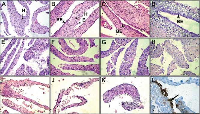
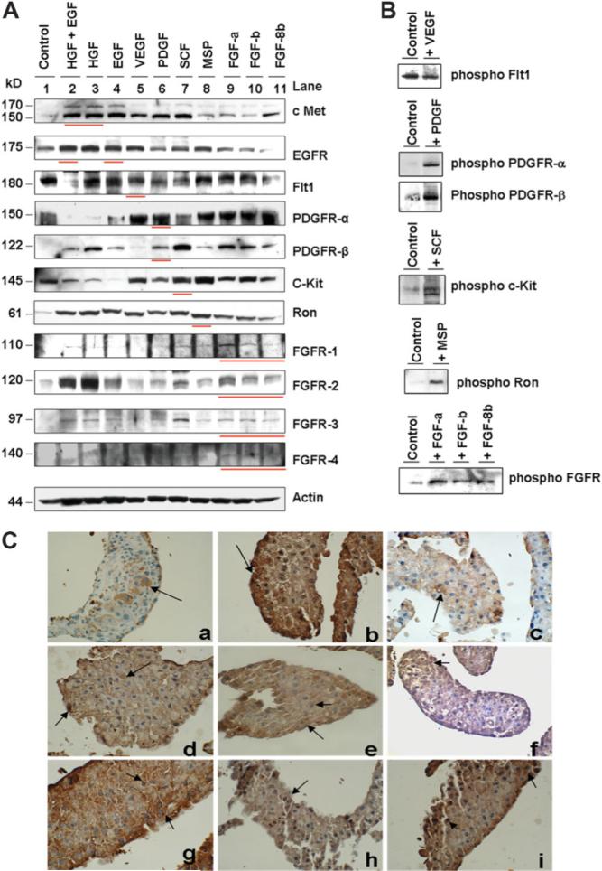
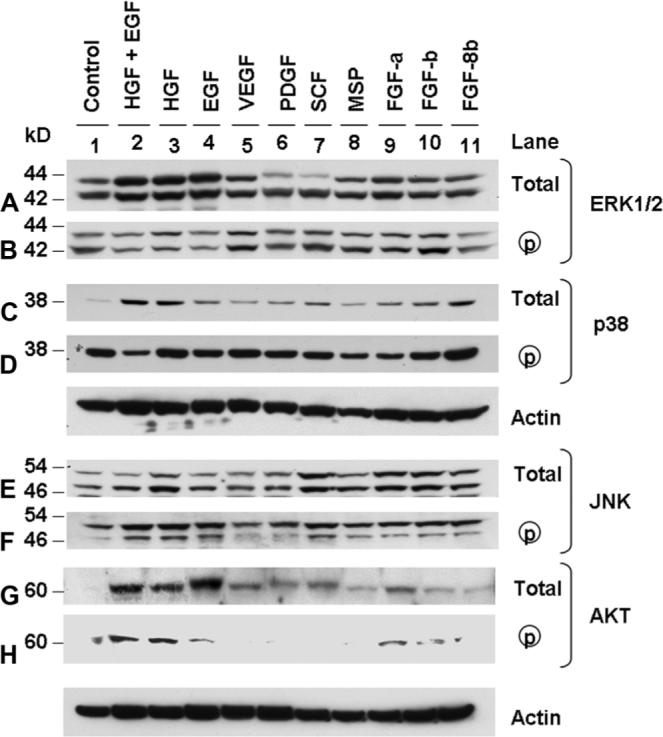
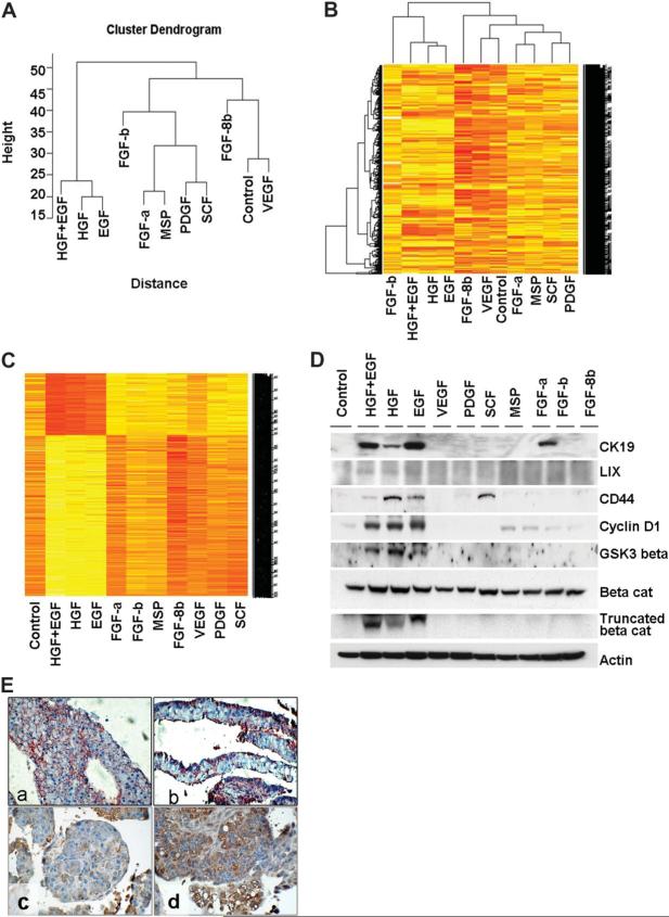
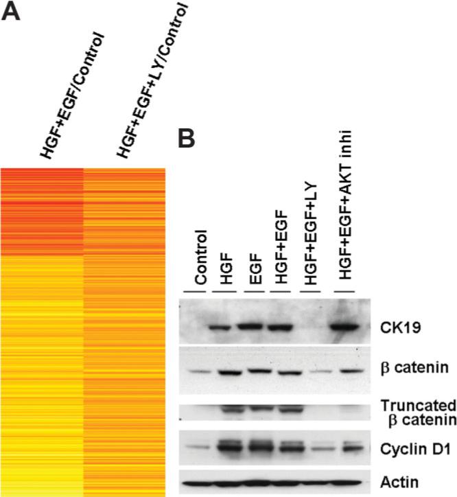
Previous studies from our laboratory have demonstrated that hepatocytes can transdifferentiate into biliary epithelium (BE) both in vivo and in vitro; however, the mechanisms are unclear. The current study was designed to investigate the mechanisms of hepatocyte transdifferentiation in vitro. Rat hepatocytes were cultured in roller bottles to obtain hepatocyte organoid cultures, which were stimulated with various growth factors (GFs) including hepatocyte growth factor (HGF), epidermal growth factor (EGF), vascular endothelial growth factor (VEGF), platelet-derived growth factor (PDGF), stem cell factor (SCF), macrophage-stimulating protein (MSP), fibroblast growth factor-a (FGF-a), fibroblast growth factor-b (FGF-b), and fibroblast growth factor-8b (FGF-8b). Only the cultures treated with HGF, EGF, and their combination exhibited formation of hepatocyte-derived biliary epithelium (BE) despite the presence and activation of all the pertinent cognate membrane receptors of the rest of the GFs. Microarray analysis of the organoid cultures identified specific up-regulation of approximately 500 target genes induced by HGF and EGF, including members of the extracellular matrix (ECM) protein family, Wnt/beta-catenin pathway, transforming growth factor beta (TGF-beta)/bone morphogenetic protein (BMP) pathway, and CXC (cysteine-any amino acid-cysteine) chemokines. To investigate the downstream signaling involved in hepatocyte to biliary epithelial cell (BEC) transdifferentiation, we investigated expression and activities of mitogen-activated protein (MAP) kinases [extracellular signal-regulated kinase (ERK)1/2, p38, and c-Jun N-terminal kinase (JNK)/stress-activated protein kinase (SAPK)] as well as serine/threonine kinase AKT. The analysis indicated that AKT phosphorylation was particularly increased in cultures treated with HGF, EGF, and their combination. Whereas phosphatidylinositol 3-kinase (PI3K) inhibitor LY294002 completely inhibited biliary epithelium formation, AKT inhibitor could only moderately reduce formation of BE in the organoid cultures treated with HGF+EGF. Most of the HGF+EGF target genes were altered by LY294002.

Conclusion: Taken together, these data indicate that hepatocyte to BE transdifferentiation is regulated by HGF and EGF receptors and that PI3 kinase-mediated signaling independent of AKT is a crucial component of the transdifferentiation process.


Figures

Fig. 1
Fig. 1
Histology of the hepatocyte organoid tissues at day 14 of the culture treated with different GFs. Hematoxylin-eosin–stained sections of cultures treated with HGF (B), EGF (C), HGF+EGF (D), VEGF (E), PDGF (F), SCF (G), MSP (H), FGF-a (I), FGF-b (J), and FGF-8b (K). Biliary epithelium (BE) developed in HGF and EGF treatment (arrowhead). H, hepatocytes; E, endothelial cell. Biliary marker AE1/AE3–stained section of the hepatocyte organoid tissue at day 14 of the culture treated with HGF+EGF (1m). AE1/AE3 positive BE (arrow).
Fig. 2
Fig. 2
(A) Western blot analysis of different membrane receptors (met, EGFR, Flt1, PDGFR, c-kit, ron, and FGFRs) in hepatocyte organoid culture lysates obtained at day 14 from various GF treatments–HGF, EGF, VEGF, PDGF, SCF, MSP, and FGFs–as described in Materials and Methods. Pertinent ligand and the receptor pairs are marked with a red underline. Actin blot was used as a loading control. Forty micrograms protein was separated on 4% to 12% gradient gel. (B) Phosphorylated form of the receptors detected by western blotting with and without the respective ligand in the hepatocyte organoid cultures. (C) Immunohistochemical localization of the membrane receptors in the organoid cultures developed in the presence of ITS+Dex (control). (A) Flt1, (B) PDGFR, (C) PDGFR, (D) c-kit, (E) Ron, (F) FGFR1, (G) FGFR2, (H) FGFR3, and (I) FGFR4.
Fig. 3
Fig. 3
Western blot analysis of total and activated protein kinases (ERK1/2, p38, JNK/SAPK, and AKT) in hepatocyte organoid culture lysates obtained from various GF treatments–HGF, EGF, VEGF, PDGF, SCF, MSP, and FGFs–as described in Materials and Methods. Forty micrograms protein was separated on 4% to 12% gradient gel. Actin blot used as a loading control.
Fig. 4
Fig. 4
(A) Hierarchical clustering of unsupervised gene array data obtained from Affymetrix Rat genome 2.0 analysis of hepatocyte organoid cultures treated with different GFs as labeled. (B) Heatmap of the genes statistically significant resulting after the dChip preprocessing (2667 genes). (C) Heatmap of the tightly clustered genes in the HGF+EGF, HGF, and EGF treatments after SAM package was applied. Yellow, up-regulated (512 genes); red, down-regulated (192 genes). (D) Western blot analysis of representative proteins whose genes were up-regulated in the gene array after HGF and EGF treatments. Forty micrograms protein was separated on 4% to 12% gradient gel. Actin blot was used as a loading control. (E) Immunohistochemical localization of HGF+EGF target genes. (A and B) Beta-catenin staining in control (A) and after HGF+EGF treatment (B). Beta-catenin staining increased in the biliary epithelium, whereas there was less hepatocyte membranous staining in HGF+EGF treatment. TGF-1 staining in control (C) and after HGF+EGF treatment (D). TGF-1 staining was higher in the hepatocytes underlying the biliary epithelium in HGF+EGF treatment.
Fig. 5
Fig. 5
Effect of PI3K inhibitor LY294002 on the HGF+EGF target genes. (A) Heatmap of the HGF+EGF modulated genes with and without LY compared with the control. Gene expression is largely reversed in the presence of LY. Yellow, up-regulated (approximately 500 genes); red, down-regulated (approximately 125 genes). (B) Reversal of the HGF and EGF mediated up-regulation of the genes at the protein level on addition of PI3 kinase inhibitor LY294002 detected by western blot. AKT inhibitor was ineffective in preventing HGF and EGF–mediated up-regulation of target genes.

Similar articles

Cited by

References

    1. Sirica AE, Williams TW. Appearance of ductular hepatocytes in rat liver after bile duct ligation and subsequent zone 3 necrosis by carbon tetrachloride. Am J Pathol. 1992;140:129–136. - PMC - PubMed
    1. Desmet V, Roskams T, Van Eyken P. Ductular reaction in the liver. Pathol Res Pract. 1995;191:513–524. - PubMed
    1. Nishikawa Y, Doi Y, Watanabe H, Tokairin T, Omori Y, Su M, et al. Transdifferentiation of mature rat hepatocytes into bile duct-like cells in vitro. Am J Pathol. 2005;166:1077–1088. - PMC - PubMed
    1. Cable EE, Isom HC. Exposure of primary rat hepatocytes in long-term DMSO culture to selected transition metals induces hepatocyte proliferation and formation of duct-like structures. Hepatology. 1997;26:1444–1457. - PubMed
    1. Michalopoulos GK, Barua L, Bowen WC. Transdifferentiation of rat hepatocytes into biliary cells after bile duct ligation and toxic biliary injury. Hepatology. 2005;41:535–544. - PMC - PubMed

Publication types

MeSH terms