. 2008 Apr 8;6(4):e84.
doi: 10.1371/journal.pbio.0060084.

Control of apoptosis by asymmetric cell division

Affiliations

Control of apoptosis by asymmetric cell division

Julia Hatzold et al. PLoS Biol. .

Abstract

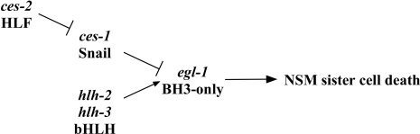
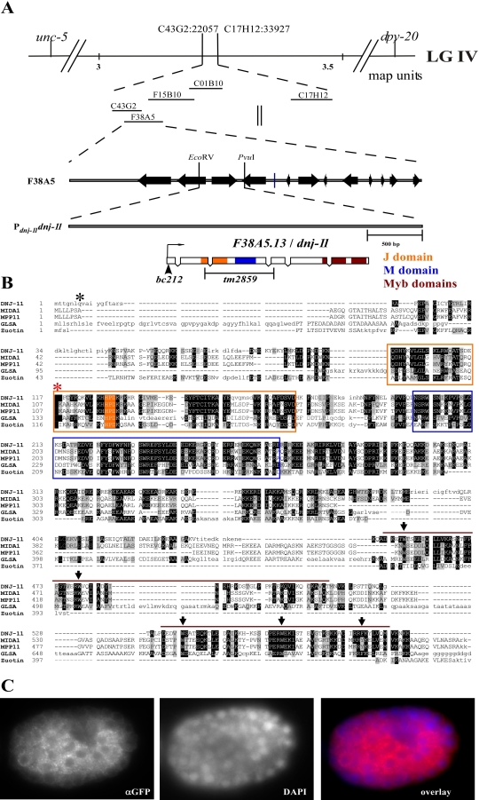
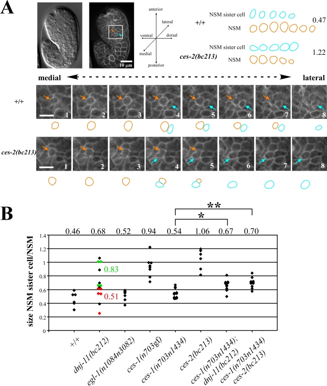
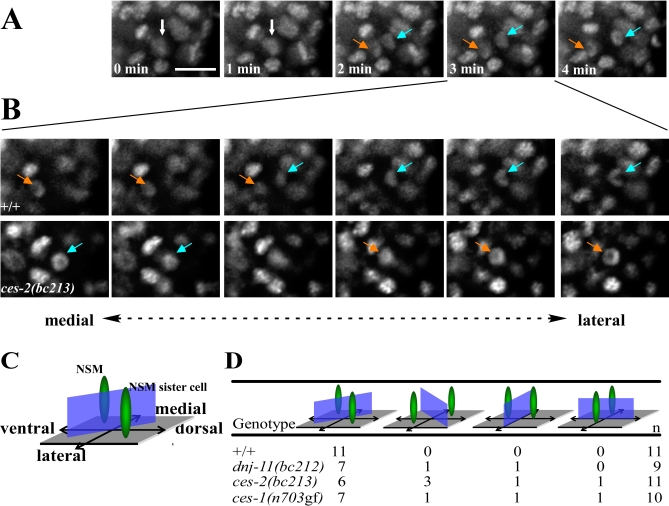
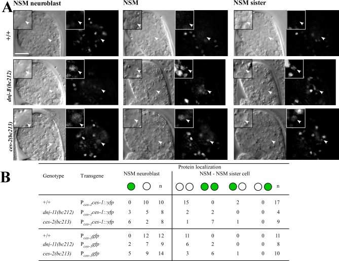
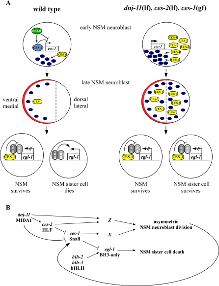
Asymmetric cell division and apoptosis (programmed cell death) are two fundamental processes that are important for the development and function of multicellular organisms. We have found that the processes of asymmetric cell division and apoptosis can be functionally linked. Specifically, we show that asymmetric cell division in the nematode Caenorhabditis elegans is mediated by a pathway involving three genes, dnj-11 MIDA1, ces-2 HLF, and ces-1 Snail, that directly control the enzymatic machinery responsible for apoptosis. Interestingly, the MIDA1-like protein GlsA of the alga Volvox carteri, as well as the Snail-related proteins Snail, Escargot, and Worniu of Drosophila melanogaster, have previously been implicated in asymmetric cell division. Therefore, C. elegans dnj-11 MIDA1, ces-2 HLF, and ces-1 Snail may be components of a pathway involved in asymmetric cell division that is conserved throughout the plant and animal kingdoms. Furthermore, based on our results, we propose that this pathway directly controls the apoptotic fate in C. elegans, and possibly other animals as well.


Conflict of interest statement

Competing interests. The authors have declared that no competing interest exists.

Figures

Figure 1
Figure 1. The Genetic Pathway of the NSM Sister Cell Death
egl-1 is required for the death of the NSM sister cell. egl-1 activity in the NSM sister cell is positively regulated by the genes hlh-2 and hlh-3 and negatively regulated by the gene ces-1. The activity of ces-1 is negatively regulated by the gene ces-2. See text for details.
Figure 2
Figure 2. dnj-11 Encodes a Member of the Family of MIDA1-Like Proteins
(A) (Top) Genes and single-nucleotide polymorphisms (SNPs) used for mapping bc212 are indicated. (Middle) Cosmids assayed for rescue of dnj-11(bc212) are shown. (Bottom) Pdnj-11dnj-11 is a subclone of F38A5 and represents the minimal dnj-11(bc212) rescue fragment. The J domain, M domain, and Myb domains of the dnj-11 ORF are indicated in orange, blue, and maroon, respectively. (B) The alignment of the DNJ-11 sequence with the sequence of mouse MIDA1, human MPP11, V. cateri GlsA, and the S. cerevisiae Zuotin was performed using the DIALIGN algorithm. Identical amino acids and similar amino acids are shaded in black and gray, respectively. The orange box marks the J domain and the conserved tripeptide is shaded in orange. The blue box indicates the M domain. The maroon bars above the sequences represent the Myb domains. Arrows point to conserved aromatic residues in the Myb domains. bc212 results in the truncation of the DNJ-11 protein after aa 6 (asterisk). tm2859 results in a frame shift after aa 116 (red asterisk) and a stop after an additional 67 aas in a different frame. (C) Embryos transgenic for the functional Pdnj-11dnj-11::gfp transgene were fixed as described in Materials and Methods and stained with an anti-GFP antibody (αGFP) to detect DNJ-11::GFP and with DAPI (DAPI) to detect nuclei.
Figure 3
Figure 3. dnj-11, ces-1, and ces-2 Are Involved in Asymmetric NSM Neuroblast Division
(A) (Left top) Nomarski and epifluorescence image of a wild-type embryo transgenic for the transgene Ppie-1gfp::ph(PLC1δ1) immediately after the NSM neuroblast divided. The orange arrow points to the NSM, the blue arrow points to the NSM sister cell. (Bottom) Series of eight consecutive focal planes (0.5 μm distance) (1–8) through the NSM and NSM sister cell of a wild-type (upper panel, +/+) or ces-2(bc213) (lower panel, ces-2(bc213)) embryo. (Right top) Summary of schematic representations of the outlines. Numbers on the right indicate the ratios between the sizes of the NSM sister cells and the NSMs in the Z-series shown. (B) The ratio of the sizes of the NSM sister cell and the NSM was determined as described in Materials and Methods. The NSM sister cell was defined as the lateral cell of the two daughter cells. Each diamond represents the ratio of a single, independent NSM neuroblast division. The numbers above the diamonds represent the average ratio obtained for a given genotype. The apoptotic fate of the NSM sister cells was determined in a subset of dnj-11(bc212) embryos (red/died, green/survived). The complete genotypes of the embryos analyzed from left to right were: tIs38 (Ppie-1gfp::ph(PLC1δ1)), dnj-11(bc212) bcIs25; tIs38, egl-1(n1048n3082); tIs38, ces-1(n703gf); tIs38, ces-1(n703n1434); tIs38, ces-2(bc213); bcIs25; tIs38, ces-1(n703n1434); dnj-11(bc212) bcIs25; tIs38, and ces-1(n703n1434) ces-2(bc213); bcIs25; tIs38. p-Values were determined by the Student's t-test. *ces-1(n703n1434) compared to ces-1(n703n1434); dnj-11(bc212): p<0.002, **ces-1(n703n1434) compared to ces-1(n703n1434) ces-2(bc213): p<0.001.
Figure 4
Figure 4. dnj-11, ces-1, and ces-2 Are Involved in Orienting the Mitotic Spindle of the NSM Neuroblasts
(A) Epifluorescent images shown are maximum projections of Z-series of a wild-type embryo carrying the transgene Phis-24his-24::gfp. The scale bar represents 5 μm. Ventral is to the left and dorsal to the right. The Z-series were taken every 60 s during a 4-min time period. Chromosomal DNA in the NSM neuroblast prior to the division is indicated by white arrows. Between minute 1 and minute 2, the NSM neuroblast starts dividing. The chromatids of the NSM (indicated by orange arrows) move toward the ventral side, the chromatids of the NSM sister cell (indicated by blue arrows) move to the dorsal side. (B) (Upper panel). Epifluorescence images of six consecutive focal planes of the Z-series at minute 3 shown in (A) as maximum projection. Medial to lateral planes are shown from left to right. The chromatids of the ventral NSM are located medially. The chromatids of the dorsal NSM sister cell are located laterally. (Lower panel). Six consecutive focal planes of a Z-series of a ces-2(bc213) embryo. Medial to lateral planes are shown from left to right. The chromatids of the ventral NSM are located laterally. The chromatids of the dorsal NSM sister cell are located medially. (C) Schematic representation of the ventral/dorsal and medial/lateral positions of the chromatids of NSM and NSM sister cell (indicated in green) in wild-type embryos. The blue rectangle indicates the cleavage plane of the NSM neuroblast. (D) Quantification of the orientation of the cleavage planes observed. The complete genotypes of the embryos analyzed from top to bottom were: dtIs372; dnj-11(bc212) bcls25; dtls372, ces-2(bc213); bcls25; dtls372, and ces-1(n703); bcIs25; dtIs372.
Figure 5
Figure 5. The Loss of dnj-11 or ces-2 Function Results in Increased Levels of CES-1 Protein and Increased ces-1 Transcription in the NSM Lineage
(A) Nomarski and epifluorescence images of wild-type (+/+), dnj-11(bc212), and ces-2(bc213) embryos transgenic for a stable Pces-1ces-1::yfp transgene. White arrowheads indicate NSM neuroblasts (left column), NSMs (middle column), and NSM sister cells (right column). The scale bar represents 10 μm. Insets in the upper left corner of each image show the respective cells at a higher magnification. (B) Summary of the data on the presence of CES-1 protein in the NSM lineage using the Pces-1ces-1::yfp transgene and ces-1 expression using the Pces-1gfp transgene. Green circles represent cells in which CES-1::YFP or GFP were detected. White circles represent cells in which CES-1::YFP or GFP were not detected. The complete genotypes of the embryos analyzed from top to bottom were: bcIs58 (Pces-1ces-1::yfp), dnj-11(bc212) bcIs25; bcIs58, ces-2(bc213); bcIs25; bcIs58, bcEx619 (Pces-1gfp), dnj-11(bc212) bcIs25; bcEx619, and ces-2(bc213); bcIs25; bcEx619.
Figure 6
Figure 6. Regulation of Asymmetric NSM Neuroblast Division and the Apoptotic Death of the NSM sister Cell
(A) Molecular models. (Wild type) By repressing the transcription of the Snail-related gene ces-1, the proteins CES-2 HLF and DNJ-11 MIDA1 ensure that the CES-1 Snail protein is present at an appropriate, low level in the unpolarized, early NSM neuroblast. Blue oval indicates polarity factor that is required to establish polarity in the late NSM neuroblast and the synthesis of which is promoted by CES-1. Indicated in red is a complex that restricts the polarity factor to the ventral/medial side of the NSM neuroblast. Once localized to the ventral/medial side, the polarity factor is involved in restricting CES-1 to the ventral/medial side of the late NSM neuroblast. See text for details. (dnj-11(lf), ces-2(lf), ces-1(gf)) Loss-of-function mutations of dnj-11 or ces-2, or a gain-of-function mutation of ces-1 result in an increased level of CES-1 Snail protein in the early NSM neuroblast. This increased level of CES-1 protein results in a level of polarity factor too high to establish asymmetry and restrict CES-1 protein to the ventral/medial side. See text for details. (B) Genetic pathway. dnj-11 acts upstream of or in parallel to ces-2 to negatively regulate the function of ces-1, thereby causing asymmetric NSM neuroblast division and NSM sister cell death. ces-1 function can affect asymmetric NSM neuroblast division by regulating the function of a gene or genes required for the process. By negatively regulating egl-1, ces-1 function can also prevent the hlh-2, hlh-3–dependent death of the NSM sister cells. ces-1 function in NSM sister cell death is affected by the process of asymmetric NSM neuroblast division, which causes the asymmetric distribution of the product of the ces-1 gene. See text for details.

References

    1. Horvitz HR, Herskowitz I. Mechanisms of asymmetric cell division: two Bs or not two Bs, that is the question. Cell. 1992;68:237–255. - PubMed
    1. Sulston JE, Horvitz HR. Post-embryonic cell lineages of the nematode, Caenorhabditis elegans . Dev Biol. 1977;56:110–156. - PubMed
    1. Sulston JE, Schierenberg E, White JG, Thomson JN. The embryonic cell lineage of the nematode Caenorhabditis elegans . Dev Biol. 1983;100:64–119. - PubMed
    1. Frank CA, Hawkins NC, Guenther C, Horvitz HR, Garriga G. C. elegans HAM-1 positions the cleavage plane and regulates apoptosis in asymmetric neuroblast divisions. Dev Biol. 2005;284:301–310. - PubMed
    1. Cordes S, Frank CA, Garriga G. The C. elegans MELK ortholog PIG-1 regulates cell size asymmetry and daughter cell fate in asymmetric neuroblast divisions. Development. 2006;133:2747–2756. - PubMed

Publication types

MeSH terms

Substances

Associated data