Comparative Study
. 2008 Apr 10;58(1):26-33.
doi: 10.1016/j.neuron.2008.01.007.

The cadherin Flamingo mediates level-dependent interactions that guide photoreceptor target choice in Drosophila

Affiliations
Comparative Study

The cadherin Flamingo mediates level-dependent interactions that guide photoreceptor target choice in Drosophila

Pei-Ling Chen et al. Neuron. .

Abstract

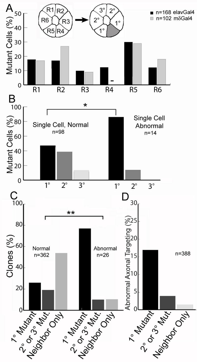
Quantitative differences in cadherin activity have been proposed to play important roles in patterning connections between pre- and postsynaptic neurons. However, no examples of such a function have yet been described, and the mechanisms that would allow such differences to direct growth cones to specific synaptic targets are unknown. In the Drosophila visual system, photoreceptors are genetically programmed to make a complex, stereotypic set of synaptic connections. Here we show that the atypical cadherin Flamingo functions as a short-range, homophilic signal, passing between specific R cell growth cones to influence their choice of postsynaptic partners. We find that individual growth cones are sensitive to differences in Flamingo activity through opposing interactions between neighboring cells and require these interactions to be balanced in order to extend along the appropriate trajectory.


Figures

Figure 1
Figure 1. Flamingo is not required cell-autonomously in R cell axons
(A). Schematic representation of R cells and their axons. In each ommatidium, in the retina, each R cell occupies an invariant relative position; this arrangement is preserved in the axon fascicle projecting into the brain, even as the bundle of axons twists 180° (curved line). In the brain, within the lamina plexus, each R cell axon (pink) then extends to a target (red), arranged in an invariant position relative to the fascicle. R cells axons from neighboring ommatidia (blue) choose an overlapping set of targets. The axons of R7 and R8 extend into the medulla, and are not shown. (B) In the retina each R cell sub-type is uniquely identifiable by its characteristic position and morphology (B, G, L, Q). Using the MARCM method, single R cells (green) are made homozygous for either a control chromosome (C-E, H-J, M-O, R-T) or a chromosome bearing a null allele in flamingo (F, K, P, U). During pupal development, R cell axons extend away from bundles of axons containing all R cells from a single ommatidium (D, I, N, S). Each R cell can be identified in the retina, and traced down into the brain, where it extends to a specific target located in an invariant position relative to the ommatial bundle. Arrowheads demark where each R cell axon starts its lateral extension across the lamina plexus; arrows demark the target cartridge.
Figure 2
Figure 2. Flamingo acts non-autonomously to control the targeting of neighboring photoreceptors
(A-C, F-H, K-M) Retina. Homozygous wild type cells (green) abutting flamingo null mutant cells (black) are generated at random, and are uniquely 23 identified by their morphology (red) (B, G, L). Retina. (C, H, M) Flamingo null mutant cells (chevrons) arise within ommatidia containing wild-type neighbors. Mutant cells can be identified by the absence of GMR-RFP expression (white), which is particularly noticeable in the rhabdome. (D, I, N) Schematic images of the labeled R cell axons (green), homozygous mutant axons (black) and heterozygous axons (gray) extending to their targets (red). (E, J, O) Confocal images of labeled R cell projections (green) extending to their targets (red). Normally extending axons (arrows) extend to specific targets; mistargeting axons extend two growth cones, innervating inappropriate targets (arrowheads). Inset panels depict single axons at high magnification. In (E), neither growth cone innervated the appropriate target (denoted * in D, E). Scale bar 10μm.
Figure 3
Figure 3. Flamingo mediated interactions between immediately neighboring R cells are critical for target selection
(A) The distribution of flamingo mutant R cells in two variations of reverse marcm. Flamingo mutant R cells, elavGal4 (black bars), mδGal4 (grey bars, n=102). In mδGal4 clones, all labeled wild-type axons are R4, so none are ever mutant (-). Inset panel designates the neighbor relationships between R cells relative to a single wild type cell (grey). (B) The distribution of projection defects in ommatidia containing only 1 mutant cell, and 1-3 wild-type cells, pooled between elavGal4 and mδGal4 datasets. The data is divided into those cases where the wild-type axons targeted normally (left bars), and abnormally (right bars). The fraction of clones is plotted as a function of the separation between the mutant cell and the wild-type cell (* p<0.01, χ2 test). (C) Plot of the fraction of clones of each type, dividing the data into two groups. Left bars: normal targeting. Right bars: abnormal targeting. These distributions are different (p<.001; χ2 test). “Neighbor Only” denotes clones in which the only mutant cells were in the neighboring ommatidium (185 cases), or clones in which there were no mutant neighbors at all (12 cases). (D) The frequency of abnormal axon targeting as a function of the neighbor relationship between wild-type and mutant cells.
Figure 4
Figure 4. Overexpression of Flamingo in R4 is sufficient to cause mistargeting of neighboring axons
(A) The expression pattern of mδGal4 (green) and Flamingo protein (red) in the lamina plexus. (B) The GcmGal4 expression pattern (green), and Flamingo (red). (C) The pattern of cartridges (red) in the adult lamina of animals overexpressing flamingo using mδGal4. Single cartridges are inset. (D) The pattern of cartridges (red) in the adult lamina of animals overexpressing Flamingo using GcmGal4. Scale bar, 20μm. (E-H). The pattern of cartridges in the adult lamina overexpressing either 2 copies (X2) or 1 copy (X1) of either full-length Flamingo (E-G) or a truncated form of Flamingo lacking the intracellular domain (ΔC, H). Single cartridges are inset. (I-L) Dye injection into single ommatidia (red). Panels contain all of the axons from one ommatidium; insets display the corresponding retinal image.(I)Control. (J-H)Flamingo overexpression in R4 using mδGal4. Arrows indicate R4 axons, arrowheads denote R cell growth cones that have targeted inappropriately. Scale bar, 10 μm.

Comment in

References

    1. Barth M, Hirsch HV, Meinertzhagen IA, Heisenberg M. Experience-dependent developmental plasticity in the optic lobe of Drosophila melanogaster. J Neurosci. 1997;17:1493–1504. - PMC - PubMed
    1. Chae J, Kim MJ, Goo JH, Collier S, Gubb D, Charlton J, Adler PN, Park WJ. The Drosophila tissue polarity gene starry night encodes a member of the protocadherin family. Development. 1999;126:5421–5429. - PubMed
    1. Clandinin TR, Zipursky SL. Afferent growth cone interactions control synaptic specificity in the Drosophila visual system. Neuron. 2000;28:427–436. - PubMed
    1. Clandinin TR, Zipursky SL. Making connections in the fly visual system. Neuron. 2002;35:827–841. - PubMed
    1. Das G, Reynolds-Kenneally J, Mlodzik M. The atypical cadherin Flamingo links Frizzled and Notch signaling in planar polarity establishment in the Drosophila eye. Dev Cell. 2002;2:655–666. - PubMed

Publication types

MeSH terms

LinkOut - more resources