Comparative Study
. 2008 Apr 10;58(1):42-51.
doi: 10.1016/j.neuron.2008.02.003.

Endocytosis is required for synaptic activity-dependent release of amyloid-beta in vivo

Affiliations
Comparative Study

Endocytosis is required for synaptic activity-dependent release of amyloid-beta in vivo

John R Cirrito et al. Neuron. .

Abstract

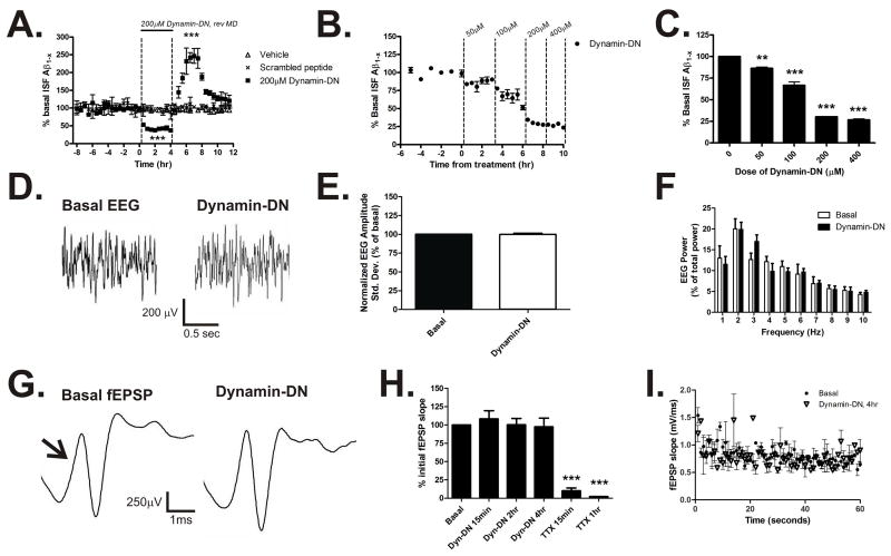
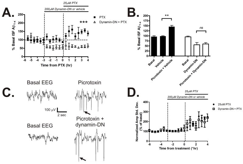
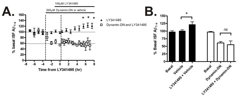
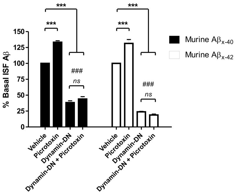
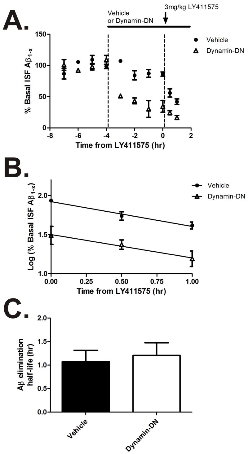
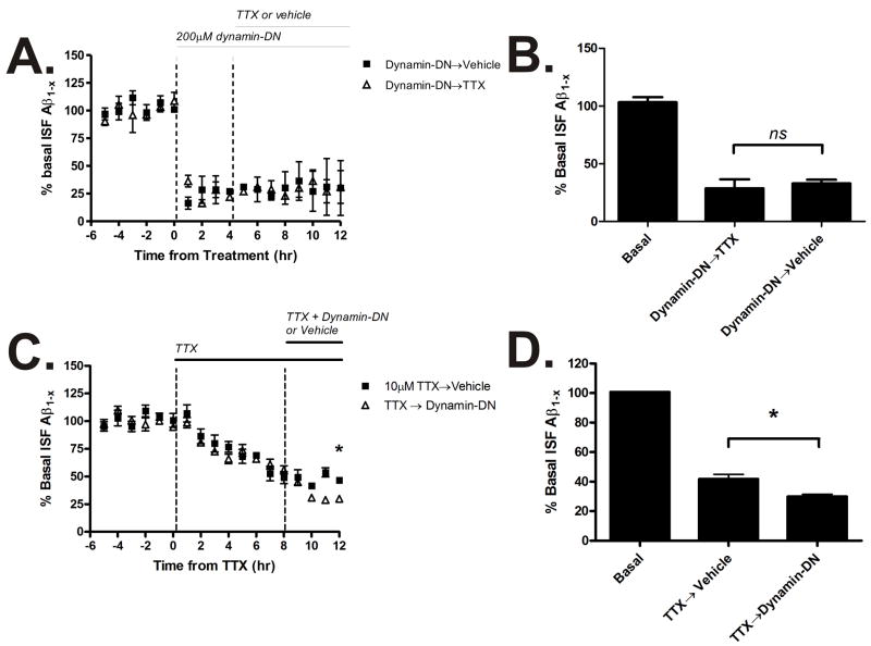
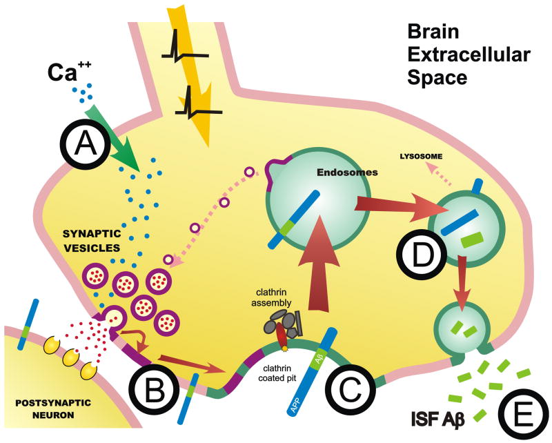
Aggregation of amyloid-beta (Abeta) peptide into soluble and insoluble forms within the brain extracellular space is central to the pathogenesis of Alzheimer's disease. Full-length amyloid precursor protein (APP) is endocytosed from the cell surface into endosomes where it is cleaved to produce Abeta. Abeta is subsequently released into the brain interstitial fluid (ISF). We hypothesized that synaptic transmission results in more APP endocytosis, thereby increasing Abeta generation and release into the ISF. We found that inhibition of clathrin-mediated endocytosis immediately lowers ISF Abeta levels in vivo. Two distinct methods that increased synaptic transmission resulted in an elevation of ISF Abeta levels. Inhibition of endocytosis, however, prevented the activity-dependent increase in Abeta. We estimate that approximately 70% of ISF Abeta arises from endocytosis-associated mechanisms, with the vast majority of this pool also dependent on synaptic activity. These findings have implications for AD pathogenesis and may provide insights into therapeutic intervention.


Figures

Figure 1
Figure 1. Inhibition of endocytosis reduces ISF Aβ levels
(A) In vivo measurement of ISF Aβ levels during inhibition of endocytosis in 3 month old Tg2576 mice. Infusion of a dynamin-DN inhibitory peptide (200μM) into the hippocampus via reverse microdialysis reduced ISF Aβ1-x levels to 36.1 ± 4.2% of baseline levels over fours hours (p<0.0001, n=9 per group). When dynamin-DN was removed from the microdialysis perfusion buffer, ISF Aβ levels increased transiently by 246.7 ± 20.7% compared to baseline (p<0.0001) and returned to basal levels by eight hours of washout. ISF Aβ levels did not change significantly over the course of the study in either untreated mice or in mice treated with 200μM scrambled peptide. (B) A dose escalation of dynamin-DN demonstrated a maximum reduction of ISF Aβ levels with a dose of 200μM of inhibitory peptide (p<0.0001, n=3). (C) shows final Aβ levels achieved during each epoch depicted in B. (D–F) EEG recordings during basal and dynamin-DN administration. (D) Representative EEG traces during basal and dynamin-DN treatment. EEG amplitude standard deviation (E) and frequency (F) were not significantly different when endocytosis was inhibited (n=3). (G–I) Evoked fEPSPs from dentate gyrus during basal and dynamin-DN treatment. (G) Representative fEPSP traces elicited by medial perforant pathway stimulation. Arrow denotes initial fEPSP slope. (H) The initial slope of the fEPSP did not change during 4 hours of dynamin-DN treatment; however when the perfusion buffer was changed to contain 10μM TTX the fEPSP was significantly reduced by 89.8 ± 3.8% within 15 minutes of treatment (p<0.0001, n = 3). (I) Likewise, fEPSP slope was similar under basal and dynamin-DN treatment during 1 Hz trains of stimuli for 60 seconds (n=3). Data presented as mean ± SEM. ** represent p<0.001, *** represent p<0.0001.
Figure 2
Figure 2. Activity-dependent release of Aβ requires endocytosis
(A) ISF A levels in Tg2576 mice treated with vehicle or 200μM dynamin-DN followed by 25μM picrotoxin via reverse microdialysis to increase neuronal activity (n = 4 per group). (B) Picrotoxin alone increased ISF A levels by 145.5 ± 6.8% averaged over 4 hours of administration (p<0.01). Pretreatment with 200μM dynamin-DN prevented the increase in ISF A levels caused by picrotoxin. (C) Representative EEG traces during basal conditions and during picrotoxin treatment with and without dynamin-DN administration. This low dose of picrotoxin did not generate seizures in any of the mice tested, but consistently caused synchronous spikes in EEG activity (arrows) in the presence and absence of dynamin-DN. (D) EEG amplitude increased similarly in picrotoxin-treated mice administered vehicle or dynamin-DN. Data presented as mean ± SEM. ** represents p<0.001, *** represents p<0.0001.
Figure 3
Figure 3. Increased glutamate release elevates ISF Aβ levels which requires endocytosis
(A) ISF A levels in Tg2576 mice treated with dynamin-DN or vehicle followed by LY341495, an mGluR2/3 antagonist that increases pre-synaptic glutamate release. (B) LY341495 increased ISF A levels by 122.2 ± 8.4% during hours 6-8 of treatment (p<0.05, n = 4). The increase in A levels was prevented by pretreatment with dynamin-DN (n = 4). Data presented as mean ± SEM. * represents p<0.05.
Figure 4
Figure 4. Endocytosis is required for activity-dependent release of endogenous murine
Aβ. In C57Bl6 mice, 25μM picrotoxin increased ISF murine A 40 and Aβ42 levels to 133.4 ± 2.3 and 131.5 ± 6.1% respectively (p<0.0001) of baseline over 12 hours. 200μM dynamin-DN reduced murine A 40 levels to 38.3 ± 2.8% (p<0.0001) and Aβ42 levels to 23.8 ± 0.8% of baseline (p<0.0001). Aβ42 levels were significantly more reduced than Aβ40 levels (p<0.0001; noted by ###). Pretreatment with dynamin-DN for 1 hour then co-administration of dynamin-DN and picrotoxin caused ISF A 40 and A 42 levels to decrease to 43.9 ± 3.7% and 18.9 ± 1.5% of baseline which was not statistically different from dynamin-DN treatment alone. n = 6 per group. Data presented as mean ± SEM. *** represents p<0.0001.
Figure 5
Figure 5. Dynamin-DN does not affect ISF Aβ elimination
(A) Hippocampal ISF A levels were measured in Tg2576 mice that were treated with vehicle or 200μM dynamin-DN, followed by 3mg/kg LY411575 s.c. to inhibit -secretase activity (n = 4-6 per group). (B) Plots of log % basal ISF A levels were linear in both vehicle and dynamin-DN treated mice, demonstrating first-order kinetics of ISF A elimination in both groups. (C) The elimination half-life of ISF A (calculated from the slope in panel B) was the same in vehicle and dynamin-DN treated mice (1.1 ± 0.24 hr and 1.2 ± 0.27 hr, respectively). Data presented as mean ± SEM.
Figure 6
Figure 6. Distinct pathways for Aβ generation
(A) Tg2576 mice were either treated with 200μM dynamin-DN for 12 hours or pre-treated with 200μM dynamin-DN for 4 hours followed by dynamin-DN and 10μM TTX together for an additional 8 hours. Hippocampal ISF A levels were measured by microdialysis. (B) Dynamin-DN reduced ISF A levels to 33.0 ± 3.2% of baseline over 12 hours (p<0.0001) and co-administration with TTX did not additionally change A levels compared to dynamin-DN alone (n=4 per group). (C) Mice were either treated with 10μM TTX alone for 12 hours or with TTX for 8 hours followed by TTX and 200μM dynamin-DN together for an additional 4 hours. 10μM TTX alone for 12 hours reduced ISF Aβ levels to 41.8 ± 3.2% of baseline (p<0.0001, n=8). When TTX was co-administered with dynamin-DN, ISF Aβ levels decreased to 30.0 ± 1.4% of baseline (p<0.0001, n=5). The 7% difference between TTX and TTX with dynamin-DN was significant (p<0.05). Data presented as mean ± SEM. * represents p<0.05.
Figure 7
Figure 7. Model of synaptic-dependent release of Aβ
(A) Depolarization of the synaptic terminal causes calcium influx leading to synaptic vesicle release. (B) Synaptic vesicle membrane recycling from the cell surface via clathrin-mediated endocytosis causes more APP to internalized (C). Within endosomes, BACE and β-secretase cleave APP to produce Aβ (D) which is secreted from the neuron into the brain ISF (E).

Similar articles

Cited by

References

    1. Andersen OM, Reiche J, Schmidt V, Gotthardt M, Spoelgen R, Behlke J, von Arnim CA, Breiderhoff T, Jansen P, Wu X, et al. Neuronal sorting protein-related receptor sorLA/LR11 regulates processing of the amyloid precursor protein. Proc Natl Acad Sci U S A. 2005;102:13461–13466. - PMC - PubMed
    1. Buckner RL, Snyder AZ, Shannon BJ, LaRossa G, Sachs R, Fotenos AF, Sheline YI, Klunk WE, Mathis CA, Morris JC, Mintun MA. Molecular, structural, and functional characterization of Alzheimer's disease: evidence for a relationship between default activity, amyloid, and memory. J Neurosci. 2005;25:7709–7717. - PMC - PubMed
    1. Busciglio J, Gabuzda DH, Matsudaira P, Yankner BA. Generation of beta-amyloid in the secretory pathway in neuronal and nonneuronal cells. Proc Natl Acad Sci U S A. 1993;90:2092–2096. - PMC - PubMed
    1. Caccamo A, Oddo S, Billings LM, Green KN, Martinez-Coria H, Fisher A, LaFerla FM. M1 receptors play a central role in modulating AD-like pathology in transgenic mice. Neuron. 2006;49:671–682. - PubMed
    1. Cam JA, Zerbinatti CV, Knisely JM, Hecimovic S, Li Y, Bu G. The low density lipoprotein receptor-related protein 1B retains beta-amyloid precursor protein at the cell surface and reduces amyloid-beta peptide production. J Biol Chem. 2004;279:29639–29646. - PubMed

Publication types

Substances