Review
. 2008 Apr 10;58(1):65-77.
doi: 10.1016/j.neuron.2008.01.037.

Regulation of motor neuron specification by phosphorylation of neurogenin 2

Affiliations
Review

Regulation of motor neuron specification by phosphorylation of neurogenin 2

Yong-Chao Ma et al. Neuron. .

Abstract

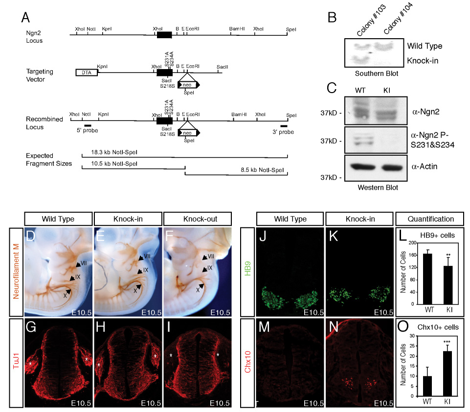
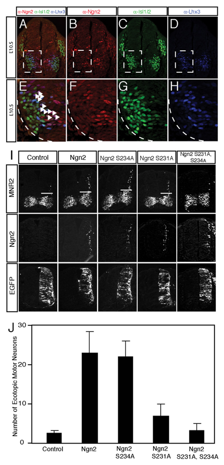
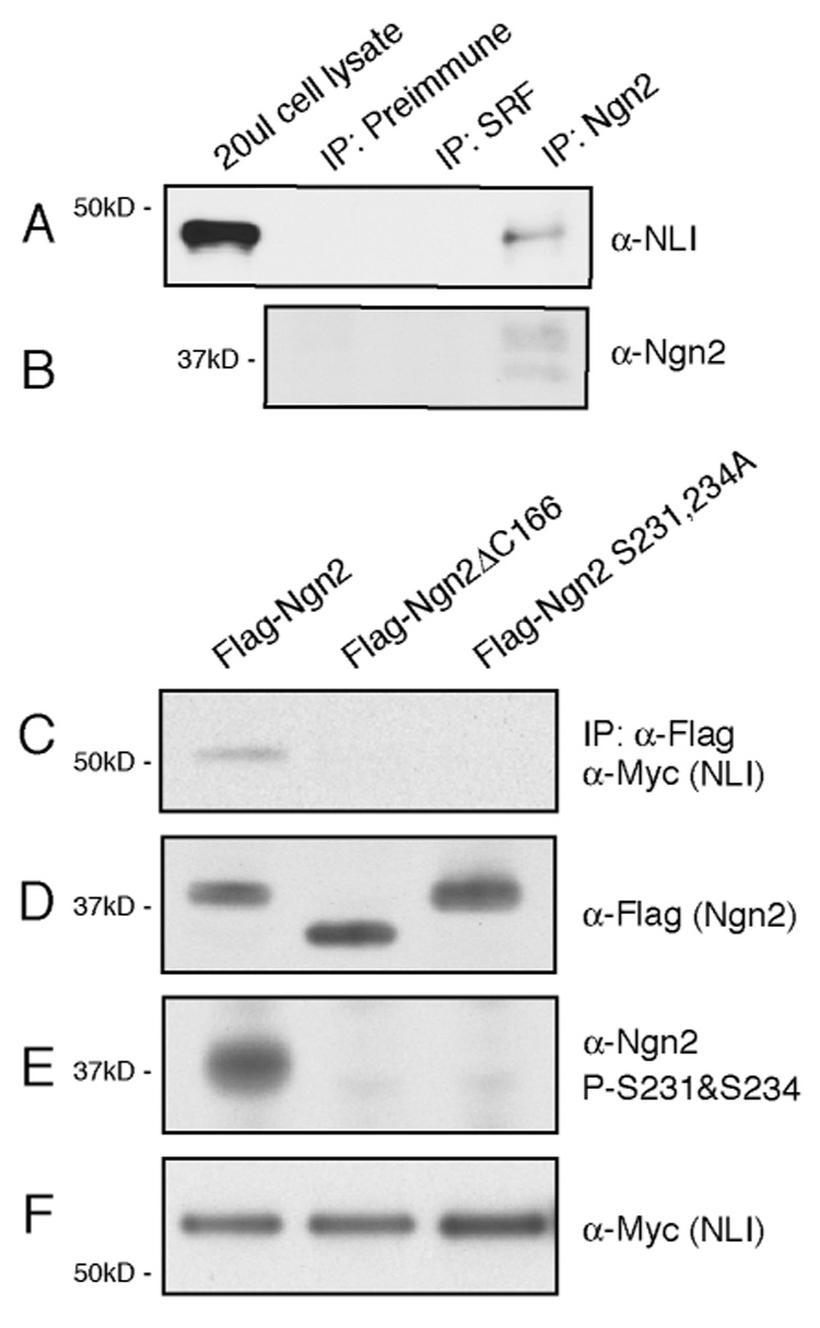
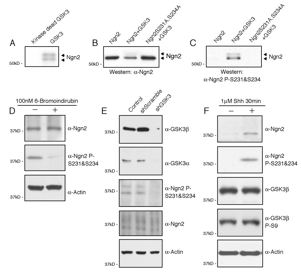
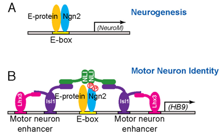
The mechanisms by which proneural basic helix-loop-helix (bHLH) factors control neurogenesis have been characterized, but it is not known how they specify neuronal cell-type identity. Here, we provide evidence that two conserved serine residues on the bHLH factor neurogenin 2 (Ngn2), S231 and S234, are phosphorylated during motor neuron differentiation. In knockin mice in which S231 and S234 of Ngn2 were mutated to alanines, neurogenesis occurs normally, but motor neuron specification is impaired. The phosphorylation of Ngn2 at S231 and S234 facilitates the interaction of Ngn2 with LIM homeodomain transcription factors to specify motor neuron identity. The phosphorylation-dependent cooperativity between Ngn2 and homeodomain transcription factors may be a general mechanism by which the activities of bHLH and homeodomain proteins are temporally and spatially integrated to generate the wide diversity of cell types that are a hallmark of the nervous system.


Figures

Figure 1
Figure 1. Neurogenin 2 is Phosphorylated on S231 and S234 During Spinal Motor Neuron Differentiation
(A) Sequence alignment showing S231 and S234 located near the C-terminus of Ngn2 are highly conserved across species. (B) Endogenous Ngn2 is phosphorylated on S231 and S234. Endogenous Ngn2 was immunoprecipitated from E11.5 mouse neural tube and telencephalon nuclear lysates with an anti-Ngn2 antibody crosslinked to protein A beads. The immunoprecipitaed proteins were left untreated or treated with alkaline phosphatase, separated on SDS-PAGE and blotted with the anti-Ngn2 or the anti-Ngn2 P-S231&S234 antibodies. (C-E) Endogenous Ngn2 is phosphorylated on S231 and S234 in spinal motor neuron progenitors. Coronal neural tube sections from E10 mouse embryos were stained with the anti-Ngn2 P-S231&S234 antibodies (red, C) and a monoclonal antibody recognizing motor neuron progenitor-specific transcription factor Olig2 (green, E). Merged image of anti-olig antibody and anti-Ngn2 P-S231&S234 antibody staining appears as yellow (D). (F-H) Ngn2 is phosphorylated on S231 and S234 in a subset of Ngn2-positive motor neuron progenitors. Coronal neural tube sections from E10 mouse embryos were stained with the anti-Ngn2 P-S231&S234 antibodies (red, F) and a rat anti-Ngn2 antibody (blue, H). Merged image of anti-Ngn2 P-S231&S234 antibody and anti- Ngn2 antibody staining appears as pink (G). Panels F through H correspond to the boxed area in panel C.
Figure 2
Figure 2. Motor Neuron Differentiation Is Defective in the Ngn2S231A&S234A Knock-in Mice
(A) Schematic representation of the mouse genomic region bearing the Ngn2 gene (top), the Ngn2S231A&S234A knock-in targeting vector (middle), and the Ngn2 locus after homologous recombination (bottom) with the targeting vector. The solid block represents the single exon encoding Ngn2, in which S231 and S234 have been mutated to alanines in the Ngn2S231A&S234A knock-in mice. 5’ and 3’ external probes are indicated and were used to detect the presence or absence of the recombined locus by Southern hybridization analysis of NotI plus SpeI digested genomic DNA. The expected NotI-SpeI restriction endonuclease fragments revealed by Southern blot analysis of wild type and targeted genomic DNA are shown. The positions of relevant restriction endonuclease sites are shown (B: BamHI; E: EcoRI). (B) Southern blot analysis showing successful targeting of the Ngn2S231A&S234A knock-in allele in mouse ES cells. Southern blot analysis of genomic DNA from targeted ES cell clones #103 and #104 digested with NotI and SpeI, and then probed with a 5’ external probe that recognizes an 18.3 kb wild type DNA fragment and a 10.5 kb DNA fragment that would only be present if the homologous recombination targeting event has occurred. (C) Absence of S231 and S234 phosphorylation in the Ngn2S231A&S234A knock-in mice. Lysates prepared from E11.5 neural tubes and telencephalons of wild type or Ngn2S231A&S234A knock-in mice were analyzed by immunoblotting using the anti-Ngn2 or the anti-Ngn2 P-S231&S234 antibodies. Note that the expression level of the Ngn2 protein is not altered in the Ngn2S231A&S234A knock-in mice compared to wild type mice. Bottom panel shows immunoblot using antibodies to actin as a loading control. (D-I) In the distal cranial ganglia (indicated by the arrows in D-F) and dorsal root ganglia (indicated by the stars in G-I), neurogenesis is defective in Ngn2 knock-out embryos, but not in wild type or Ngn2S231A&S234A knock-in embryos. E10.5 wild type, Ngn2S231A&S234A knock-in or Ngn2 knock-out embryos were subjected to whole-mount immunostaining with a monoclonal anti-neurofilament M antibody (D-F). Neurofilament M staining is detected in the anlagen and the nerve roots of the three distal cranial ganglia, including geniculate (VII) and petrosal (IX) and nodose (X) ganglia, in wild type (panel D) and Ngn2S231A&S234A knock-in embryos (panel E). In comparison neurofilament M staining is absent in the geniculate (VII) and petrosal (IX) ganglia in Ngn2 knock-out embryos (panel F). Neural tube sections at the hind limb bud level from E10.5 wild type (panel G), Ngn2S231A&S234A knock-in (panel H) and Ngn2 knock-out (panel I) embryos were stained with the anti-TuJ1 antibodies recognizing a pan-neuronal marker β-III tubulin. No TuJ1 staining was detected in the DRG in Ngn2 knock-out embryos, whereas in wild type and Ngn2S231A&S234A knock-in embryos TuJ1 staining was present in the DRG. The number of TuJ1 positive neurons present in the neural tubes of wild type and Ngn2S231A&S234A knock-in mice is not significantly different: wild type: 248.2±30.1; knock-in: 237.9±36.8. To assess the number of TuJ1-positive neurons in the neural tubes, Hoechst and TuJ1 staining of the same section was overlayed, and the number of TuJ1-positive nuclei was counted. (J-O) The number of spinal motor neurons is reduced and the number of V2 interneurons is significantly increased in the Ngn2S231A&S234A knock-in mice compared to wild type mice. Immunostaining of E10.5 neural tube sections from wild type (J, M) and Ngn2S231A&S234A knock-in (K,N) mice with antibodies that detect the motor neuron markers HB9 (green, J,K) or the V2 interneuron marker Chx10 (red, M,N). For the quantification five brachial level coronal neural tube sections from each pair of wild type or homozygous Ngn2S231A&S234A knock-in embryos were analyzed. Data are from at least four pairs of embryos with the similar number (30–32 pairs) of somites, and are means ± standard error of the mean (SEM). HB9 (panel L): wild type: 164.5±13.3; knock-in: 124.2±28.7, **: P<0.001, T-test. Chx10 (panel O): wild type: 9.8±4.7; knock-in: 22.3±3.2, ***: P<2.0E-5, T-test.
Figure 3
Figure 3. The Ngn2S231A&S234A Mutant does not cooperate with LIM-HD Factors to Specify Motor Neuron Identity
(A–H) Endogenous Ngn2, Isl1/2 and Lhx3 are coexpressed in differentiating motor neuron progenitors. Coronal sections from E10.5 mouse neural tube were stained with antibodies recognizing Ngn2 (red, B), Isl1/2 (green, C) and Lhx3 (blue, D). The overlap of staining by all three antibodies is shown in panel A. The boxed areas in panels A through D are enlarged in panels E through H, respectively. The white arrowheads in E correspond to the differentiating motor neuron progenitors. (I) The Ngn2S231A&S234A mutant is deficient in cooperating with LIM-HD factors to promote motor neuron differentiation in the chick neural tube. Embryonic chick neural tubes were electroporated with wild type or phosphorylation-deficient Ngn2 constructs together with LIM-HD complex NLI-Isl1-Lhx3 and pCS2-EGFP. Cross sections of electroporated chick neural tubes were stained with a monoclonal anti-MNR2 antibody or the rabbit anti-Ngn2 antibodies to detect ectopic motor neuron differentiation (above the white bars) or the expression of Ngn2, respectively. EGFP expression was measured as a control for the efficiency of electroporation in the assays. Images shown are representative from at least three experiments. (J) Quantitative analysis of MNR2-expressing motor neurons induced by wild type Ngn2 or phosphorylation-deficient Ngn2 mutants expressed together with LIM-HD factors. Data are from at least three experiments and are means ± SEM.
Figure 4
Figure 4. Phosphorylation of Ngn2 on S231 and S234 Affects the Interaction between Ngn2 and the LIM-HD Transcription Complex
(A, B) Co-immunoprecipitation of endogenous Ngn2 and NLI from lysates prepared from E11.5 mouse neural tube and telencephalon using the anti-Ngn2 antibodies. NLI can be immunoprecipitated by the anti-Ngn2 antibodies but not by the preimmune serum or control antibodies that recognize serum responsive factor (SRF). (C-F) Phosphorylation of Ngn2 at S231 and S234 facilitates the interaction between Ngn2 and NLI. HEK293T cells were transfected with Myc-tagged NLI together with either Flag-tagged full-length Ngn2, Flag-tagged Ngn2ΔC166, in which the C-terminus of Ngn2 beyond the bHLH domain was removed, or Flag-tagged phosphorylation-deficient Ngn2S231&S234A. The transfected cells were lysed and the lysates were subject to immunoprecipitation with an anti-Flag antibody. The precipitated complexes were analyzed by immunoblotting using an anti-Myc antibody to detect the co-immunoprecipitated NLI protein (C). The expression levels of the various forms of Ngn2 were similar in 293T cells as shown by immunoblotting of the cell lysates using the anti-Flag (D). The phosphorylation of Ngn2 at S231 and S234 in 293T cells was confirmed by immunoblotting of the cell lysates with the anti-Ngn2 P-S231&S234 antibodies (E). The expression level of NLI in each sample was similarly indicated by western blotting with anti-Myc antibodies (F).
Figure 5
Figure 5. GSK3 Phosphorylates Ngn2 on S231 and S234, and Sonic Hedgehog Induces Ngn2 Expression during Motor Neuron Differentiation
(A) GSK3 phosphorylates Ngn2 in vitro. Wild type recombinant Ngn2 protein generated in bacteria was incubated with purified active or heat-inactivated GSK3 kinase and γ-32P-ATP. Samples were separated on SDS-PAGE and radiolabeled Ngn2 was detected by autoradiography. (B–C) GSK3 phosphorylates Ngn2 on S231 and S234 in vitro. Recombinant wild type Ngn2 and the Ngn2S231&S234A mutant were in vitro phosphorylated by GSK3, and immunoblotted with the anti-Ngn2 (B) or anti-Ngn2 P-S231&S234 (C) antibodies. (D) The phosphorylation of endogenous Ngn2 on S231 and S234 is reduced by the GSK3 inhibitor 6-bromoindirubin in ES cell-derived motor neuron progenitors. ES cells were differentiated into motor neuron progenitors in the absence or presence of 100nM GSK3 inhibitor 6-Bromoindirubin. The expression level and phosphorylation status of Ngn2 in these ES cell-derived motor neuron progenitors were analyzed by Western blotting with the anti-Ngn2 or the anti-Ngn2 P-S231&S234 antibodies. (E) The phosphorylation of Ngn2 on S231 and S234 was significantly decreased in ES cell-derived motor neuron progenitors expressing GSK3 shRNA. ES cells were transfected with a shRNA construct that targets both GSK3α and GSK3β, or a construct encoding a scrambled hairpin, and then differentiated into motor neuron progenitors. The expression level of GSK3, Ngn2 and the phosphorylation status of Ngn2 were analyzed by immunoblotting with anti-GSK3α, anti-GSK3β, anti-Ngn2 or anti-Ngn2 P-S231&S234 antibodies. (F) Shh induces Ngn2 expression in ES cell-derived motor neuron progenitors and the induced Ngn2 is phosphorylated at S231 and S234. ES cell-derived motor neuron progenitors were treated with 1µM Shh for 30 minutes or left untreated. The expression level and phosphorylation status of Ngn2 and GSK3β were assessed by Western blotting with anti-Ngn2, anti-Ngn2 P-S231&S234, anti-GSK3β and anti-GSK3β P-S9 antibodies, respectively. Bottom panel shows Western blot with antibodies to actin as a loading control.
Figure 6
Figure 6. Model for Phosphorylation-Dependent Cooperativity between Ngn2 and LIM-HD Transcription Factors to Specify Motor Neuron Identity
(A) To promote neurogenesis, Ngn2 dimerizes with E proteins to bind to consensus DNA motif E boxes in target promoters, and thereby activate the expression of genes such as NeuroM that induce neurogenesis. (B) During motor neuron identity specification, Ngn2 is phosphorylated on S231 and S234 by GSK3. These phosphorylation events facilitate the interaction between Ngn2 and LIM-HD transcription complexes to activate the expression of motor neuron-specific genes such as HB9. The Ngn2 S231&234 phosphorylation events are not required for Ngn2 induction of neurogenesis.

Comment in

References

    1. Agulnick AD, Taira M, Breen JJ, Tanaka T, Dawid IB, Westphal H. Interactions of the LIM-domain-binding factor Ldb1 with LIM homeodomain proteins. Nature. 1996;384:270–272. - PubMed
    1. Andersson E, Jensen JB, Parmar M, Guillemot F, Bjorklund A. Development of the mesencephalic dopaminergic neuron system is compromised in the absence of neurogenin 2. Development. 2006;133:507–516. - PubMed
    1. Arber S, Han B, Mendelsohn M, Smith M, Jessell TM, Sockanathan S. Requirement for the homeobox gene Hb9 in the consolidation of motor neuron identity. Neuron. 1999;23:659–674. - PubMed
    1. Bach I, Carriere C, Ostendorff HP, Andersen B, Rosenfeld MG. A family of LIM domain-associated cofactors confer transcriptional synergism between LIM and Otx homeodomain proteins. Genes Dev. 1997;11:1370–1380. - PubMed
    1. Bertrand N, Castro DS, Guillemot F. Proneural genes and the specification of neural cell types. Nat Rev Neurosci. 2002;3:517–530. - PubMed

Publication types

MeSH terms

Substances