Comparative Study
. 2008 Apr 10;58(1):89-103.
doi: 10.1016/j.neuron.2008.01.033.

Repeated exposure to methamphetamine causes long-lasting presynaptic corticostriatal depression that is renormalized with drug readministration

Affiliations
Comparative Study

Repeated exposure to methamphetamine causes long-lasting presynaptic corticostriatal depression that is renormalized with drug readministration

Nigel S Bamford et al. Neuron. .

Abstract

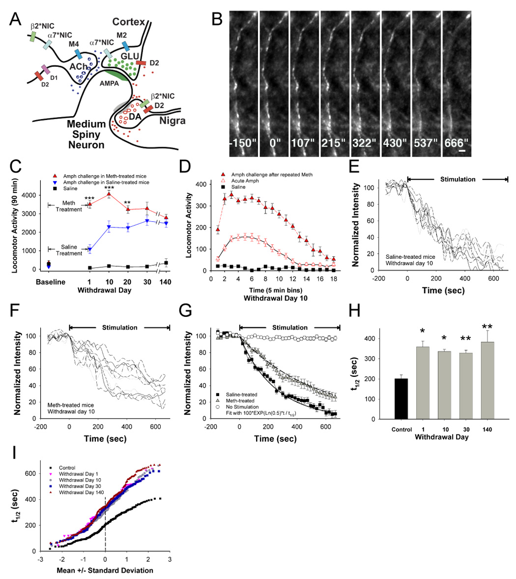
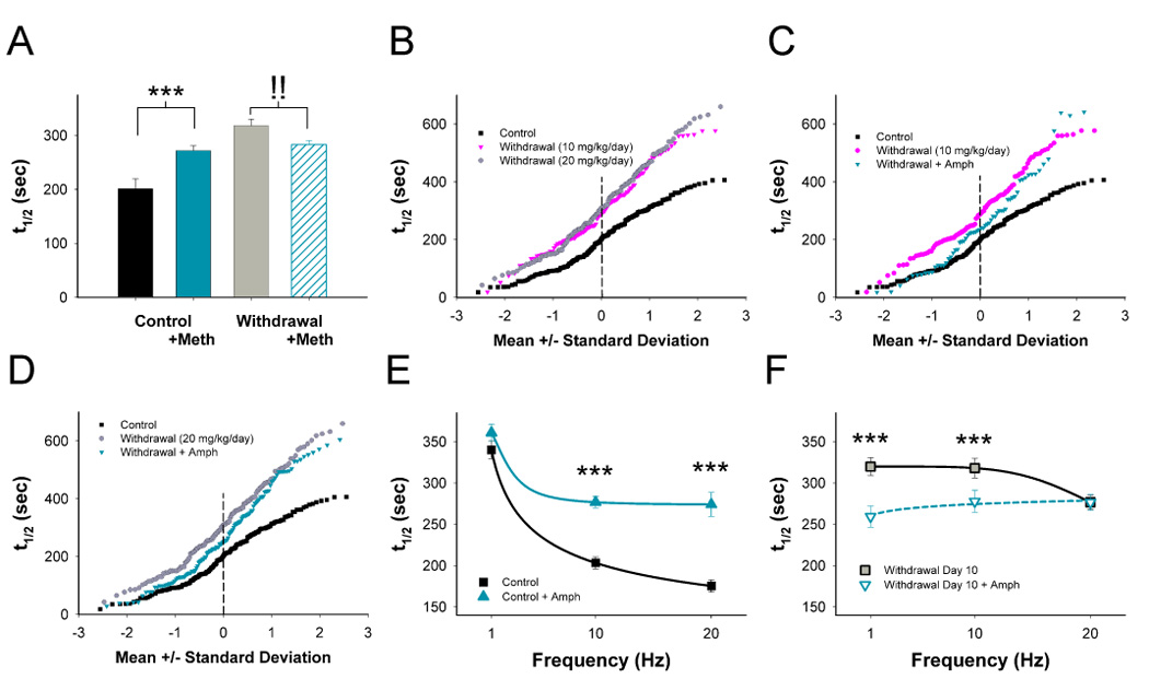
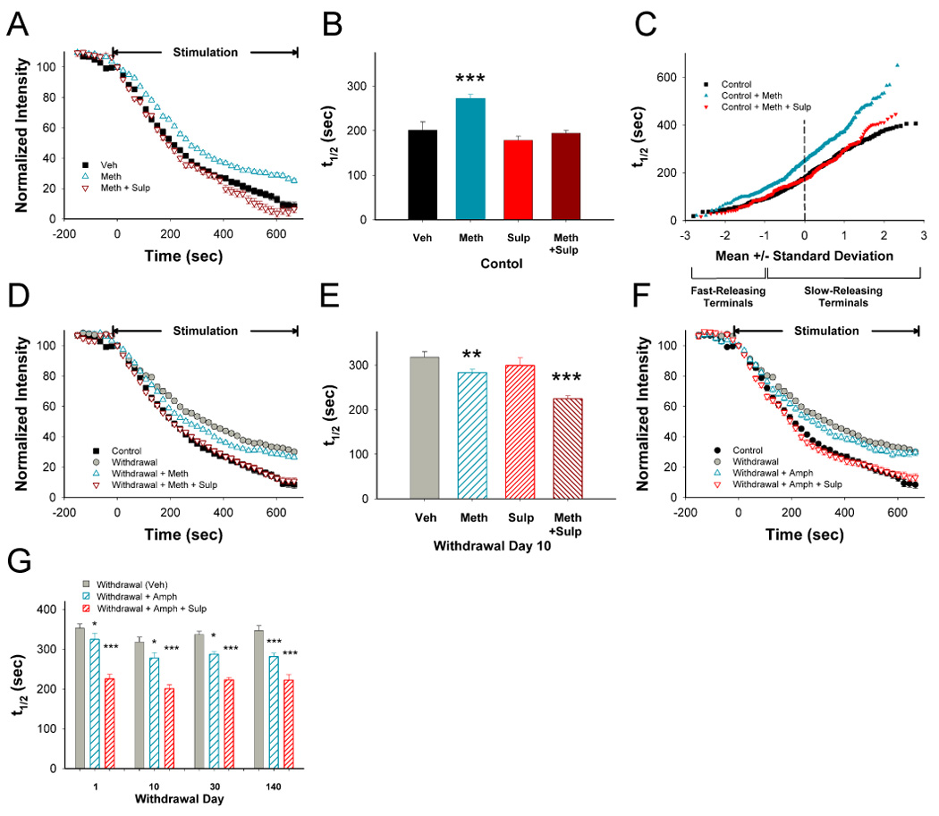
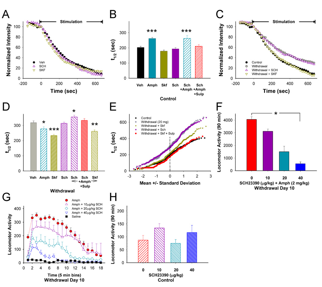
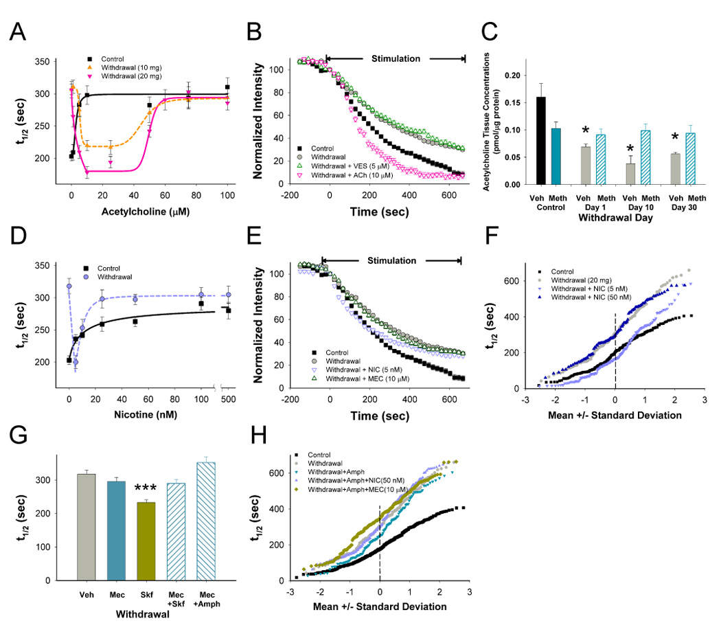
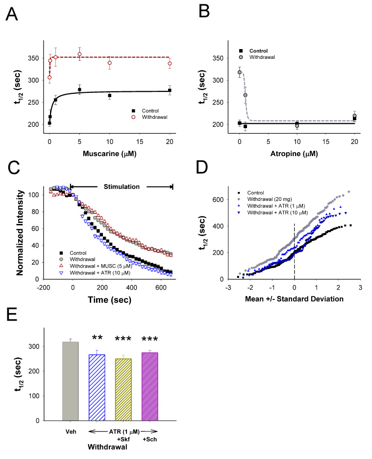
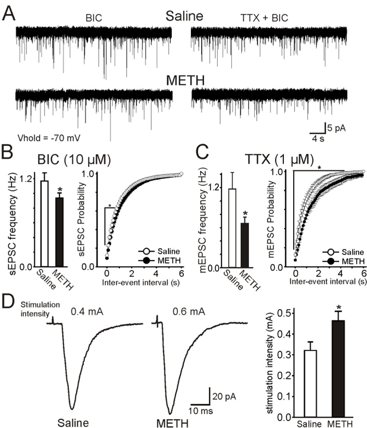
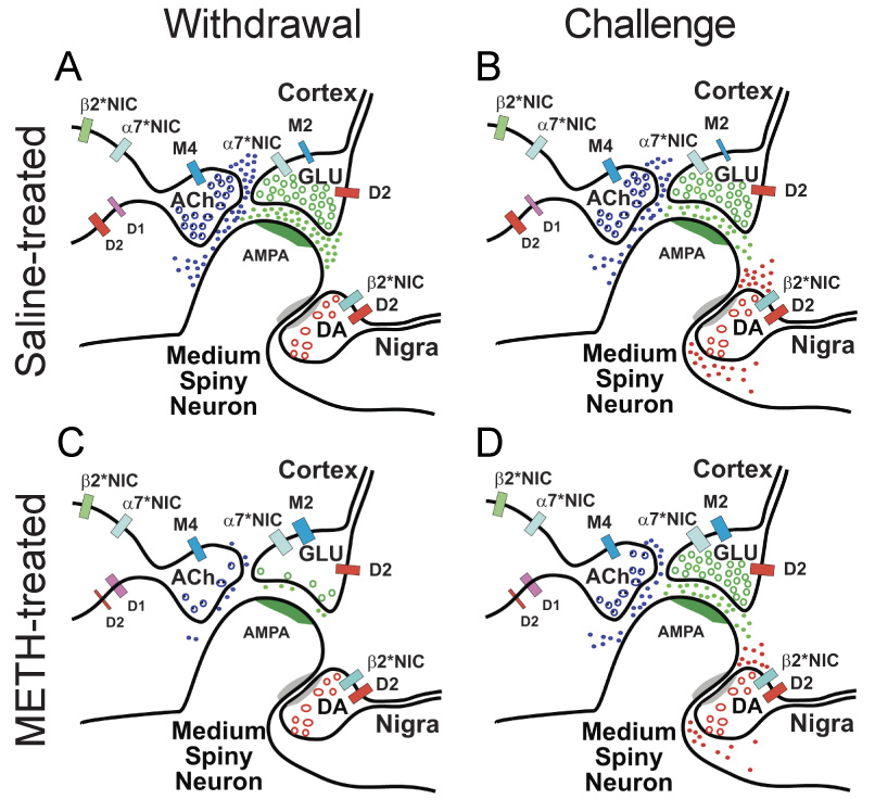
Addiction-associated behaviors such as drug craving and relapse are hypothesized to result from synaptic changes that persist long after withdrawal and are renormalized by drug reinstatement, although such chronic synaptic effects have not been identified. We report that exposure to the dopamine releaser methamphetamine for 10 days elicits a long-lasting (>4 month) depression at corticostriatal terminals that is reversed by methamphetamine readministration. Both methamphetamine-induced chronic presynaptic depression and the drug's selective renormalization in drug-experienced animals are independent of corresponding long-term changes in synaptic dopamine release but are due to alterations in D1 dopamine and cholinergic receptor systems. These mechanisms might provide a synaptic basis that underlies addiction and habit learning and their long-term maintenance.


Figures

Figure 1
Figure 1
Chronic presynaptic depression (CPD). (A) In this simplified striatal microcircuit, dopaminergic (DA) nigrostriatal fibers and cholinergic (ACh) interneurons modulate excitatory glutamatergic (GLU) corticostriatal projections on medium spiny neurons. Neurotransmitter release is modified by D1 and D2 DA receptors, M2 and M4 muscarinic receptors and α7*- and β2*-nicotinic receptors. (B) Multiphoton images of corticostriatal terminals obtained from the forelimb motor striatum, located 1.0 – 1.5 mm from the site of cortical stimulation. Images captured every 21.5 seconds reveal en passant arrays of corticostriatal terminals. Restimulation at t=0 with 10 Hz pulses shows activity-dependent destaining of fluorescent puncta. Bar: 2 µm. (C) Amphetamine (AMPH; 2 mg/kg i.p.) -elicited locomotor activity measured by ambulation summed over 90 min was determined in mice following treatment with repeated saline or methamphetamine (METH) for 10 days. Repeated METH produced a 1370%–1970% increase in AMPH-elicited ambulation through 140 days of withdrawal (p<0.001, t-test with Bonferroni correction), significantly higher than saline-treated mice challenged with saline (F(5,70)=19; n=8 mice per condition; p<0.001). Repeated METH also produced a 12%–219% increase in ambulations compared to saline-treated mice also receiving AMPH challenges (F(5,70)=8.5; p<0.001, repeated measures ANOVA), although the difference between the two treatments narrowed after withdrawal day 20 (**p<0.01, ***p<0.001, ANOVA). All values are mean±SE. (D) AMPH-elicited locomotor activity 10 days following repeated METH was higher and of longer duration, when compared with responses from saline-treated mice challenged with AMPH (F(17,238)=9.1; n=8 mice per condition; p<0.001, repeated measures ANOVA). (E) Time-intensity analysis of FM1–43 destaining from individual puncta (n=8) in slices from saline-treated mice. Stimulation begins at t=0 sec. (F) FM1-43 destaining is depressed 10 days following repeated METH. (G) Mean±SE florescence intensity of puncta shown in panel E and F demonstrates preservation of 1st order release kinetics following repeated saline or METH. The plateau line represents fluorescence measurements in the absence of stimulation. (H) Repeated METH inhibits corticostriatal release halftimes (t1/2) over 140 days of withdrawal. n=4 mice per condition; *p<0.05, **p<0.01, t-test with Bonferroni correction. (I) Individual terminal responses from panel H are represented in a normal probability plot. All terminals were depressed during withdrawal.
Figure 2
Figure 2
Paradoxical presynaptic potentiation (PPP). (A) A METH challenge in vivo decreases corticostriatal release in saline-treated controls (higher destaining halftime) but increases release on withdrawal day 10 following repeated METH. n=185–325 puncta per condition; ***p<0.01 compared to control without METH, !! p<0.01 compared to withdrawal without METH, Mann-Whitney. (B) Repeated METH at 10 and 20 mg/kg/day inhibits individual terminal responses on withdrawal day 10. An AMPH challenge 10 days following repeated METH at 10- (C) and 20 mg/kg/day (D) potentiated release from all terminals. Release half-times (t1/2) in slices from control (E) and METH–treated mice (F) on withdrawal day 10 following cortical stimulation at 1 Hz, 10 Hz and 20 Hz in the presence and absence of AMPH in vitro. ***p<0.001, Mann-Whitney.
Figure 3
Figure 3
D2 receptors (D2R) remain inhibitory following repeated METH. (A) In slices prepared from mice treated with repeated saline, a METH challenge in vivo produced inhibition of FM1-43 destaining that was reversed by the D2R antagonist sulpiride (SULP) in vitro. (B) Distribution of mean t1/2 of release for FM1-43 destaining curves shown in panel A. n=188–325 puncta; ***p<0.001 compared to control (Veh), Mann-Whitney. (C) Individual terminal responses in saline-treated controls following a challenge with METH in vivo with and without SULP. Repeated methamphetamine produced more inhibition at the slowest-releasing terminals (greater t1/2). (D) On withdrawal day 10 following repeated METH, a METH challenge in vivo accelerated corticostriatal release. The addition of SULP in vitro further accelerated release to control halftimes. (E) Distribution of mean t1/2 for destaining curves shown in panel D. n=149–362 puncta; **p<0.01, ***p<0.001 compared to vehicle (Veh), Mann-Whitney. (F) On withdrawal day 10 following repeated METH, AMPH in vitro induced PPP while AMPH in combination with SULP normalized release. (G) Following repeated METH, AMPH in vitro induced PPP over 140 days of withdrawal while AMPH in combination with SULP normalized release. n=167–368 puncta for each condition; *p<0.05, **p<0.01 compared to Veh from the same withdrawal day, Mann-Whitney.
Figure 4
Figure 4
D1 receptor (D1R) stimulation reverses CPD. (A) Compared to untreated sections (Veh), the D1R agonist SKF38393 (SKF; n=169 puncta) and antagonist SCH23390 (SCH; n=386 puncta) in vitro had no effect on release in controls following repeated saline. (B) Distribution of mean t1/2 of release for destaining curves shown in panel A with additional experimental groups from controls. Compared to untreated sections (Veh; n=188 puncta), AMPH (n=305 puncta) inhibited release, but the D1R agonist SKF (n=169 puncta) and antagonist SCH (n=386 puncta) had no effect. In the presence of AMPH, SCH had no effect with (n=116 puncta) or without sulpiride (SULP; n=151 puncta). ***p<0.001 compared to Veh, Mann-Whitney. (C) 10 days following repeated METH (withdrawal), SKF accelerated release whereas SCH had no effect. (D) Distribution of mean t1/2 of release for destaining curves shown in panel C with additional experimental groups from withdrawal. AMPH in vitro (n=128 puncta) boosted release to elicit PPP. SKF (n=247 puncta) increased release to a greater extent than AMPH whereas SCH (n=266 puncta) had no effect. SCH (n=212 puncta) blocked the potentiating effect of AMPH. SCH in combination with SULP (n=161 puncta) also blocked accelerated release by AMPH whereas SKF (n=168 puncta) had little effect. *p<0.05, **p<0.01; ***p<0.001 compared to Veh (n=149 puncta), Mann-Whitney. (E) Individual terminal responses to D1 and D2R manipulation in withdrawal. (F) Mice were treated with METH (20 mg/kg/day i.p.) for 10 days. An AMPH challenge (2 mg/kg i.p.) on withdrawal day 10 induced sensitized locomotor ambulations summed over 90 min. The D1R antagonist SCH inhibited this locomotor response (*p<0.001; n=8 mice per treatment group) with a significant linear trend over dose levels (r2=0.97). (G) Interval locomotor responses for treatment groups in panel F. (H) Additional mice were treated with saline for 10 days. 10 days later, these mice were treated with the D1R antagonist SCH and challenged with saline. There were small variations in locomotor activity but at the doses used, SCH had no effect on locomotor activity (p=0.48; n=8 mice per treatment group; r2=0.01).
Figure 5
Figure 5
CPD and PPP are regulated through nAChRs. (A) Terminal release over a range of acetylcholine (ACh) concentrations 10 days following repeated saline- (control) and METH (withdrawal; 10 and 20 mg/kg/day, 10 days; n=30–381 puncta). (B) 10 days following repeated METH (20 mg/kg/day), vesamicol (VES) had little effect on CPD while ACh potentiated release to a greater extent than controls. (C) Striatal tissue concentrations of ACh, measured by HPLC, remained depressed during METH withdrawal. *p <0.01 compared to untreated control mice (Veh; n=8 slices from 4 mice; t-test). (D) In control slices, increasing concentrations of nicotine (NIC) inhibited release (t1/2=240 sec at IC50=3.52 nM; n=104–299 puncta). 10 days following repeated METH, release was accelerated at low concentrations of NIC (5 nM) but higher concentrations of NIC rapidly decreased release (IC50=12.5 nM; n=77–190 puncta). (E) On withdrawal day 10, low NIC concentrations accelerated release whereas the nAChR channel blocker mecamylamine (MEC) had little effect on CPD. (F) Individual terminal responses during withdrawal for low (5 nM) and high (50 nM) concentrations of NIC. (G) During withdrawal, MEC prevented potentiation of release by SKF and AMPH (n=149–247 puncta; ***p<0.001, Mann-Whitney). (H) Individual terminal responses during withdrawal demonstrate inhibition of AMPH-induced PPP by both NIC and MEC (n=60–188 puncta). Concentration dependence curves were fit with a Hill equation.
Figure 6
Figure 6
CPD develops through sensitized mAChRs. (A) Terminal release over a range of muscarinic (MUSC) concentrations from slices prepared from saline- (control) and METH-treated mice (withdrawal) on withdrawal day 10. MUSC inhibited release to a greater extent and at a lower dose in withdrawal (t1/2=342 sec at IC50=0.01 µM; n=57–176 puncta) than controls (t1/2=276 sec at IC50=0.38 µM; n=86–265 puncta). (B) Atropine (ATR) accelerated release (t1/2=263 sec at EC50=1.02 µM; n=55–254 puncta) in withdrawal but had no effect in controls (n=77–254 puncta). (C) MUSC inhibited release whereas ATR potentiated release in withdrawal. (D) Individual terminal responses from withdrawal mice with and without ATR (1 and 10 µM; n=55–381 puncta) are compared to controls. (E) In the presence of ATR (1 µM; n=155 puncta), SKF (n=94 puncta) and SCH (n=142 puncta) had little effect on corticostriatal release during METH withdrawal. **p<0.01, ***p<0.001 compared to Veh, Mann-Whitney.
Figure 7
Figure 7
Response to CPD in medium spiny neurons (MSNs). (A) Traces represent spontaneous (s) EPSCs in the presence of bicuculline (BIC, 10 µM, a GABAA receptor blocker) alone (left) or BIC and tetrodotoxin (TTX; right) in MSNs from saline- and METH-treated animals at a holding potential of −70 mV. (B) In the presence of BIC only, there was a small but significant reduction of sEPSCs in cells from METH- compared to saline-treated mice. Histogram on the right is a cumulative inter-event interval distribution of sEPSCs. Intervals were significantly different (p<0.05). (C) In a subset of cells TTX was added to isolate mEPSCs. After TTX, there was a significant decrease in mEPSC frequency in cells from METH- compared to saline-treated mice. Histogram on the right is a cumulative inter-event interval distribution of mEPSCs. (D) Responses evoked in MSNs by stimulation of the cortical layers in saline- and METH-treated animals. More stimulation intensity was needed to induce responses of similar amplitude in cells from METH- than in cells from salinetreated mice. Traces represent the average of 3 responses. The graph on the right indicates that the threshold current required to induce responses was significantly higher in cells from METH- compared to saline-treated mice. Student’s t-tests or ANOVAs were used for group comparisons. Asterisks indicate differences were statistically significant (p<0.05).
Figure 8
Figure 8
Proposed mechanism for METH-induced synaptic plasticity. (A) The simplified striatal circuit is composed of medium spiny neurons that receive excitatory glutamatergic (GLU) corticostriatal projections, modulatory dopaminergic (DA) nigrostriatal fibers, and tonically active acetylcholine (ACh) – releasing interneurons (TANs). ACh modulates GLU release (Malenka and Kocsis, 1988) through excitatory α7*-nicotinic (NIC) (Marchi et al., 2002; Pakkanen et al., 2005) and inhibitory M2 mAChRs (Calabresi et al., 2000) located on corticostriatal terminals (Hersch et al., 1994) and regulates its own release through M4 muscarinic (Zhang et al., 2002) and both α7*-NIC and β2*-NIC autoreceptors (Azam et al., 2003). (B) Under control conditions, DA released by a psychostimulant inhibits GLU release from a subset of cortical terminals via D2R (Bamford et al., 2004b). Although TANs possess both inhibitory D2R (Yan et al., 1997) and excitatory D1R (Le Moine et al., 1991; Yan et al., 1997), D2R responses predominate so that DA reduces ACh efflux from striatal cholinergic interneurons (DeBoer and Abercrombie, 1996). (C) Following repeated METH, a reduction in ACh availability sensitizes muscarinic and nicotinic receptors. Enhanced muscarinic inhibition and reduced nicotinic excitation promotes CPD. (D) During withdrawal, DA released by a psychostimulant challenge induces PPP. DA increases ACh efflux (Bickerdike and Abercrombie, 1997) through TAN D1R responses (Berlanga et al., 2003) to excite GLU release through α7*-nAChRs.

Comment in

References

    1. Abercrombie ED, DeBoer P. Substantia nigra D1 receptors and stimulation of striatal cholinergic interneurons by dopamine: a proposed circuit mechanism. J Neurosci. 1997;17:8498–8505. - PMC - PubMed
    1. Ahmed SH, Koob GF. Transition to drug addiction: a negative reinforcement model based on an allostatic decrease in reward function. Psychopharmacology (Berl) 2005;180:473–490. - PubMed
    1. Azam L, Winzer-Serhan U, Leslie FM. Co-expression of alpha7 and beta2 nicotinic acetylcholine receptor subunit mRNAs within rat brain cholinergic neurons. Neuroscience. 2003;119:965–977. - PubMed
    1. Bamford NS, Robinson S, Palmiter RD, Joyce JA, Moore C, Meshul CK. Dopamine modulates release from corticostriatal terminals. J Neurosci. 2004a;24:9541–9552. - PMC - PubMed
    1. Bamford NS, Zhang H, Schmitz Y, Wu NP, Cepeda C, Levine MS, Schmauss C, Zakharenko SS, Zablow L, Sulzer D. Heterosynaptic dopamine neurotransmission selects sets of corticostriatal terminals. Neuron. 2004b;42:653–663. - PubMed

Publication types

MeSH terms

Substances