. 2008 Jun;129(6):304-12.
doi: 10.1016/j.mad.2008.02.010. Epub 2008 Mar 4.

Aging-induced alterations in gene transcripts and functional activity of mitochondrial oxidative phosphorylation complexes in the heart

Affiliations

Aging-induced alterations in gene transcripts and functional activity of mitochondrial oxidative phosphorylation complexes in the heart

Claudia C Preston et al. Mech Ageing Dev. 2008 Jun.

Abstract

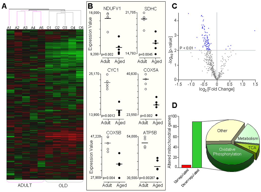
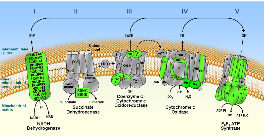
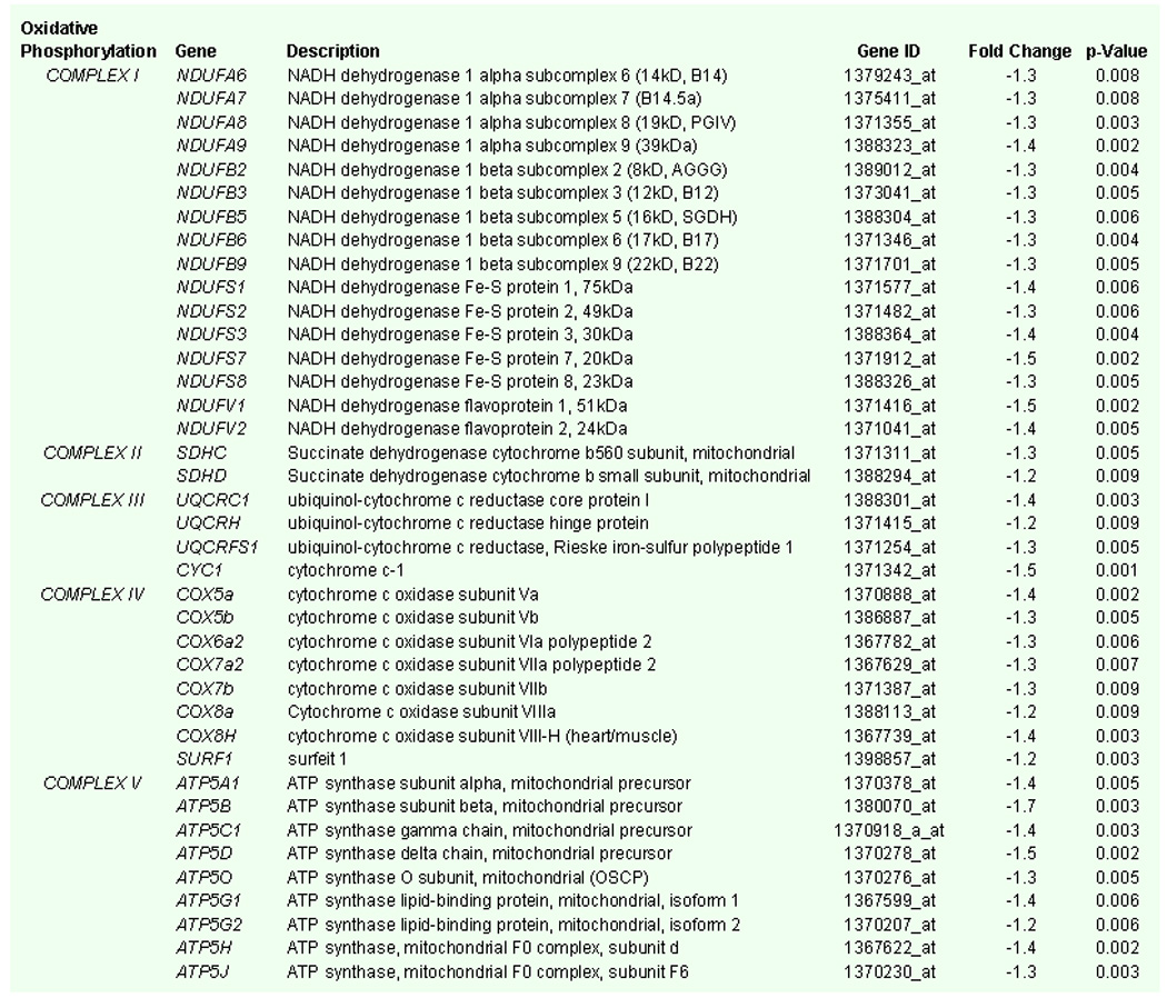
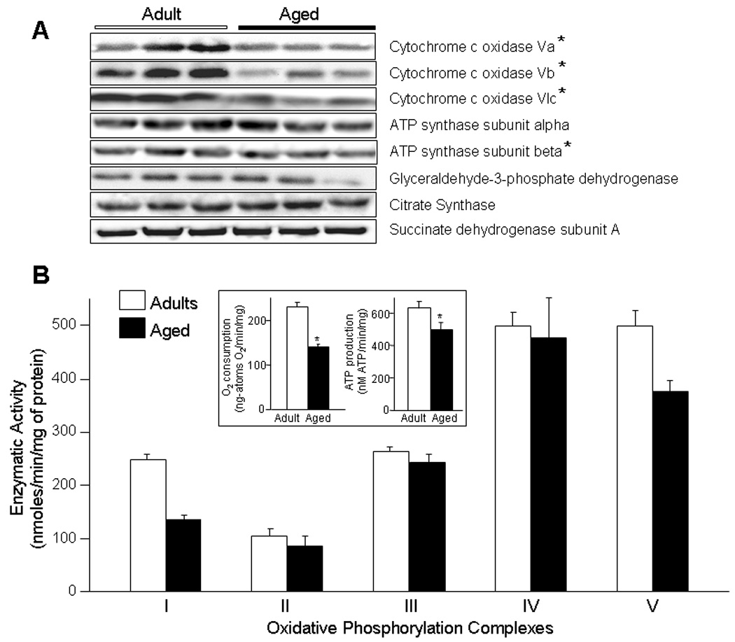
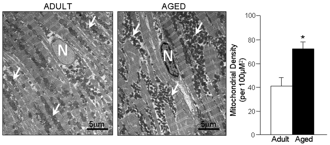
Aging is associated with progressive decline in energetic reserves compromising cardiac performance and tolerance to injury. Although deviations in mitochondrial functions have been documented in senescent heart, the molecular bases for the decline in energy metabolism are only partially understood. Here, high-throughput transcription profiles of genes coding for mitochondrial proteins in ventricles from adult (6-months) and aged (24-months) rats were compared using microarrays. Out of 614 genes encoding for mitochondrial proteins, 94 were differentially expressed with 95% downregulated in the aged. The majority of changes affected genes coding for proteins involved in oxidative phosphorylation (39), substrate metabolism (14) and tricarboxylic acid cycle (6). Compared to adult, gene expression changes in aged hearts translated into a reduced mitochondrial functional capacity, with decreased NADH-dehydrogenase and F(0)F(1) ATPase complex activities and capacity for oxygen-utilization and ATP synthesis. Expression of genes coding for transcription co-activator factors involved in the regulation of mitochondrial metabolism and biogenesis were downregulated in aged ventricles without reduction in mitochondrial density. Thus, aging induces a selective decline in activities of oxidative phosphorylation complexes I and V within a broader transcriptional downregulation of mitochondrial genes, providing a substrate for reduced energetic efficiency associated with senescence.


Figures

Fig. 1
Fig. 1
Aging-associated alterations in mitochondrial gene transcripts. A) Hierarchical clustering and heat map of samples from adult (6 month old, n=5) and aged (24 month old, n=5) rat ventricles. Shown are the expression pattern of 614 genes coding for mitochondrial proteins where Green color indicates downregulation, Red color upregulation, and Black color no change in the level of expression of genes. Samples from adult (Pink line) and aged (Gray line) animals clustered together as illustrated by condition tree (A1–A5 adult, O1–O5 aged samples). B) Expression values for selected genes coding for subunits of the mitochondrial OxPhos complexes from adult (n=5, White dots) and aged (n=5, Black dots) ventricles are shown as dot-plots highlighting the clustering of gene expression within each age group with the statistical significance of age-related changes between the two groups shown with their p values. C) Volcano plot illustrating the statistical distribution of 614 genes in aged compared with adult ventricles. There are 94 genes that were significantly different (p < 0.01) in the aged compared to adults (Blue dots). D) Number of significantly downregulated (89 genes, Green bar) and upregulated genes (5 genes, Red bar) in the aged heart and distribution according to their molecular function; 65% changes in genes coding for mitochondrial energetic pathways (41% for oxidative phosphorylation, 18% for substrate metabolism pathways and 6% for TCA cycle) and remaining 35% for other mitochondrial functions.
Fig. 2
Fig. 2
Schematic representation of genes coding for subunits of mitochondrial oxidative phosphorylation complexes. Genes coding for subunits of the electron transport chain (Complexes I – IV) and F0F1 ATP synthase (Complex V) that are significantly downregulated in the aging ventricle are colored Green, while subunits not altered with aging are shown in Gray. None of the genes coding for subunits of the oxidative phosphorylation pathway were upregulated in the aged hearts.
Fig. 3
Fig. 3
List of genes coding for mitochondrial oxidative phosphorylation pathway that are significantly (p < 0.01) downregulated in the aged rat heart with gene name, description, gene identification code (ID), fold change and p-values.
Fig. 4
Fig. 4
Comparison of aging associated reduction in expression of selected genes coding for mitochondrial oxidative phosphorylation subunits determined by microarray and Real Time-Polymerase Chain Reaction (RT-PCR) techniques; CS=Citrate Synthase, COX5a=Cytochrome c oxidase subunit 5a, COX5b=Cytochrome c oxidase subunit 5b and ATP5B=ATP synthase beta.
Fig. 5
Fig. 5
A) Western blots of selected protein subunits of the mitochondrial electron transport chain and ATP synthase complexes from adult (6 month old) and aged (24 month old) rat ventricles. Cytochrome c oxidase subunits Va, Vb, VIc and ATP synthase subunit beta are decreased with aging by 20%, 29%, 40% and 20% respectively (p < 0.01). There was no significant difference between adult and aged in expression of ATP synthase subunit alpha, glyceraldehyde-3-phosphate dehydrogenase, citrate synthase and succinate dehydrogenase subunit A. B) Enzymatic activities of Complexes I to V of the mitochondrial oxidative phosphorylation pathway in adult (n=6) and aged (n=6) ventricles. Compared to adults, functional activities of Complexes I and V are significantly decreased in the aged mitochondria (p < 0.01), while no significant differences were observed in activities of Complexes II, III and IV. All activities are expressed as nmoles/min/mg of mitochondrial protein; Inset, the rate of oxygen consumption and adenosine triphosphate (ATP) production in mitochondria from aged (n=6) ventricle is significantly decreased compared to adult (n=6). White bars = adult, black bars = aged, * p < 0.01.
Fig. 6
Fig. 6
List of genes coding for transcription co-activator factors that are significantly (p < 0.01) altered with aging with gene name, description, gene identification code (ID), fold change and p-values.
Fig. 7
Fig. 7
Electron microscopy of ventricular tissue and numeric density of mitochondria in adult (6 month old, n=8) and aged (24 month old, n=8) rat ventricles. N=nucleus, White arrows point to mitochondrial clusters, White bars=adult, Black bars=aged, * p < 0.01.

References

    1. Abete P, Ferrara N, Cioppa A, Ferrara N, Bianco S, Calabrese C, Cacciatore F, Longobardi G, Rengo F. Preconditioning does not prevent post-ischemic dysfunction in aging heart. J Am Coll Cardiol. 1996;27:1777–1786. - PubMed
    1. Benit P, Beugnot R, Chretien D, Giurgea I, De Lonlay-Debeney P, Issartel JP, Corral-Debrinski M, Kerscher S, Rustin P, Rotig A, Munnich A. Mutant NDUFV2 subunit of mitochondrial complex I causes early onset hypertrophic cardiomyopathy and encephalopathy. Hum Mutat. 2003;21:582–586. - PubMed
    1. Benzi G, Pastoris O, Marzatico F, Villa RF, Dagani F, Curti D. The mitochondrial electron transfer alteration as a factor involved in the brain aging. Neurobiol Aging. 1992;13:361–368. - PubMed
    1. Brandt U. Energy Converting NADH:Quinone Oxidoreductase (Complex I) Annu Rev Biochem. 2006 - PubMed
    1. Capaldi RA, Halphen DG, Zhang YZ, Yanamura W. Complexity and tissue specificity of the mitochondrial respiratory chain. J Bioenerg Biomembr. 1988;20:291–311. - PubMed

Publication types

Substances