. 2008 Jul;36(7):832-44.
doi: 10.1016/j.exphem.2008.02.009. Epub 2008 Apr 8.

A cascade of Ca(2+)/calmodulin-dependent protein kinases regulates the differentiation and functional activation of murine neutrophils

Affiliations

A cascade of Ca(2+)/calmodulin-dependent protein kinases regulates the differentiation and functional activation of murine neutrophils

Peter Gaines et al. Exp Hematol. 2008 Jul.

Abstract

Objective: The function of neutrophils as primary mediators of innate immunity depends on the activity of granule proteins and critical components of the nicotinamide adenine dinucleotide phosphate (NADPH) oxidase complex. Expression of their cognate genes is regulated during neutrophil differentiation by a complex network of intracellular signaling pathways. In this study, we have investigated the role of two members of the calcium/calmodulin-dependent protein kinase (CaMK) signaling cascade, CaMK I-like kinase (CKLiK) and CaMKKalpha, in regulating neutrophil differentiation and functional activation.

Materials and methods: Mouse myeloid cell lines were used to examine the expression of a CaMK cascade in developing neutrophils and to examine the effects of constitutive activation vs inhibition of CaMKs on neutrophil maturation.

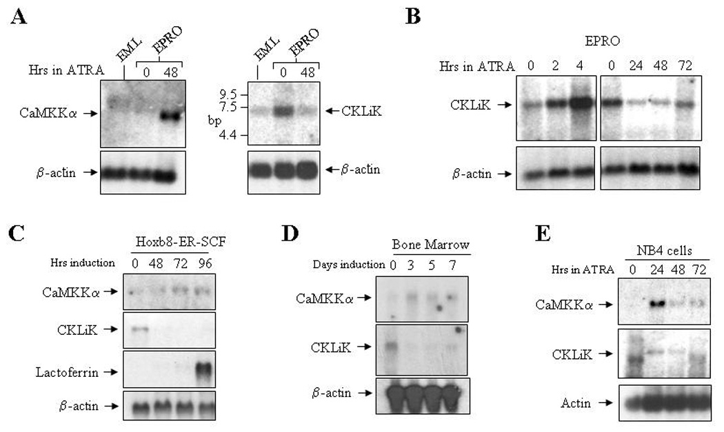
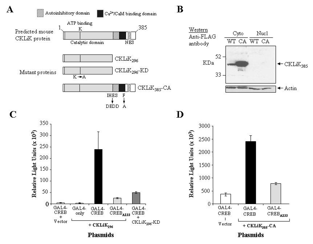
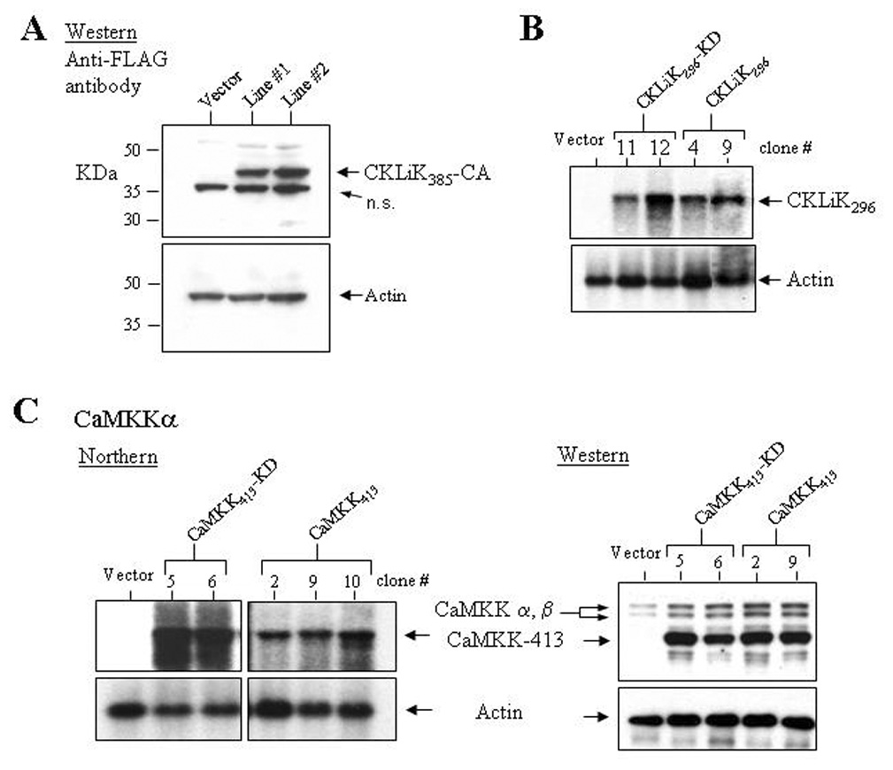
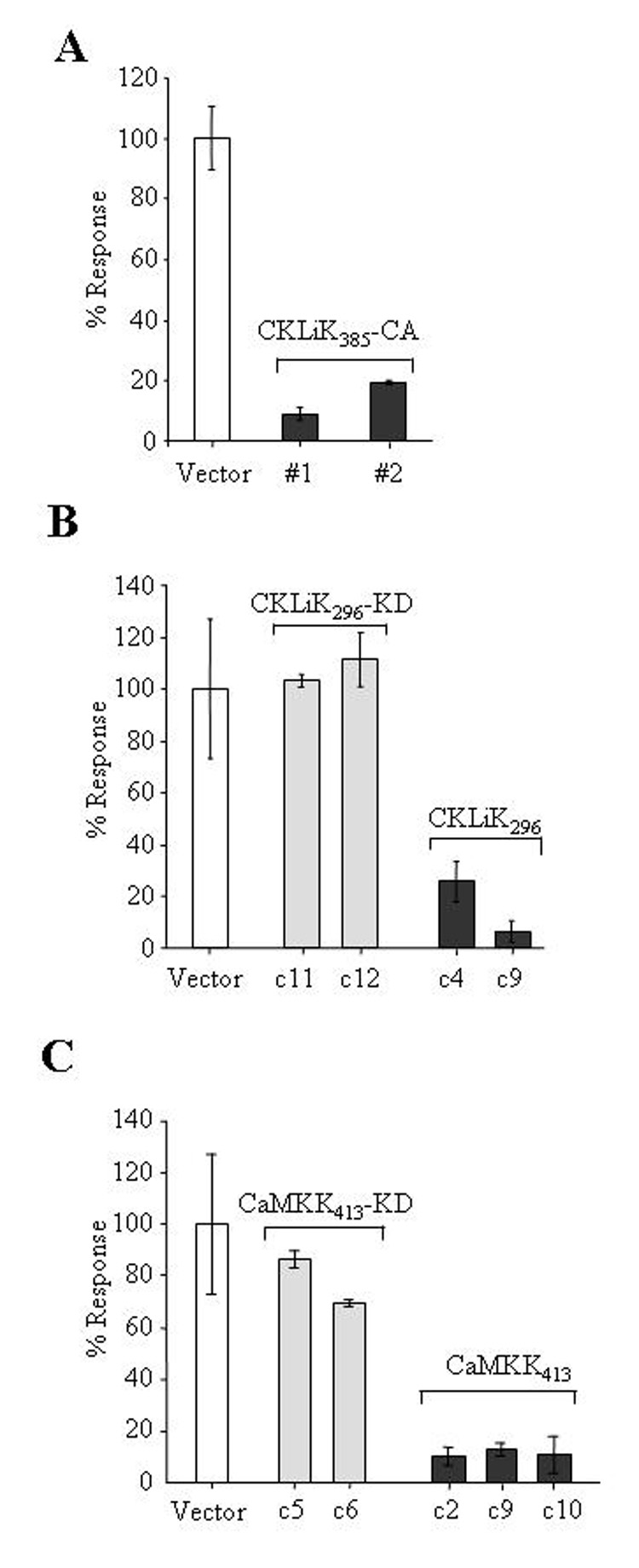
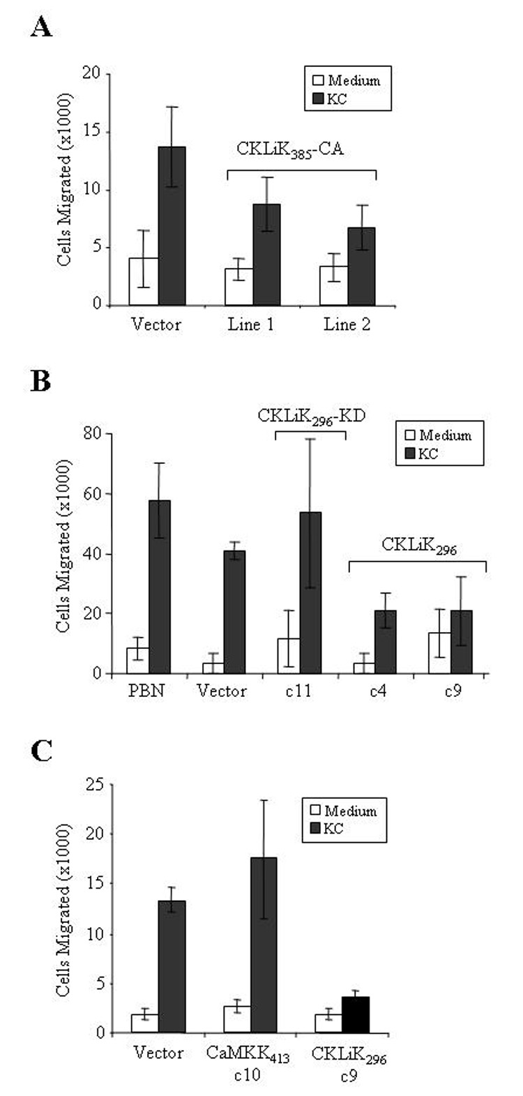
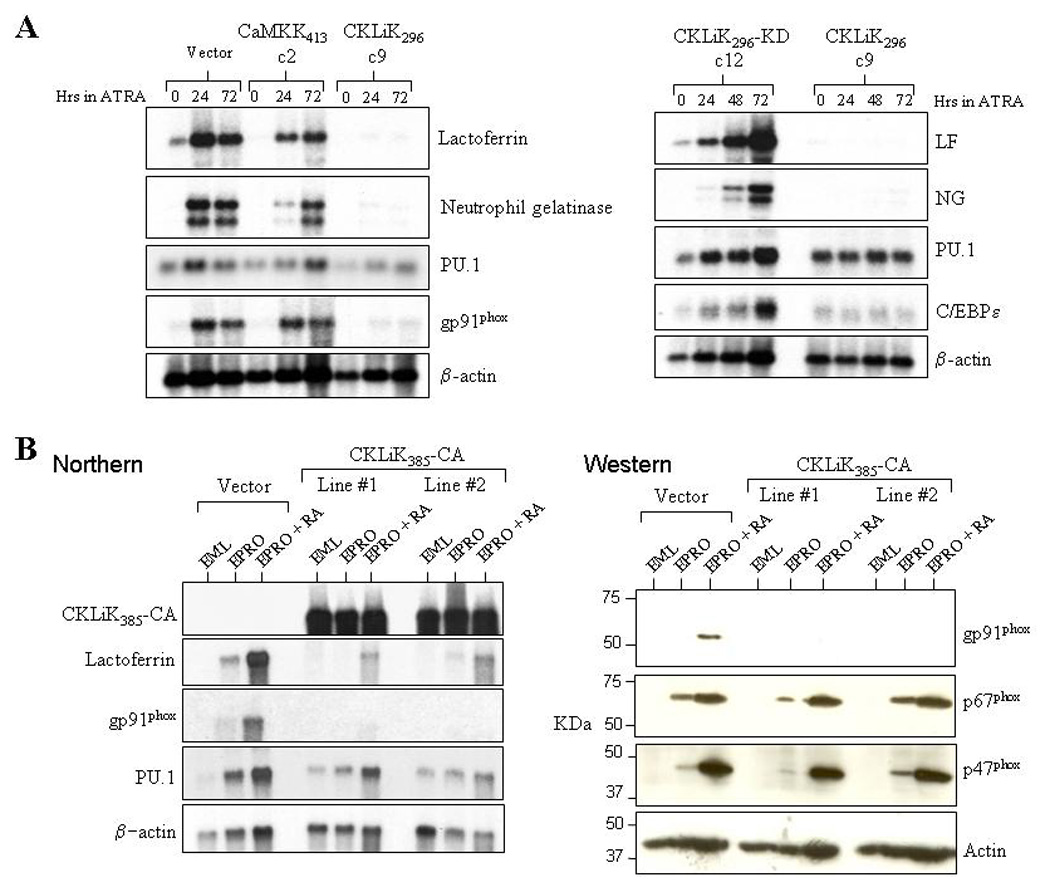
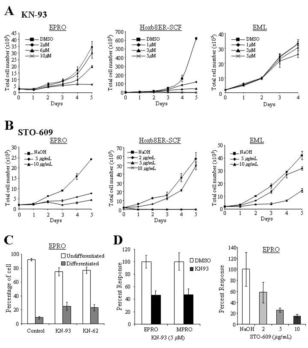
Results: Expression of CaMKKalpha was shown to increase during neutrophil differentiation in multiple cell lines, whereas expression of CKLiK increased as multipotent progenitors committed to promyelocytes, but then decreased as cells differentiated into mature neutrophils. Expression of constitutively active CKLiKs did not affect morphologic maturation, but caused dramatic decreases in both respiratory burst responses and chemotaxis. This loss of neutrophil function was accompanied by reduced secondary granule and gp91(phox) gene expression. The CaMK inhibitor KN-93 attenuated cytokine-stimulated proliferative responses in promyelocytic cell lines, and inhibited the respiratory burst. Similar data were observed with the CaMKKalpha inhibitor, STO-609.

Conclusions: Overactivation of a cascade of CaMKs inhibits neutrophil maturation, suggesting that these kinases play an antagonistic role during neutrophil differentiation, but at least one CaMK is required for myeloid cell expansion and functional activation.


Figures

Figure 1
Figure 1. Expression of CaM kinase transcripts in multiple models of neutrophil differentiation
(A) Northern blots were generated using total RNA (5 µg) from uninduced EML and EPRO cells, and EPRO cells induced with 10 µM ATRA for 48 hours. Blots were sequentially hybridized with 32P-labelled cDNAs for the kinase shown and mouse β-actin as a control. (B) Northern analyses were performed using total RNAs from uninduced EPRO cells and cells induced with ATRA for 2 and 4 hours (left panels), or for 24, 48 and 72 hours (right panels). (C) A northern blot was generated using total RNA from SCF ER-Hoxb8 cells grown in the presence of β-estrodiol (1 µM, time 0), and cells grown after β-estrodiol withdrawal to induce neutrophil differentiation (48–96 hours). The blot was sequentially hybridized with 32P-labelled cDNA probes for the two kinases, lactoferrin (to demonstrate neutrophil differentiation) and β-actin. (D) Expression of CaMKKα and CKLiK were assessed in bone marrow-derived stem cells induced with SCF, IL-3 and G-CSF for 3 and 5 days, and then cultured in G-CSF alone. (E) A northern analysis identifies the expression of human CaMKKα and CKLiK in ATRA-induced NB-4 cells.
Figure 2
Figure 2. Expression and constitutive activity of mutant CKLiK proteins
(A) Depicted are the wild-type form of CKLiK and the two constitutively active forms, CKLiK296 (truncated at Gln296) and CKLiK385-CA (internal mutations that mimic Ca2+/CaM binding). Also shown is the “kinase dead” form of the truncated CKLiK (CKLiK296-KD) that contains a mutation in the ATP binding site (Lys52). (B) Results of a Western blot containing cytoplasmic versus nuclear extracts from HEK293 cells transfected with wild-type (WT) or constitutively active (CA) versions of CKLiK demonstrate abundant expression in the cytoplasm, whereas nuclear expression was undetectable. The blot was probed with an anti-FLAG antibody to detect CKLiK expression and with an anti-actin antibody to demonstrate amounts of protein in each lane. (C and D) Activation of CREB by each mutant form of CKLiK was tested by transfecting COS-1 cells with the expression vectors for each mutant kinase together with the two reporter plasmids, pCMV-GAL4-CREBΔb-zip (GAL4-CREB) and p5XGAL4-E1b-luciferase. Control vectors also used were one lacking the CREB activation domain (GAL4-only) and one with a Ser133 to Ala mutation in the CREB motif (GAL4-CREBA133). Levels of luciferase activity were normalized to levels of β-galactosidase expressed from co-transfected plasmids. Values shown are the averages ± SD from triplicate transfections performed in parallel and are representative of at least 3 independent experiments.
Figure 3
Figure 3. Ectopic expression of mutant CKLiK and CaMKKα in EML cell lines
(A) A Western blot containing protein lysates from two independently generated EML cell lines stably transduced with CKLiK385-CA or an empty vector was probed with an anti-FLAG antibody. Below is shown the same blot probed with an anti-actin antibody. (B) Ectopic expression of CKLiK296 and CKLiK296-KD in clonal sublines of transduced EML cells was demonstrated by northern analyses. (C) Ectopic expression of constitutively active and kinase dead forms of CaMKKα (CaMKK413 and CaMKK413-KD, respectively) in EML cells was confirmed by northern assays and a Western blot probed with anti-CaMKK. Shown in the Western blot is expression of the endogenous CaMKKα and β isoforms, and the shorter, truncated forms. Levels of actin expression are shown below each blot.
Figure 4
Figure 4. Expression of constitutively active CaM kinases inhibits the respiratory burst in differentiated EPRO cells
(A) EPRO cells expressing CKLiK385-CA demonstrate reduced levels of the respiratory burst as compared to cells expressing the empty vector. Assays were performed on ATRA-induced cells stimulated with PMA (4 µg/mL) using an enhanced luminol reagent to detect O2 production. Shown are the percentages of peak light units emitted over 10 sec. during a 5 min. period of analysis as compared to control cells expressing the empty vector, set at a 100% response. (B) Expression of the truncated CKLiK (CKLiK296) but not the kinase dead form (CKLiK296-KD) inhibits the respiratory burst. Shown are the responses of two clonal sublines of each type of transduced EPRO cells as compared to cells expressing the empty vector. (C) Expression of the constitutively active CaMKKα (CaMKK413) inhibits the respiratory burst to levels similar to that observed by the constitutively active CKLiKs. Data shown for all assays are given as averages ± SD from triplicate samples analyzed in parallel and are representative of at least three independent experiments.
Figure 5
Figure 5. Chemotaxis is inhibited by overexpression of constitutively active CKLiK
(A) Expression of the full-length, constitutively active CKLiK inhibits chemotaxis. Chemotaxis assays were performed using ATRA induced cells and transwell plates with 3 µm membranes. Graphed are the average total number of cells ± SD that migrated after a 2 hour incubation into the bottom chamber containing medium only or KC. P values generated by the Student’s two sample t-test (Excel, Microsoft Corporation, Redmond, WA) for the differences between chemotaxis by cells expressing the vector versus CKLiK385-CA are as follows: vector vs. line #1, p = 0.004; vector vs. line #2, p = 0.0006. Data shown are from triplicate experiments performed in parallel. (B) EPRO cells that express CKLiK296 exhibit reduced chemotaxis as compared to cells expressing the empty vector or CKLiK296-KD, or to peripheral blood neutrophils (PBN). Statistical analyses yielded the following results: vector vs. CKLiK296-KD c11, p = 0.15 (not statistically different); vector vs. CKLiK296 c4, p = 0.005; vector vs. CKLiK296 c9, p = 0.009. (C) Ectopic expression of CaMKK413 does not disrupt chemotaxis, as demonstrated by levels of cell migration similar to that observed by cell expressing the empty vector. P values: vector vs. CaMKK413 c10, p = 0.23 (not statistically different), vector vs. CKLiK296 c9, p = 0.00008.
Figure 6
Figure 6. Neutrophil-specific gene expression is inhibited by constitutively active CKLiK
(A) Shown are the results of two northern assays that were performed using total RNAs from uninduced and ATRA-induced EPRO cells expressing the empty vector, constitutively active versions of CaMKKα (CaMKK413, clone 2) or CKLiK (CKLiK296, clone 9), or the kinase dead version of CKLiK296 (CKLiK296-KD, clone 12). The blots were sequentially probed with 32P-labelled cDNAs for the secondary granule genes lactoferrin (LF) and neutrophil gelatinase (NG), the transcriptional regulator PU.1, and either the NADPH oxidase component gp91phox (left panel) or the neutrophil-specific transcriptional regulator C/EBPε (right panel). A β-actin probe was also used to demonstrate amounts of RNA in each lane. (B) A northern assay (left panel) demonstrates that two independently generated lines of EPRO cells that express CKLiK385-CA exhibit reduced lactoferrin and gp91phox transcript expression as compared to cells expressing the empty vector. A Western assay was also performed (right panel), which demonstrates that expression of gp91phox proteins are undetectable in EPRO-CKLiK385-CA cells whereas expression of either p47phox or p67phox is normally upregulated. Levels of actin expression are shown in the bottom panels of each figure.
Figure 7
Figure 7. Effects of CaM kinase inhibitors on neutrophil growth and functional activation
(A) KN93 inhibited the proliferation of both EPRO and SCF ER-Hoxb8 cell lines, whereas the inhibitor did not affect the growth of EML cells. For each assay, cells were diluted to 2 × 10⁵ cells/mL and total numbers of cells that excluded trypan blue after the indicated times were counted using a hemacytometer. Note levels of KN93 used with EPRO cells were two-fold higher than those used in SCF ER-Hoxb8 and EML cells. (B) The CaMKK inhibitor STO-609 significantly reduced the growth profiles of both EPRO and SCF ER-Hoxb8 cells, and inhibited the growth of EML cells. (C) Morphologic maturation of EPRO cells exposed to the CaMK inhibitors KN-62 or KN-93 was assessed by examining cells after 5 days of treatment and counting the number undifferentiated cells with promyelocyte characteristics (e.g. high nuclear to cytoplasmic ratios) versus differentiated cells with kidney shaped or lobulated nuclei. Shown are the results of three independent experiments. P values: KN-62 vs. control, p = 0.001; KN-93 vs. control, p = 0.001. (D) The respiratory burst exhibited by ATRA-induced EPRO or MPRO cells upon PMA stimulation was inhibited by the CaM kinase inhibitor KN-93 (left panel); STO-609 also inhibited the oxidative burst produced by EPRO cells in a dose-dependent fashion. Shown are data from differentiated cells that were incubated with the inhibitors for 24 hours prior to each assay, with data sets given as percentages of peak light units emitted as compared to cells incubated with the diluting reagent. Data shown are the average responses ± SD from three independent experiments.

Similar articles

Cited by

References

    1. Liu H, Pope RM. Phagocytes: Mechanisms of inflammation and tissue destruction. Rheum. Dis. Clin. North Am. 2004;30:19–39. - PubMed
    1. Frangogiannis NG, Smith CW, Entman ML. The inflammatory response in myocardial infarction. Cardiovasc. Res. 2002;53:31–47. - PubMed
    1. Soderling TR. The ca-calmodulin-dependent protein kinase cascade. Trends Biochem. Sci. 1999;24:232–236. - PubMed
    1. Corcoran EE, Means AR. Defining Ca2+/calmodulin-dependent protein kinase cascades in transcriptional regulation. J. Biol. Chem. 2001;276:2975–2978. - PubMed
    1. Goldberg J, Nairn AC, Kuriyan J. Structural basis for the autoinhibition of calcium/calmodulin-dependent protein kinase I. Cell. 1996;84:875–887. - PubMed

Publication types

MeSH terms